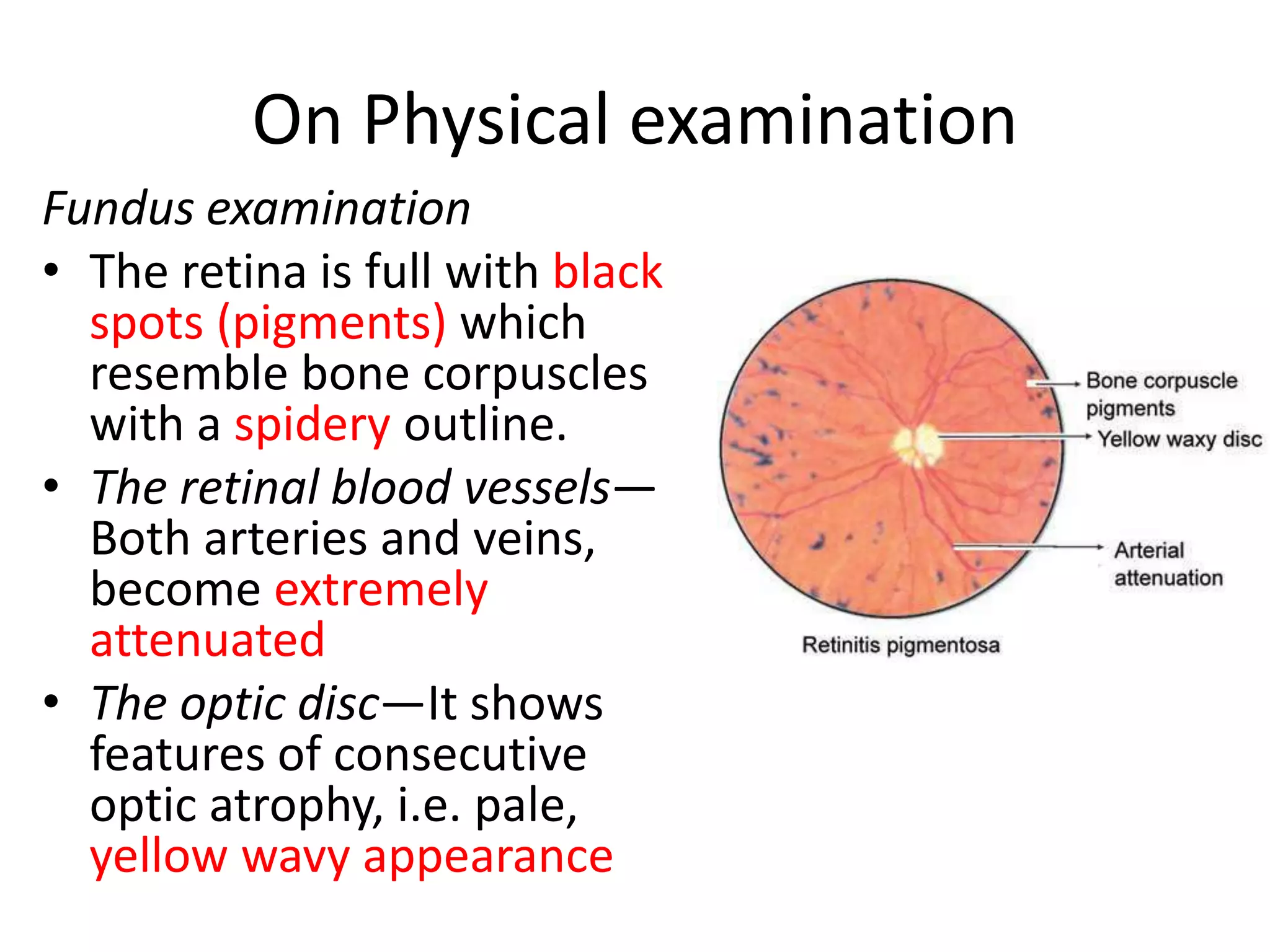
RETINITIS
PIGMENTOSA
by Muhd Ariff B Mahdzub
• It is slow degenerative, hereditary disease of
the retina, involving the rods and cones.
• It appears as a recessive trait and usually
occurs due to consanguinity of the parents.
On History taking
• Age: it begins in childhood and causes
blindness in middle and advanced age.
• Side: It is a bilateral affection usually.
• The course is slow, chronic and progressive.
Cont.
• Patients complain of night blindness (early
sign),
• tunnel vision (peripheral visual loss) or
• central visual loss and photopsia (seeing
flashes of light). *advance case
On Physical examination
Fundus examination
• The retina is full with black
spots (pigments) which
resemble bone corpuscles
with a spidery outline.
• The retinal blood vessels—
Both arteries and veins,
become extremely
attenuated
• The optic disc—It shows
features of consecutive
optic atrophy, i.e. pale,
yellow wavy appearance
gradual increase in density of the pigment with
anterior and posterior spread, (advance case)
-Kanski page:647
• Peripheral pigmentation may become severe,
with marked arteriolar narrowing and disc
pallor
• -Kanski page:647
cont
• Dark adaptation is increased due to rods
dysfunction.
• The electroretinogram (ERG) and electro-
oculogram (EOG) are markedly subnormal or
completely extinguished early in the disease.
Complications
• Posterior subcapsular cataract (common in all
forms of RP),
• open-angle glaucoma (3%),
• keratoconus (uncommon)
• posterior vitreous detachment
Kanski page:646
Treatment
• No specific treatment is yet commercially
available
• Cataract surgery is generally benefical
• Regular follow-up to detect treatable
complications
• rehabilitation and social service support
• Smoking should be avoided.
Kanski page:646

Retinitis pigmentosa (opthalmology)

  • 1.
  • 2.
    • It isslow degenerative, hereditary disease of the retina, involving the rods and cones. • It appears as a recessive trait and usually occurs due to consanguinity of the parents.
  • 3.
    On History taking •Age: it begins in childhood and causes blindness in middle and advanced age. • Side: It is a bilateral affection usually. • The course is slow, chronic and progressive.
  • 4.
    Cont. • Patients complainof night blindness (early sign), • tunnel vision (peripheral visual loss) or • central visual loss and photopsia (seeing flashes of light). *advance case
  • 5.
    On Physical examination Fundusexamination • The retina is full with black spots (pigments) which resemble bone corpuscles with a spidery outline. • The retinal blood vessels— Both arteries and veins, become extremely attenuated • The optic disc—It shows features of consecutive optic atrophy, i.e. pale, yellow wavy appearance
  • 6.
    gradual increase indensity of the pigment with anterior and posterior spread, (advance case) -Kanski page:647
  • 7.
    • Peripheral pigmentationmay become severe, with marked arteriolar narrowing and disc pallor • -Kanski page:647
  • 8.
    cont • Dark adaptationis increased due to rods dysfunction. • The electroretinogram (ERG) and electro- oculogram (EOG) are markedly subnormal or completely extinguished early in the disease.
  • 9.
    Complications • Posterior subcapsularcataract (common in all forms of RP), • open-angle glaucoma (3%), • keratoconus (uncommon) • posterior vitreous detachment Kanski page:646
  • 10.
    Treatment • No specifictreatment is yet commercially available • Cataract surgery is generally benefical • Regular follow-up to detect treatable complications • rehabilitation and social service support • Smoking should be avoided. Kanski page:646