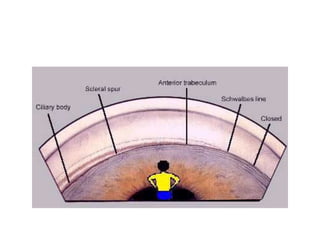
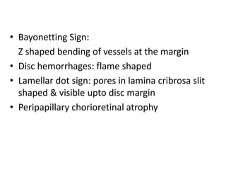
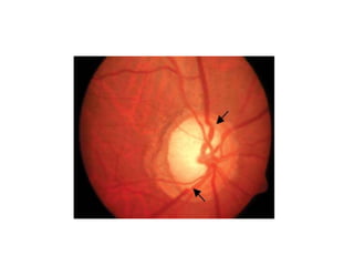
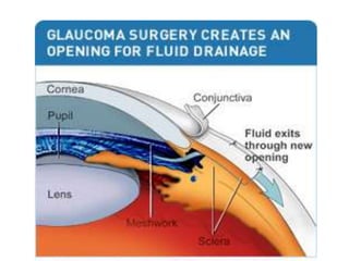
PRIMARY ANGLE CLOSURE
GLAUCOMA
SARAL LAMICHHANE
MBBS IV Year/Roll-61
Introduction
Glaucoma refers to a group of disorders
characterized by progressive optic neuropathy
resulting to characteristic visual field defects;
raised Intraocular Pressure(IOP) being the most
important risk factor.
 Normal Tension Glaucoma(NTG/LTG) also exist
 Normal IOP = 10 – 21 mmHg
(Mean: 162.5 mmHg)
Classification:
1) Congenital & Developmental Glaucoma
2) Primary Adult Glaucoma:
(No obvious systemic/ocular cause of rise in IOP)
a) Primary Open Angle Glaucoma (POAG)
b) Primary Angle Closure Glaucoma (PACG)
c) Primary Mixed Mechanism Glaucoma
3) Secondary Glaucoma:
(Rise of IOP with some primary ocular or systemic disease)
Primary Angle Closure Glaucoma
Primary glaucoma in which rise in Intraocular
Pressure (IOP) occurs due to blockage of aqueous
humor outflow by closure or narrower angle of
the anterior chamber.
Predisposing Risk Factors:
1) Anatomic Factors:
- Hyperopic eye
- Iris-lens diaphragm placed anteriorly
- Narrow angle of anterior chamber
- Plateau Iris Configuration
2) General Factors:
- Age: 5th Decade onwards
- Sex: Females > Males
- Rainy Season
- Family History
- Race (Caucasians, South East Asians)
- Heredity: Family history
Precipitating factors:
- Dim illumination
- Emotional Stress
- Use of mydriatics: Atropine, Tropicamide, Cyclopentolate
• Aqueous humor secretion from Non-pigmented
epithelium of Ciliary processes Posterior Chamber
Pupil Anterior Chamber:
• Conventional (Trabecular) Pathway: 90%
Trabecular Meshwork Canal of Schlemm
Collector Channels Episcleral Vessels
• Uveoscleral Pathway:
Trabecular Meshwork Ciliary body
Suprachoroidal space Venous Drainage
Mechanism:
 Relative Pupil Block
- Iris bombe formation
- Appositional Angle Closure
- Synechial angle closure
Plateau Iris Configuration
- Anteriorly positioned ciliary
displacing the iris anteriorly
Phacomorphic mechanism
- Abnormal lens
Symptoms:
• Eye redness
• Eyeache
• Headache
• Nausea/Vomiting
• Blurring of vision
• Colored halos
Signs:
• Circumcorneal congestion
• Corneal edema, hazy cornea
• Very shallow anterior chamber
(Eclipse Sign)
• Iridocorneal angle closure as
seen in gonioscopy
• Raised Intraocular pressure
• Reduced visual acuity
• Vertically oval & mid-dilated
pupil
Progression
 Primary Angle Closure Suspect:
• No clinical signs/symptoms
• Irido-trabecular contact in greater than 2700
• Intraocular Pressure (IOP) Normal
• No Peripheral Anterior Synechiae (PAS)
• Optic disc & Visual Field Normal
Angle Closure:
• Irido-trabecular contact in greater than 2700
• Mild clinical symptoms/signs
• IOP may or may not be raised
• PAS may or may not be present
• Optic disc & Visual Field Normal
Acute Angle Closure:
• Typical clinical symptoms/signs present
• Irido-trabecular contact greater than 2700
• IOP raised
• PAS present
• Optic disc & Visual field normal
Angle Closure Glaucoma:
• Typical clinical symptoms/signs present
• Irido-trabecular contact greater than 2700
• IOP raised, PAS formed
• Optic Nerve shows glaucomatous damage
• Characteristic visual field defect
Optic Nerve Changes
• Glaucomatous cupping:
Increase in Cup : Disc ratio  0.6
Normal CDR = 0.3-0.4
• Cup Asymmetry:
CDR difference > 0.2 in two eyes
• Neuroretinal rim thinning: Cup to Disc distance
ISNT Rule- Thickness of Inferior rim> Superior rim
> Nasal rim> Temporal rim
Thinning usually in same sequence
• Bayonetting Sign:
Z shaped bending of vessels at the margin
• Disc hemorrhages: flame shaped
• Lamellar dot sign: pores in lamina cribrosa slit
shaped & visible upto disc margin
• Peripapillary chorioretinal atrophy
Visual Field Defect
• Isopter contraction
• Baring of blind spot
• Paracentral Scotoma
• Siedel’s Scotoma
• Superior Arcuate or Bjerrum’s Scotoma
• Inferior Arcuate or Bjerrum’s Scotoma
• Ring or Double Arcuate Scotoma
• Roenne’s central nasal step
• Tubular Vision
Diagnosis
• History: Symptoms
• Clinical Examination: Signs
• Diagnostic tests:
–Tonometry: IOP measurement
–Gonioscopy: Angle structures
–Slit lamp biomicroscopy
–Ultrasonic biomicroscopy
–Fundoscopy
–Visual Field Analysis
Management
Principle:
Immediate medical therapy (Lower IOP)
Definitive treatment
Prophylaxis of fellow eye
Long term glaucoma surveillance and IOP
management of both eyes
Immediate medical therapy:
• Intravenous line access
• Systemic hyperosmotic agents:
Mannitol 20% iv 1g/kg within 30-45 mins
Glycerol oral 1g/kg of 50% solution in lemon juice
• Systemic Carbonic Anhydrase Inhibitors:
Acetazolamide 500mg oral stat followed by 250 mg tds
• Analgesics/Antiemetics
• Topical Antiglaucoma medication:
Beta- Blockers:
Timolol 0.5%, Betaxolol 0.5%, Levobunalol 0.5%
Prostaglandin analogue:
Latanoprost (0.005%), Bimatoprost
Alpha-1 Adrenergic agonist:
Brimonidine (0.1-0.2%)
Pilocarpine 2% qid after 1 hour
• Steroid eye drops: Anti inflammatory
Definitive treatment:
Peripheral iridotomy
LASER Iridotomy
Trabeculectomy (Filtration Surgery)
Fellow Eye Prophylaxis:
Pilocarpine eye drops 1drop QID
Prophylactic LASER Iridotomy
Take home message…..
 Glaucoma as a disease affecting optic nerve,
Raised IOP as a risk factor only.
 Pupillary block mechanism of PACG.
4 stages of progression: PACS, AC, AAC, PACG.
Optic Nerve Damage & Visual Field Defects must
be present in PACG.
Immediate therapy with Mannitol and Topical
Antiglaucoma: B-blockers, PG Analog, Pilocarpine
Definitive treatment- Iridotomy, Trabeculectomy
Thank You!!!!

Primary Angle Closure Glaucoma- Saral

  • 1.
    PRIMARY ANGLE CLOSURE GLAUCOMA SARALLAMICHHANE MBBS IV Year/Roll-61
  • 2.
    Introduction Glaucoma refers toa group of disorders characterized by progressive optic neuropathy resulting to characteristic visual field defects; raised Intraocular Pressure(IOP) being the most important risk factor.  Normal Tension Glaucoma(NTG/LTG) also exist  Normal IOP = 10 – 21 mmHg (Mean: 162.5 mmHg)
  • 3.
    Classification: 1) Congenital &Developmental Glaucoma 2) Primary Adult Glaucoma: (No obvious systemic/ocular cause of rise in IOP) a) Primary Open Angle Glaucoma (POAG) b) Primary Angle Closure Glaucoma (PACG) c) Primary Mixed Mechanism Glaucoma 3) Secondary Glaucoma: (Rise of IOP with some primary ocular or systemic disease)
  • 4.
    Primary Angle ClosureGlaucoma Primary glaucoma in which rise in Intraocular Pressure (IOP) occurs due to blockage of aqueous humor outflow by closure or narrower angle of the anterior chamber. Predisposing Risk Factors: 1) Anatomic Factors: - Hyperopic eye - Iris-lens diaphragm placed anteriorly - Narrow angle of anterior chamber - Plateau Iris Configuration
  • 5.
    2) General Factors: -Age: 5th Decade onwards - Sex: Females > Males - Rainy Season - Family History - Race (Caucasians, South East Asians) - Heredity: Family history Precipitating factors: - Dim illumination - Emotional Stress - Use of mydriatics: Atropine, Tropicamide, Cyclopentolate
  • 8.
    • Aqueous humorsecretion from Non-pigmented epithelium of Ciliary processes Posterior Chamber Pupil Anterior Chamber: • Conventional (Trabecular) Pathway: 90% Trabecular Meshwork Canal of Schlemm Collector Channels Episcleral Vessels • Uveoscleral Pathway: Trabecular Meshwork Ciliary body Suprachoroidal space Venous Drainage
  • 9.
    Mechanism:  Relative PupilBlock - Iris bombe formation - Appositional Angle Closure - Synechial angle closure Plateau Iris Configuration - Anteriorly positioned ciliary displacing the iris anteriorly Phacomorphic mechanism - Abnormal lens
  • 10.
    Symptoms: • Eye redness •Eyeache • Headache • Nausea/Vomiting • Blurring of vision • Colored halos Signs: • Circumcorneal congestion • Corneal edema, hazy cornea • Very shallow anterior chamber (Eclipse Sign) • Iridocorneal angle closure as seen in gonioscopy • Raised Intraocular pressure • Reduced visual acuity • Vertically oval & mid-dilated pupil
  • 11.
    Progression  Primary AngleClosure Suspect: • No clinical signs/symptoms • Irido-trabecular contact in greater than 2700 • Intraocular Pressure (IOP) Normal • No Peripheral Anterior Synechiae (PAS) • Optic disc & Visual Field Normal
  • 12.
    Angle Closure: • Irido-trabecularcontact in greater than 2700 • Mild clinical symptoms/signs • IOP may or may not be raised • PAS may or may not be present • Optic disc & Visual Field Normal
  • 13.
    Acute Angle Closure: •Typical clinical symptoms/signs present • Irido-trabecular contact greater than 2700 • IOP raised • PAS present • Optic disc & Visual field normal
  • 14.
    Angle Closure Glaucoma: •Typical clinical symptoms/signs present • Irido-trabecular contact greater than 2700 • IOP raised, PAS formed • Optic Nerve shows glaucomatous damage • Characteristic visual field defect
  • 15.
    Optic Nerve Changes •Glaucomatous cupping: Increase in Cup : Disc ratio  0.6 Normal CDR = 0.3-0.4 • Cup Asymmetry: CDR difference > 0.2 in two eyes • Neuroretinal rim thinning: Cup to Disc distance ISNT Rule- Thickness of Inferior rim> Superior rim > Nasal rim> Temporal rim Thinning usually in same sequence
  • 19.
    • Bayonetting Sign: Zshaped bending of vessels at the margin • Disc hemorrhages: flame shaped • Lamellar dot sign: pores in lamina cribrosa slit shaped & visible upto disc margin • Peripapillary chorioretinal atrophy
  • 22.
    Visual Field Defect •Isopter contraction • Baring of blind spot • Paracentral Scotoma • Siedel’s Scotoma • Superior Arcuate or Bjerrum’s Scotoma • Inferior Arcuate or Bjerrum’s Scotoma • Ring or Double Arcuate Scotoma • Roenne’s central nasal step • Tubular Vision
  • 26.
    Diagnosis • History: Symptoms •Clinical Examination: Signs • Diagnostic tests: –Tonometry: IOP measurement –Gonioscopy: Angle structures –Slit lamp biomicroscopy –Ultrasonic biomicroscopy –Fundoscopy –Visual Field Analysis
  • 27.
    Management Principle: Immediate medical therapy(Lower IOP) Definitive treatment Prophylaxis of fellow eye Long term glaucoma surveillance and IOP management of both eyes
  • 28.
    Immediate medical therapy: •Intravenous line access • Systemic hyperosmotic agents: Mannitol 20% iv 1g/kg within 30-45 mins Glycerol oral 1g/kg of 50% solution in lemon juice • Systemic Carbonic Anhydrase Inhibitors: Acetazolamide 500mg oral stat followed by 250 mg tds • Analgesics/Antiemetics
  • 29.
    • Topical Antiglaucomamedication: Beta- Blockers: Timolol 0.5%, Betaxolol 0.5%, Levobunalol 0.5% Prostaglandin analogue: Latanoprost (0.005%), Bimatoprost Alpha-1 Adrenergic agonist: Brimonidine (0.1-0.2%) Pilocarpine 2% qid after 1 hour • Steroid eye drops: Anti inflammatory
  • 30.
    Definitive treatment: Peripheral iridotomy LASERIridotomy Trabeculectomy (Filtration Surgery) Fellow Eye Prophylaxis: Pilocarpine eye drops 1drop QID Prophylactic LASER Iridotomy
  • 33.
    Take home message….. Glaucoma as a disease affecting optic nerve, Raised IOP as a risk factor only.  Pupillary block mechanism of PACG. 4 stages of progression: PACS, AC, AAC, PACG. Optic Nerve Damage & Visual Field Defects must be present in PACG. Immediate therapy with Mannitol and Topical Antiglaucoma: B-blockers, PG Analog, Pilocarpine Definitive treatment- Iridotomy, Trabeculectomy
  • 34.