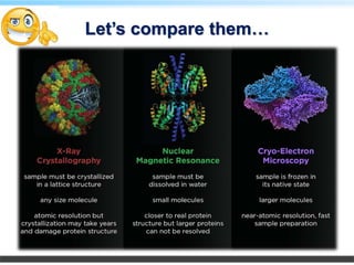
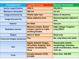
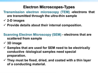
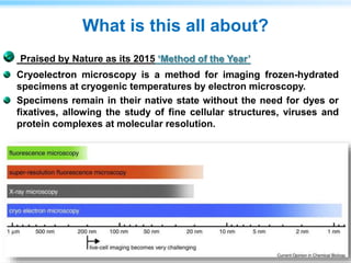
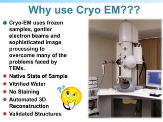
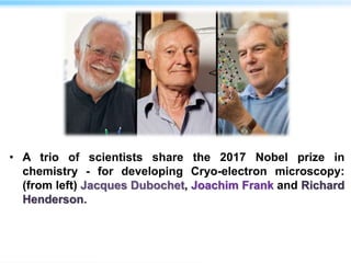
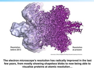
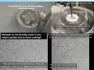
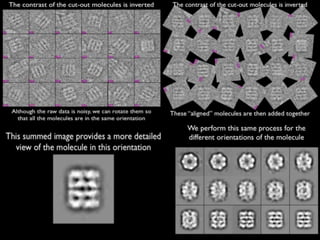
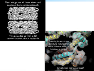
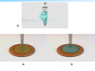
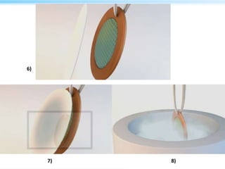
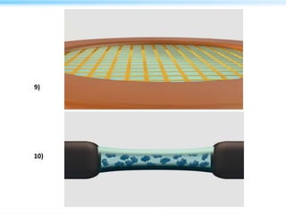
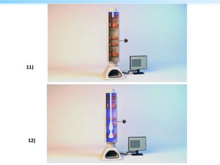
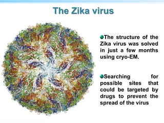
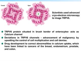
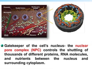
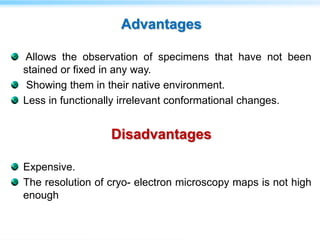
By,
Aiswariya S,
Akash Mahadev Iyer &
Lifa Prathapan
S M.Sc Biochemistry
Department of Biochemistry
University of Kerala
Is the human
body made
up of cells or
molecules or
atoms?
Does knowing the structure of a molecule matters?
As Francis Crick, one of Britain's great scientists, once said:
"If you want to understand function, study structure."
Molecular structure - key to understanding Nature's intricate design
mechanisms and blueprints.
Chemical structure determines the molecular geometry and physical
properties of a compound by portraying the spatial arrangement of
atoms and chemical bonds in the molecule.
Important visual representation of a chemical formula- Elemental
composition
Higher resolution instrumentation allows us to study single molecules
For eg; Chemical structure of amino acids influences three
dimensional structures of proteins..
Structural biology explores the 3-D shapes of biological
macromolecules and their complexes at atomic
resolution.
Approaches such as X-ray crystallography, NMR, ESR,
Electron microscopy etc. are used to determine the
myriad physical forms that proteins and nucleic acids
adopt
Let’s compare them…
a)Rhodospirillum rubrum in light microscope
b)A thin section of R.rubrum in TEM
Scanning Tunneling
Microscopic Image of DNA
double helix
Membrane protein Aquaporin visualized by Atomic Force Microscopy
Characteristic Light Microscope Electron microscope
Useful magnification 2000 X 500,000 X
Maximum resolution 200 nm 0.2 nm
Image produced by Visible light rays Beam of electrons
Image focused by
Glass objective lens Electromagnetic objective
lens
Interior Air filled Vaccum
Radiation source
Mercury Lamps, Xenon
Lamps, Lasers or Light-
emitting Diodes
High voltage Tungsten
Lmp (50kV)
Object Alive or Dead Dead only
Coloured images produced Yes No, only black and white
Use
Highly detailed images,
3D surface imaging, Sub-
cellular visualization,
Virus
Reasonable details-
morphology. Visualize
small invertebrates, whole
cells etc.
Cost
Usually between $150-
$15,000
More than $50,000
Electron Microscopes-Types
Transmission electron microscopy (TEM) -electrons that
are transmitted through the ultra-thin sample
 2-D images
 Provide details about their internal composition.
Scanning Electron Microscopy (SEM) - electrons that are
scattered from sample
 3D image
 Samples that are used for SEM need to be electrically
conductive -biological samples need special
preparation.
 They must be fixed, dried, and coated with a thin layer
of a conducting material.
Instrumentation
TEM parts
Electron column
with specimen
chamber- BODY
Electron source
Thermionic el-
gun
Field
emission gunSeveral lenses
that focus el
beam
Imaging system
Auxiliary
components
Vacuum system
High vacuum
minimizes collision
frequency of electrons
with gas atoms and
ensures stable
operation
Eg; LaB6
crystal,
Tungsten
filament
Sharp
pointed
cathode
Sample
Primary Backscattered electrons
Atomic number and
Topographical information
Cathodoluminescence
Electrical information
Secondary electrons
Topographical Information
X- rays
Thickness
Composition information
Auger electrons
Surface sensitive
Compositional information
Incident Beam
 Lenses in electron
microscopes are grouped
into three systems ;
The condenser lens - directs the
el- beam into the sample
 Controls the beam intensity and
beam coherence
The objective lens - produces a
magnified real image of the
sample and is used for focusing.
The projector lens -allow easier
control of the magnification and
projects the magnified image onto
a detector.
Detectors
• Several types of detectors can be used in TEM .
• Detectors use the information contained in the electron
waves exiting from the sample to form an image.
• CCDs
• Hybrid pixel detectors
• MAPS-Monolithic Active Pixel Sensors
• DEDs-Direct Electron Detectors
In CCD detectors, electrons are
converted to photons upon hitting
scintillating plate, and photons are
recorded by CCDs detectors.
Recorded signal had a poor signal-
to-noise ratio due to scattering of
electrons on the scintillating plate
DEDs -directly acquire
electrons without the need of
converting them to photons-
Improved resolution, better
signal-to-noise ratio, faster
readout.
Utility of TEM
use a beam of electrons
to examine the structures of molecules and materials at the atomic
scale.
As the beam passes through very thin sample, it interacts with the
molecules, which projects an image of the sample onto the detector
(CCD).
-thus, it can reveal much finer details than light
microscopy.
Biomolecules – incompatible with the high-vacuum conditions and
intense electron beams used in traditional TEMs.
High energy electrons cause severe radiation damage to delicate
samples by breaking the atomic bonds, loss of water and secondary
damage that is caused by the radicals that are generated.
What is this all about?
Praised by Nature as its 2015 ‘Method of the Year’
Cryoelectron microscopy is a method for imaging frozen-hydrated
specimens at cryogenic temperatures by electron microscopy.
Specimens remain in their native state without the need for dyes or
fixatives, allowing the study of fine cellular structures, viruses and
protein complexes at molecular resolution.
Why use Cryo EM???
Cryo-EM uses frozen
samples, gentler
electron beams and
sophisticated image
processing to
overcome many of the
problems faced by
TEMs.
Native State of Sample
Vitrified Water
No Staining
Automated 3D
Reconstruction
Validated Structures
• A trio of scientists share the 2017 Nobel prize in
chemistry - for developing Cryo-electron microscopy:
(from left) Jacques Dubochet, Joachim Frank and Richard
Henderson.
The electron microscope’s resolution has radically improved in the last
few years, from mostly showing shapeless blobs to now being able to
visualise proteins at atomic resolution…
Steps in Cryo EM
SAMPLE PREPARATION
DATA COLLECTION and
ANALYSIS
Cryo-EM samples -purified to perfect homogeneity
Heterogeneity- assessed by SDS-PAGE and size-
exclusion chromatography.
Native gel electrophoresis and static and DLS techniques
can also be used to assess the polydispersity
(characterized as particles of varied sizes in the
dispersed phase) of more complex samples.
The best way to assess quality of sample -directly
visualize it by EM.
All samples for cryo-EM are first analyzed by negative
staining electron microscopy.
SAMPLE PREPARATION
 Negative-stain EM images provide -information on a sample-
presence of contaminants or aggregates, size, shape etc.
Three major steps:
Adsorption of the sample on carbon-coated grid,
Blotting the excess sample and washing the grid with deionized
water, and
Staining with a heavy metal solution
 Heavy metal stains- uranyl acetate, phosphotungstic acid,
ammonium molybdate, methylamine vanadate.
 Heavy ions -readily interact with the electron beam and produce
phase contrast.
Vitrification- Ice free solidification of an aqueous solution
Particles are kept in a native state by embedding them in a thin layer
of vitrified water- very weak contrast.
Imaging at cryo temperatures- reduces specimen movement and
radiation damage.
Quick freezing solidifies the water without allowing ice crystals to
form (ice crystal formation damages biological molecules and
generate strong contrast that obscures molecular features.)
Vitrification
Vitrification
Liquid nitrogen has a significantly lower
boiling point (-196o C) compared to liquid
ethane (-88.6o C).
When sucrose is cooled slowly it results in
crystal sugar (or rock candy), but when cooled
rapidly it can form syrupy cotton candy
(candyfloss).
Nitrogen is not directly used; It can
make crystals due to slow cooling!!
1)
2)
Summarize….
3)
4) 5)
v
6)
7) 8)
9)
10)
11)
12)
13)
14.a) 14.b)
14.c)
14.d)
• Richard Henderson –was first to actually figure out the 3D structure
of a protein using an electron microscope.
• Jacques Dubochet found a way of freezing aqueous samples fast
enough to preserve molecules’ shape in a glassy ice matrix.
• Joachim Frank’s major contribution to the field was in processing
and analysing cryo-EM images.
• Developed computational methods for taking images of multiple,
randomly-oriented proteins and compiling them into sets of similar
orientations to obtain sharper 2D images- 3D image could be
constructed from these 2D projections.
It was James Dubochet who put the ‘cryo’ into cryo-EM.
He developed a method for freezing water so rapidly that the water
forms a disordered glass, rather than crystalline ice.
This is important because ordered ice crystals would strongly
diffract the microscope’s electron beam, obscuring any information
about the molecules being studied.
Catapulting the sample into a bath of liquid nitrogen-cooled ethane
freezes the thin film of water on a TEM sample so quickly that the
water molecules don’t have time to arrange into a crystalline lattice.
In this ‘vitrified’ sample, the water is disordered but the 3D structure
of the biomolecules in the sample is retained
Spliceosome
Spliceosome-cellular
machine that performs
pruning of mRNA.
Chops out unnecessary
pieces, called introns,
and joins together the
leftover, essential
sequences, called exons.
The edited mRNA is then
exported to the cell’s
cytoplasm, where it gets
translated into protein.
The structure of the
Zika virus was solved
in just a few months
using cryo-EM.
Searching for
possible sites that
could be targeted by
drugs to prevent the
spread of the virus
The Zika virus
TRPV6 protein situated in brush border of enterocytes- acts as
Calcium channel
Deviations in TRPV6 channels - advancement of malignancy by
upsetting the control of cell multiplication and cell demise.
Drug development to correct abnormalities in calcium uptake, which
have been linked to cancers of the breast, endometrium, prostate,
and colon.
Scientists used advanced
cryo-electron microscopy
to image TRPV6.
Gatekeeper of the cell’s nucleus- the nuclear
pore complex (NPC) controls the shuttling of
thousands of different proteins, RNA molecules,
and nutrients between the nucleus and
surrounding cytoplasm.
Advantages
Allows the observation of specimens that have not been
stained or fixed in any way.
Showing them in their native environment.
Less in functionally irrelevant conformational changes.
Disadvantages
Expensive.
The resolution of cryo- electron microscopy maps is not high
enough
Applications
• Studying the Molecular mechanisms of various life
processes
• Cryo-EM is used to characterize the structure and
mechanism of action of macromolecular complexes, -
spliceosome and snRNP complexes, anaphase-
promoting complex, ion channels, and human γ-
secretase.
• Nano particle research
• Structure based Drug design
• Material Characterization
 ’Cryo-electron microscopy of vitrified specimens.’ -Dubochet J, Adrian M, Chang JJ,
McDowall AW, Schultz PQuarterly Reviews of Biophysics.
 Methods in Molecular Biophysics- By Nathan R. Zaccai, Igor N. Serdyuk, Joseph Zaccai,
Cambridge University Press
 ‘Cryo electron microscopy to determine the structure of macromolecular complexes’-
Marta Carroni and Helen R. Saibil- Methods - Journal – Elsevier.
 ‘Cryo-electron tomography: The challenge of doing structural biology in situ’Vladan
Lučić, Alexander Rigort, Wolfgang Baumeister- Journal of Cell Biology
 ‘Introduction to high-resolution cryo-electron microscopy’- Mariusz Czarnocki-Cieciura
Marcin Nowotny- Postępy Biochemii
 ‘Electronic detectors for electron microscopy’ A.R Faruqi and R. Henderson- Current
Opinion in Structural Biology
 Biophysical Techniques in Drug Discovery- edited by Angeles Canales, Royal Society of
Chemistry.
 ‘Move over X-ray crystallography. Cryo-electron microscopy is kicking up a storm by
revealing the hidden machinery of the cell.’- Ewen Callaway, NATURE .
Cryo electron microscopy

Cryo electron microscopy

  • 1.
    By, Aiswariya S, Akash MahadevIyer & Lifa Prathapan S M.Sc Biochemistry Department of Biochemistry University of Kerala
  • 2.
    Is the human bodymade up of cells or molecules or atoms?
  • 3.
    Does knowing thestructure of a molecule matters? As Francis Crick, one of Britain's great scientists, once said: "If you want to understand function, study structure." Molecular structure - key to understanding Nature's intricate design mechanisms and blueprints. Chemical structure determines the molecular geometry and physical properties of a compound by portraying the spatial arrangement of atoms and chemical bonds in the molecule. Important visual representation of a chemical formula- Elemental composition Higher resolution instrumentation allows us to study single molecules For eg; Chemical structure of amino acids influences three dimensional structures of proteins..
  • 4.
    Structural biology exploresthe 3-D shapes of biological macromolecules and their complexes at atomic resolution. Approaches such as X-ray crystallography, NMR, ESR, Electron microscopy etc. are used to determine the myriad physical forms that proteins and nucleic acids adopt
  • 5.
  • 7.
    a)Rhodospirillum rubrum inlight microscope b)A thin section of R.rubrum in TEM Scanning Tunneling Microscopic Image of DNA double helix Membrane protein Aquaporin visualized by Atomic Force Microscopy
  • 8.
    Characteristic Light MicroscopeElectron microscope Useful magnification 2000 X 500,000 X Maximum resolution 200 nm 0.2 nm Image produced by Visible light rays Beam of electrons Image focused by Glass objective lens Electromagnetic objective lens Interior Air filled Vaccum Radiation source Mercury Lamps, Xenon Lamps, Lasers or Light- emitting Diodes High voltage Tungsten Lmp (50kV) Object Alive or Dead Dead only Coloured images produced Yes No, only black and white Use Highly detailed images, 3D surface imaging, Sub- cellular visualization, Virus Reasonable details- morphology. Visualize small invertebrates, whole cells etc. Cost Usually between $150- $15,000 More than $50,000
  • 9.
    Electron Microscopes-Types Transmission electronmicroscopy (TEM) -electrons that are transmitted through the ultra-thin sample  2-D images  Provide details about their internal composition. Scanning Electron Microscopy (SEM) - electrons that are scattered from sample  3D image  Samples that are used for SEM need to be electrically conductive -biological samples need special preparation.  They must be fixed, dried, and coated with a thin layer of a conducting material.
  • 10.
    Instrumentation TEM parts Electron column withspecimen chamber- BODY Electron source Thermionic el- gun Field emission gunSeveral lenses that focus el beam Imaging system Auxiliary components Vacuum system High vacuum minimizes collision frequency of electrons with gas atoms and ensures stable operation Eg; LaB6 crystal, Tungsten filament Sharp pointed cathode
  • 11.
    Sample Primary Backscattered electrons Atomicnumber and Topographical information Cathodoluminescence Electrical information Secondary electrons Topographical Information X- rays Thickness Composition information Auger electrons Surface sensitive Compositional information Incident Beam
  • 12.
     Lenses inelectron microscopes are grouped into three systems ; The condenser lens - directs the el- beam into the sample  Controls the beam intensity and beam coherence The objective lens - produces a magnified real image of the sample and is used for focusing. The projector lens -allow easier control of the magnification and projects the magnified image onto a detector.
  • 13.
    Detectors • Several typesof detectors can be used in TEM . • Detectors use the information contained in the electron waves exiting from the sample to form an image. • CCDs • Hybrid pixel detectors • MAPS-Monolithic Active Pixel Sensors • DEDs-Direct Electron Detectors
  • 14.
    In CCD detectors,electrons are converted to photons upon hitting scintillating plate, and photons are recorded by CCDs detectors. Recorded signal had a poor signal- to-noise ratio due to scattering of electrons on the scintillating plate DEDs -directly acquire electrons without the need of converting them to photons- Improved resolution, better signal-to-noise ratio, faster readout.
  • 15.
    Utility of TEM usea beam of electrons to examine the structures of molecules and materials at the atomic scale. As the beam passes through very thin sample, it interacts with the molecules, which projects an image of the sample onto the detector (CCD). -thus, it can reveal much finer details than light microscopy. Biomolecules – incompatible with the high-vacuum conditions and intense electron beams used in traditional TEMs. High energy electrons cause severe radiation damage to delicate samples by breaking the atomic bonds, loss of water and secondary damage that is caused by the radicals that are generated.
  • 16.
    What is thisall about? Praised by Nature as its 2015 ‘Method of the Year’ Cryoelectron microscopy is a method for imaging frozen-hydrated specimens at cryogenic temperatures by electron microscopy. Specimens remain in their native state without the need for dyes or fixatives, allowing the study of fine cellular structures, viruses and protein complexes at molecular resolution.
  • 17.
    Why use CryoEM??? Cryo-EM uses frozen samples, gentler electron beams and sophisticated image processing to overcome many of the problems faced by TEMs. Native State of Sample Vitrified Water No Staining Automated 3D Reconstruction Validated Structures
  • 18.
    • A trioof scientists share the 2017 Nobel prize in chemistry - for developing Cryo-electron microscopy: (from left) Jacques Dubochet, Joachim Frank and Richard Henderson.
  • 19.
    The electron microscope’sresolution has radically improved in the last few years, from mostly showing shapeless blobs to now being able to visualise proteins at atomic resolution…
  • 20.
    Steps in CryoEM SAMPLE PREPARATION DATA COLLECTION and ANALYSIS
  • 21.
    Cryo-EM samples -purifiedto perfect homogeneity Heterogeneity- assessed by SDS-PAGE and size- exclusion chromatography. Native gel electrophoresis and static and DLS techniques can also be used to assess the polydispersity (characterized as particles of varied sizes in the dispersed phase) of more complex samples. The best way to assess quality of sample -directly visualize it by EM. All samples for cryo-EM are first analyzed by negative staining electron microscopy. SAMPLE PREPARATION
  • 22.
     Negative-stain EMimages provide -information on a sample- presence of contaminants or aggregates, size, shape etc. Three major steps: Adsorption of the sample on carbon-coated grid, Blotting the excess sample and washing the grid with deionized water, and Staining with a heavy metal solution  Heavy metal stains- uranyl acetate, phosphotungstic acid, ammonium molybdate, methylamine vanadate.  Heavy ions -readily interact with the electron beam and produce phase contrast.
  • 23.
    Vitrification- Ice freesolidification of an aqueous solution Particles are kept in a native state by embedding them in a thin layer of vitrified water- very weak contrast. Imaging at cryo temperatures- reduces specimen movement and radiation damage. Quick freezing solidifies the water without allowing ice crystals to form (ice crystal formation damages biological molecules and generate strong contrast that obscures molecular features.) Vitrification
  • 24.
    Vitrification Liquid nitrogen hasa significantly lower boiling point (-196o C) compared to liquid ethane (-88.6o C).
  • 25.
    When sucrose iscooled slowly it results in crystal sugar (or rock candy), but when cooled rapidly it can form syrupy cotton candy (candyfloss).
  • 26.
    Nitrogen is notdirectly used; It can make crystals due to slow cooling!!
  • 29.
  • 30.
  • 31.
  • 32.
  • 33.
  • 34.
  • 35.
  • 36.
  • 37.
  • 38.
    • Richard Henderson–was first to actually figure out the 3D structure of a protein using an electron microscope. • Jacques Dubochet found a way of freezing aqueous samples fast enough to preserve molecules’ shape in a glassy ice matrix. • Joachim Frank’s major contribution to the field was in processing and analysing cryo-EM images. • Developed computational methods for taking images of multiple, randomly-oriented proteins and compiling them into sets of similar orientations to obtain sharper 2D images- 3D image could be constructed from these 2D projections.
  • 39.
    It was JamesDubochet who put the ‘cryo’ into cryo-EM. He developed a method for freezing water so rapidly that the water forms a disordered glass, rather than crystalline ice. This is important because ordered ice crystals would strongly diffract the microscope’s electron beam, obscuring any information about the molecules being studied. Catapulting the sample into a bath of liquid nitrogen-cooled ethane freezes the thin film of water on a TEM sample so quickly that the water molecules don’t have time to arrange into a crystalline lattice. In this ‘vitrified’ sample, the water is disordered but the 3D structure of the biomolecules in the sample is retained
  • 41.
    Spliceosome Spliceosome-cellular machine that performs pruningof mRNA. Chops out unnecessary pieces, called introns, and joins together the leftover, essential sequences, called exons. The edited mRNA is then exported to the cell’s cytoplasm, where it gets translated into protein.
  • 42.
    The structure ofthe Zika virus was solved in just a few months using cryo-EM. Searching for possible sites that could be targeted by drugs to prevent the spread of the virus The Zika virus
  • 43.
    TRPV6 protein situatedin brush border of enterocytes- acts as Calcium channel Deviations in TRPV6 channels - advancement of malignancy by upsetting the control of cell multiplication and cell demise. Drug development to correct abnormalities in calcium uptake, which have been linked to cancers of the breast, endometrium, prostate, and colon. Scientists used advanced cryo-electron microscopy to image TRPV6.
  • 44.
    Gatekeeper of thecell’s nucleus- the nuclear pore complex (NPC) controls the shuttling of thousands of different proteins, RNA molecules, and nutrients between the nucleus and surrounding cytoplasm.
  • 45.
    Advantages Allows the observationof specimens that have not been stained or fixed in any way. Showing them in their native environment. Less in functionally irrelevant conformational changes. Disadvantages Expensive. The resolution of cryo- electron microscopy maps is not high enough
  • 46.
    Applications • Studying theMolecular mechanisms of various life processes • Cryo-EM is used to characterize the structure and mechanism of action of macromolecular complexes, - spliceosome and snRNP complexes, anaphase- promoting complex, ion channels, and human γ- secretase. • Nano particle research • Structure based Drug design • Material Characterization
  • 47.
     ’Cryo-electron microscopyof vitrified specimens.’ -Dubochet J, Adrian M, Chang JJ, McDowall AW, Schultz PQuarterly Reviews of Biophysics.  Methods in Molecular Biophysics- By Nathan R. Zaccai, Igor N. Serdyuk, Joseph Zaccai, Cambridge University Press  ‘Cryo electron microscopy to determine the structure of macromolecular complexes’- Marta Carroni and Helen R. Saibil- Methods - Journal – Elsevier.  ‘Cryo-electron tomography: The challenge of doing structural biology in situ’Vladan Lučić, Alexander Rigort, Wolfgang Baumeister- Journal of Cell Biology  ‘Introduction to high-resolution cryo-electron microscopy’- Mariusz Czarnocki-Cieciura Marcin Nowotny- Postępy Biochemii  ‘Electronic detectors for electron microscopy’ A.R Faruqi and R. Henderson- Current Opinion in Structural Biology  Biophysical Techniques in Drug Discovery- edited by Angeles Canales, Royal Society of Chemistry.  ‘Move over X-ray crystallography. Cryo-electron microscopy is kicking up a storm by revealing the hidden machinery of the cell.’- Ewen Callaway, NATURE .

Editor's Notes

  • #5 explain how the cellular machinery works -Structural biology strives to understand the processes of life at the level of single molecules and atoms. This is achieved by determining the positions of each of the thousands of atoms that comprise biological macromolecules with great precision
  • #6 First and by far most successful method that allows the determination of the exact position of each atom in a molecule is X-ray crystallography . Nuclear Magnetic Resonance (NMR) spectroscopy allows the determination of the atomic structures of proteins and other biomolecules. Electron microscopy (EM) has long remained a low-resolution technique that provides only information on the overall shape of macromolecules
  • #12 Answer: Auger electrons are electrons that are emitted when an electron from a higher energy level falls into a vacancy in an inner shell. Explanation: The process usually occurs when the atom is bombarded with high energy electrons. If the collision ejects an inner-shell electron, an electron from a higher level will quickly drop to this lower level to fill the vacancy
  • #22 characterized as particles of varied sizes in the dispersed phase of a disperse system 
  • #24 ice crystals disrupted the electron beams so much that the images were useless
  • #42 2.2 Å structure of β-galactosidase and 1.8 Å structure of glutamate dehydrogenase (currently the highest-resolution structure obtained by cryo-EM