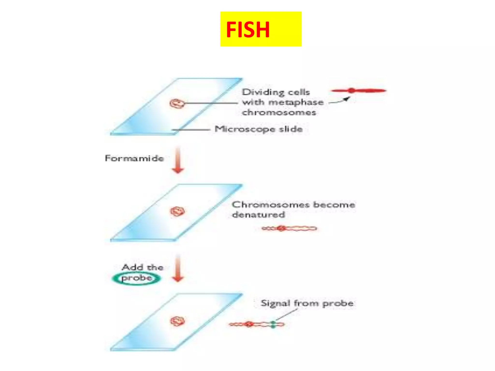
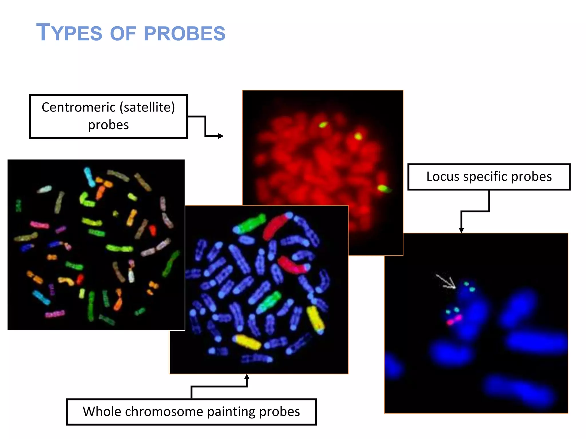
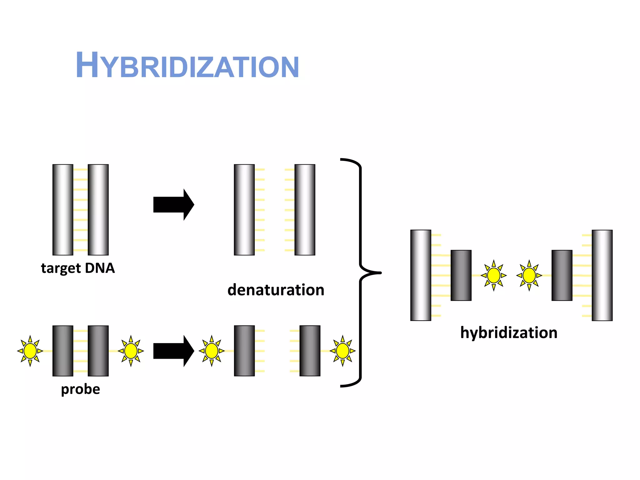
FLUORESCENT IN-SITU HYBRIDIZATION
TECHNIQUE
PRESENTED BY : DEV VRAT SHUKLA
DEPARTMENT OF BOTANY
UNIVERSITY OF ALLAHABAD
INTRODUCTION
A technique that hybridizes a DNA nucleic acid probe to a
target DNA sequence contained within a cell nucleus.
A variety of specimen types can by analyzed using FISH. The
intact cells are attached to a microscope slide using standard
cytogenetic methods.
FISH enables the position of a marker on a chromosome or
extended DNA molecule to be directly visualized
In FISH, the marker is a DNA sequence that is visualized by
hybridization with a fluorescent probe.
In situ hybridization intact chromosome is examined by probing
it with a labeled DNA molecule.
 The position on the chromosome at which hybridization occurs
provides information about the map location of the DNA
sequence used as the probe.
 DNA in the chromosome is made single stranded (‘denatured’).
 The standard method for denaturing chromosomal DNA
without destroying the morphology of the chromosome is to dry
the preparation onto a glass microscope slide and then treat with
formamide.
FISH
HISTORY OF DEVELOPMENT OF FISH
The abilities to detect specific molecular identities was first
demonstrated using antigen-antibody interactions.
 In 1977, the first antibody dependent fluorescent detection of nucleic
acid was achieved.
 The first application of fluorescent in situ detection come in 1980,
when RNA was directly labeled on the 3 end with fluorophore
PROBES
 Probe is a nucleic acid that
 can be labeled with a marker which allows
identification and quantitation
 will hybridize to another nucleic acid on the
basis of base complementarity
TYPES OF FISH PROBES
 Centromere
 Telomere
 Whole chromosome paint
 locus
TYPES OF PROBES
Centromeric (satellite)
probes
Locus specific probes
Whole chromosome painting probes
FISH PROCEDURE
Denature the chromosomes
Denature the probe
Hybridization
Fluorescence staining
Examine slides or store in the dark
Procedures Sample preparation and hybridization
Prepare slides with metaphase chromosomes or
interphase nuclei
Dehydrate in ethanol
Denature DNA at 70 oC
Denature labeled probe
Incubate at 37oC for 4-16 hours for
hybridization
HYBRIDIZATION
target DNA
probe
denaturation
hybridization
EpiFluorescent Microscope
CCD Camera
Filters
FISH Analysis
Software
ADVANTAGES OF FISH
Rapid
High efficiency of hybridization and detection
Lots of cells can be analyzed
Cells do not have to be replicating
PROBLEMS WITH IN SITU HYBRIDIZATION
 Permeabilization problems
Uneven cell penetration
 No signals
High amount of background autofluorescene
FISH using r-RNA targeted probes is the method of choice for all
studies in which exact cell numbers and cellular locations need to
be determined.
Development & design of more r-RNA targeted probes for novel
microorganisms in environment
The methodology is being continuously improved so far
however, microscopic analysis by FISH has not been automated
sufficiently which would be desirable in many investigations
Accurate quantification still remains a challenging task and
study needs careful controls.
Conclusion
REFERENCES
Elements of Biotechnology :- Gupta. P.K.
Crop improvement :- www.icar.org.in
www.dnalc.org
www.biocycleopidia.org
www.nottingham.ac
THANK YOU

Fluorescent in situ hybridization technique

  • 1.
    FLUORESCENT IN-SITU HYBRIDIZATION TECHNIQUE PRESENTEDBY : DEV VRAT SHUKLA DEPARTMENT OF BOTANY UNIVERSITY OF ALLAHABAD
  • 2.
    INTRODUCTION A technique thathybridizes a DNA nucleic acid probe to a target DNA sequence contained within a cell nucleus. A variety of specimen types can by analyzed using FISH. The intact cells are attached to a microscope slide using standard cytogenetic methods. FISH enables the position of a marker on a chromosome or extended DNA molecule to be directly visualized In FISH, the marker is a DNA sequence that is visualized by hybridization with a fluorescent probe.
  • 3.
    In situ hybridizationintact chromosome is examined by probing it with a labeled DNA molecule.  The position on the chromosome at which hybridization occurs provides information about the map location of the DNA sequence used as the probe.  DNA in the chromosome is made single stranded (‘denatured’).  The standard method for denaturing chromosomal DNA without destroying the morphology of the chromosome is to dry the preparation onto a glass microscope slide and then treat with formamide.
  • 4.
  • 5.
    HISTORY OF DEVELOPMENTOF FISH The abilities to detect specific molecular identities was first demonstrated using antigen-antibody interactions.  In 1977, the first antibody dependent fluorescent detection of nucleic acid was achieved.  The first application of fluorescent in situ detection come in 1980, when RNA was directly labeled on the 3 end with fluorophore
  • 6.
    PROBES  Probe isa nucleic acid that  can be labeled with a marker which allows identification and quantitation  will hybridize to another nucleic acid on the basis of base complementarity
  • 7.
    TYPES OF FISHPROBES  Centromere  Telomere  Whole chromosome paint  locus
  • 8.
    TYPES OF PROBES Centromeric(satellite) probes Locus specific probes Whole chromosome painting probes
  • 9.
    FISH PROCEDURE Denature thechromosomes Denature the probe Hybridization Fluorescence staining Examine slides or store in the dark
  • 10.
    Procedures Sample preparationand hybridization Prepare slides with metaphase chromosomes or interphase nuclei Dehydrate in ethanol Denature DNA at 70 oC Denature labeled probe Incubate at 37oC for 4-16 hours for hybridization
  • 12.
  • 13.
  • 14.
    ADVANTAGES OF FISH Rapid Highefficiency of hybridization and detection Lots of cells can be analyzed Cells do not have to be replicating
  • 15.
    PROBLEMS WITH INSITU HYBRIDIZATION  Permeabilization problems Uneven cell penetration  No signals High amount of background autofluorescene
  • 17.
    FISH using r-RNAtargeted probes is the method of choice for all studies in which exact cell numbers and cellular locations need to be determined. Development & design of more r-RNA targeted probes for novel microorganisms in environment The methodology is being continuously improved so far however, microscopic analysis by FISH has not been automated sufficiently which would be desirable in many investigations Accurate quantification still remains a challenging task and study needs careful controls. Conclusion
  • 18.
    REFERENCES Elements of Biotechnology:- Gupta. P.K. Crop improvement :- www.icar.org.in www.dnalc.org www.biocycleopidia.org www.nottingham.ac
  • 19.