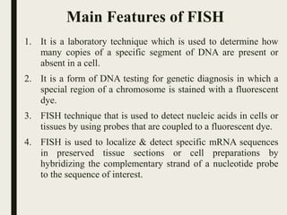
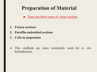
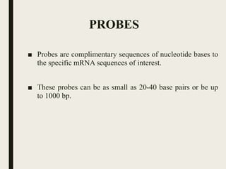
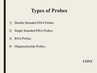
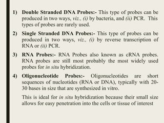
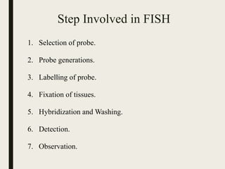
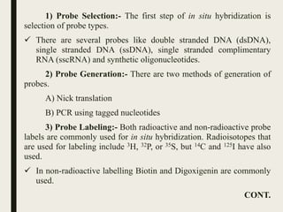
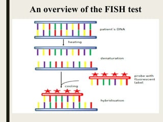
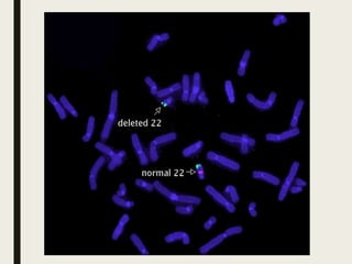
WELCOME
An Assignment On
Fluorescent in situ hybridization (FISH)
Course :- GP 502 : PRINCIPLE OF CYTOGENETICS.
Submitted To
Dr. N. B. Patel
Asso. professor,
Department of Genetics & Plant breeding,
C. P. C.A, S.D.A.U.,
Sardarkrushinagar-385 506.
Submitted by
Vaghela Gauravrajsinh K.
M.Sc. (Agri), 1st sem.,
Department of Genetics & Plant breeding,
C. P. C.A, S.D.A.U.,
Sardarkrushinagar-385 506.
FLUORESCENT IN SITU
HYBRIDIZATION (FISH)
■ Fluorescent in situ hybridization (FISH) is a molecular
cytogenetic technique that uses fluorescent probes that bind
only those parts of the chromosome with a high degree of
sequence complementarily.
OR
■ Fluorescent in situ hybridization (FISH) is a cytogenetic
technique that can be used to detect and localize the presence
or absence of specific DNA sequences on chromosomes.
Main Features of FISH
1. It is a laboratory technique which is used to determine how
many copies of a specific segment of DNA are present or
absent in a cell.
2. It is a form of DNA testing for genetic diagnosis in which a
special region of a chromosome is stained with a fluorescent
dye.
3. FISH technique that is used to detect nucleic acids in cells or
tissues by using probes that are coupled to a fluorescent dye.
4. FISH is used to localize & detect specific mRNA sequences
in preserved tissue sections or cell preparations by
hybridizing the complementary strand of a nucleotide probe
to the sequence of interest.
Preparation of Material
■ There are three types of tissue section.
1. Frozen sections
2. Paraffin embedded sections
3. Cells in suspension
 This methods are most commonly used for in situ
hybridization.
PROBES
■ Probes are complimentary sequences of nucleotide bases to
the specific mRNA sequences of interest.
■ These probes can be as small as 20-40 base pairs or be up
to 1000 bp.
Types of Probes
1) Double Stranded DNA Probes.
2) Single Stranded DNA Probes.
3) RNA Probes.
4) Oligonucleotide Probes.
CONT.
1) Double Stranded DNA Probes:- This type of probes can be
produced in two ways, viz., (i) by bacteria, and (ii) PCR. This
types of probes are rarely used.
2) Single Stranded DNA Probes:- This type of probes can be
produced in two ways, viz., (i) by reverse transcription of
RNA or (ii) PCR.
3) RNA Probes:- RNA Probes also known as cRNA probes.
RNA probes are still most probably the most widely used
probes for in situ hybridization.
4) Oligonucleotide Probes:- Oligonucleotides are short
sequences of nucleotides (RNA or DNA), typically with 20-
30 bases in size that are synthesized in vitro.
This is ideal for in situ hybridization because their small size
allows for easy penetration into the cells or tissue of interest
Step Involved in FISH
1. Selection of probe.
2. Probe generations.
3. Labelling of probe.
4. Fixation of tissues.
5. Hybridization and Washing.
6. Detection.
7. Observation.
1) Probe Selection:- The first step of in situ hybridization is
selection of probe types.
 There are several probes like double stranded DNA (dsDNA),
single stranded DNA (ssDNA), single stranded complimentary
RNA (sscRNA) and synthetic oligonucleotides.
2) Probe Generation:- There are two methods of generation of
probes.
A) Nick translation
B) PCR using tagged nucleotides
3) Probe Labeling:- Both radioactive and non-radioactive probe
labels are commonly used for in situ hybridization. Radioisotopes that
are used for labeling include 3H, 32P, or 35S, but 14C and 125I have also
used.
 In non-radioactive labelling Biotin and Digoxigenin are commonly
used.
CONT.
4) Fixation of Tissue:- There are several variables that
need to be considered when fixing tissue for in situ
hybridization.
 A) Acetic acid-alcohol mixture:- Best probe penetration, but
may permit the loss of RNA from tissue.
 B) Glutaraldehyde:- Provide best RNA retention and tissue
morphology, but because of extensive protein cross-linking
the probe penetration is low.
 C) Paraffin & Formalin:- Leads to decreased sensitivity
possibly resulting from increased cross-linking or loss of
mRNA during embedding.
 D) 4% paraformaldehyde solution:- The most widely
successful fixative solution. This provides a satisfactory
compromise between the variable and good sensitivity.
CONT.
5) Hybridization and Washing:- The hybridization is done
by simply mixing the single strand probes with the denaturated
target DNA.
 Denaturation of the DNA is obtained by heating the DNA,
which separate the two strands, and allows access of the single
strand.
 Hybridization is performed by placing a small amount of
solution containing the hybridization probe on a cover slip,
which is then placed on the slide containing tissue sections to
incubate overnight.
 The next day, washes are serially applied to the slides to
remove the probe that is not bound to target DNA/RNA.
 There are several parameters such as (i) probe construction (ii)
temperature (iii) pH (iv) formamide and (v) salt concentration
in the hybridization buffer which play a crucial role in the
specificity of target labeling during hybridization and washing.
CONT.
6) Detection:- The detection of the probes is the final step
of fluorescent in situ hybridization.
 It consists in recognizing the probes with fluorescent
antibodies corresponding antigenes incorporated in the probes.
7) Observation:- The hybridized sequences are observed
with the help of an epifluorescence microscope.
 The white light of the source lamp is filtered so that only the
relevant wavelengths for excitation of the fluorescent
molecules arrive onto the sample.
 Emission of the fluorochromes happens in general at larger
wavelengths, which allows to distinguish between excitation
and emission light by mean of another optical filter.
Control Procedure
i. Use of Alternate Sequences
ii. Use of Multiple Probes
iii. Use of non-labeled Probes
iv. Use of Northern Analysis
An overview of the FISH test
FIBER FISH
■ Fiber fish is a cytogenetic (chromosome) laboratory
technique in which FISH (fluorescence in situ
hybridization) is done on chromosomes that have been
mechanically stretched.
■ Fiber FISH provides a higher resolution of analysis than
convectional FISH.
■ Here the word fiber refers to a chromosomes fiber.
■ In fiber FISH, interphase chromosomes are attached to a
slide in such a way that they are stretched out in straight
line, rather than being tightly coiled.
 Chromosome Combing:- A technique used to produce an
array of uniformly stretched DNA that is highly suitable for
nucleic acid hybridization studies such as FISH; also
known as molecular combing or DNA combing.
Various FISH Techniques
1) M-FISH
2) Double Fusion FISH or D-FISH
3) Break apart FISH
Application of FISH
1) Applications in Medicines
2) Species identification
i. Bacterial species
ii. Genome identification
Advantages of FISH
■ FISH is able to detect many small deletions,
duplications and rearrangements that are not
visible with standard microscope analysis.
■ A diagnosis from FISH may avoid your child
having to undergo many other tests.
Benefits of FISH
■ It may help you and your doctor watch for common health
problems associated with your child’s chromosome
imbalance.
■ It may help to predict what to expect as your child gets older.
■ It may show which specific genes are included in your child’s
deletion or duplication. If the gene(s) has been associated
with a particular feature or health problem it may help to
guide management or treatment for your child.
■ It can help you to obtain specialist services for your child.
■ You can choose to join a support group to meet other parents
facing similar challenges.
■ Parents and other family members can be tested to see if they
are carriers of changes in their DNA that put them at risk of
having more children with a chromosome change.
Limitations of FISH
1. It is very expensive, hence it would be difficult for the
average laboratory to undertake this technique.
2. It is time consuming and requires detailed molecular
biological knowledge of sub cloning, in-vitro
transcription and bacterial expression.
3. FISH has limited ability to precisely define which genes
and breakpoints are involved in an imbalance.
Fluorescent in situ hybridization (FISH)

Fluorescent in situ hybridization (FISH)

  • 1.
  • 2.
    An Assignment On Fluorescentin situ hybridization (FISH) Course :- GP 502 : PRINCIPLE OF CYTOGENETICS. Submitted To Dr. N. B. Patel Asso. professor, Department of Genetics & Plant breeding, C. P. C.A, S.D.A.U., Sardarkrushinagar-385 506. Submitted by Vaghela Gauravrajsinh K. M.Sc. (Agri), 1st sem., Department of Genetics & Plant breeding, C. P. C.A, S.D.A.U., Sardarkrushinagar-385 506.
  • 3.
    FLUORESCENT IN SITU HYBRIDIZATION(FISH) ■ Fluorescent in situ hybridization (FISH) is a molecular cytogenetic technique that uses fluorescent probes that bind only those parts of the chromosome with a high degree of sequence complementarily. OR ■ Fluorescent in situ hybridization (FISH) is a cytogenetic technique that can be used to detect and localize the presence or absence of specific DNA sequences on chromosomes.
  • 4.
    Main Features ofFISH 1. It is a laboratory technique which is used to determine how many copies of a specific segment of DNA are present or absent in a cell. 2. It is a form of DNA testing for genetic diagnosis in which a special region of a chromosome is stained with a fluorescent dye. 3. FISH technique that is used to detect nucleic acids in cells or tissues by using probes that are coupled to a fluorescent dye. 4. FISH is used to localize & detect specific mRNA sequences in preserved tissue sections or cell preparations by hybridizing the complementary strand of a nucleotide probe to the sequence of interest.
  • 5.
    Preparation of Material ■There are three types of tissue section. 1. Frozen sections 2. Paraffin embedded sections 3. Cells in suspension  This methods are most commonly used for in situ hybridization.
  • 6.
    PROBES ■ Probes arecomplimentary sequences of nucleotide bases to the specific mRNA sequences of interest. ■ These probes can be as small as 20-40 base pairs or be up to 1000 bp.
  • 7.
    Types of Probes 1)Double Stranded DNA Probes. 2) Single Stranded DNA Probes. 3) RNA Probes. 4) Oligonucleotide Probes. CONT.
  • 8.
    1) Double StrandedDNA Probes:- This type of probes can be produced in two ways, viz., (i) by bacteria, and (ii) PCR. This types of probes are rarely used. 2) Single Stranded DNA Probes:- This type of probes can be produced in two ways, viz., (i) by reverse transcription of RNA or (ii) PCR. 3) RNA Probes:- RNA Probes also known as cRNA probes. RNA probes are still most probably the most widely used probes for in situ hybridization. 4) Oligonucleotide Probes:- Oligonucleotides are short sequences of nucleotides (RNA or DNA), typically with 20- 30 bases in size that are synthesized in vitro. This is ideal for in situ hybridization because their small size allows for easy penetration into the cells or tissue of interest
  • 9.
    Step Involved inFISH 1. Selection of probe. 2. Probe generations. 3. Labelling of probe. 4. Fixation of tissues. 5. Hybridization and Washing. 6. Detection. 7. Observation.
  • 10.
    1) Probe Selection:-The first step of in situ hybridization is selection of probe types.  There are several probes like double stranded DNA (dsDNA), single stranded DNA (ssDNA), single stranded complimentary RNA (sscRNA) and synthetic oligonucleotides. 2) Probe Generation:- There are two methods of generation of probes. A) Nick translation B) PCR using tagged nucleotides 3) Probe Labeling:- Both radioactive and non-radioactive probe labels are commonly used for in situ hybridization. Radioisotopes that are used for labeling include 3H, 32P, or 35S, but 14C and 125I have also used.  In non-radioactive labelling Biotin and Digoxigenin are commonly used. CONT.
  • 11.
    4) Fixation ofTissue:- There are several variables that need to be considered when fixing tissue for in situ hybridization.  A) Acetic acid-alcohol mixture:- Best probe penetration, but may permit the loss of RNA from tissue.  B) Glutaraldehyde:- Provide best RNA retention and tissue morphology, but because of extensive protein cross-linking the probe penetration is low.  C) Paraffin & Formalin:- Leads to decreased sensitivity possibly resulting from increased cross-linking or loss of mRNA during embedding.  D) 4% paraformaldehyde solution:- The most widely successful fixative solution. This provides a satisfactory compromise between the variable and good sensitivity. CONT.
  • 12.
    5) Hybridization andWashing:- The hybridization is done by simply mixing the single strand probes with the denaturated target DNA.  Denaturation of the DNA is obtained by heating the DNA, which separate the two strands, and allows access of the single strand.  Hybridization is performed by placing a small amount of solution containing the hybridization probe on a cover slip, which is then placed on the slide containing tissue sections to incubate overnight.  The next day, washes are serially applied to the slides to remove the probe that is not bound to target DNA/RNA.  There are several parameters such as (i) probe construction (ii) temperature (iii) pH (iv) formamide and (v) salt concentration in the hybridization buffer which play a crucial role in the specificity of target labeling during hybridization and washing. CONT.
  • 13.
    6) Detection:- Thedetection of the probes is the final step of fluorescent in situ hybridization.  It consists in recognizing the probes with fluorescent antibodies corresponding antigenes incorporated in the probes. 7) Observation:- The hybridized sequences are observed with the help of an epifluorescence microscope.  The white light of the source lamp is filtered so that only the relevant wavelengths for excitation of the fluorescent molecules arrive onto the sample.  Emission of the fluorochromes happens in general at larger wavelengths, which allows to distinguish between excitation and emission light by mean of another optical filter.
  • 14.
    Control Procedure i. Useof Alternate Sequences ii. Use of Multiple Probes iii. Use of non-labeled Probes iv. Use of Northern Analysis
  • 15.
    An overview ofthe FISH test
  • 17.
    FIBER FISH ■ Fiberfish is a cytogenetic (chromosome) laboratory technique in which FISH (fluorescence in situ hybridization) is done on chromosomes that have been mechanically stretched. ■ Fiber FISH provides a higher resolution of analysis than convectional FISH. ■ Here the word fiber refers to a chromosomes fiber. ■ In fiber FISH, interphase chromosomes are attached to a slide in such a way that they are stretched out in straight line, rather than being tightly coiled.  Chromosome Combing:- A technique used to produce an array of uniformly stretched DNA that is highly suitable for nucleic acid hybridization studies such as FISH; also known as molecular combing or DNA combing.
  • 18.
    Various FISH Techniques 1)M-FISH 2) Double Fusion FISH or D-FISH 3) Break apart FISH
  • 19.
    Application of FISH 1)Applications in Medicines 2) Species identification i. Bacterial species ii. Genome identification
  • 20.
    Advantages of FISH ■FISH is able to detect many small deletions, duplications and rearrangements that are not visible with standard microscope analysis. ■ A diagnosis from FISH may avoid your child having to undergo many other tests.
  • 21.
    Benefits of FISH ■It may help you and your doctor watch for common health problems associated with your child’s chromosome imbalance. ■ It may help to predict what to expect as your child gets older. ■ It may show which specific genes are included in your child’s deletion or duplication. If the gene(s) has been associated with a particular feature or health problem it may help to guide management or treatment for your child. ■ It can help you to obtain specialist services for your child. ■ You can choose to join a support group to meet other parents facing similar challenges. ■ Parents and other family members can be tested to see if they are carriers of changes in their DNA that put them at risk of having more children with a chromosome change.
  • 22.
    Limitations of FISH 1.It is very expensive, hence it would be difficult for the average laboratory to undertake this technique. 2. It is time consuming and requires detailed molecular biological knowledge of sub cloning, in-vitro transcription and bacterial expression. 3. FISH has limited ability to precisely define which genes and breakpoints are involved in an imbalance.