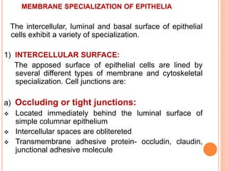
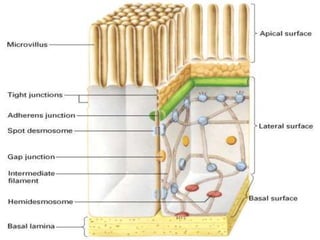
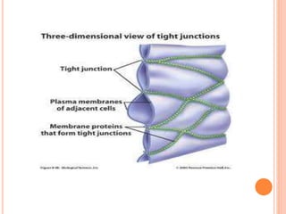
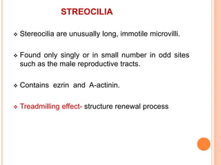
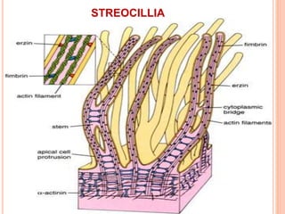
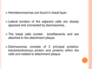
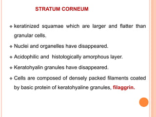
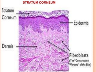
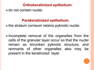
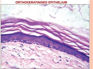
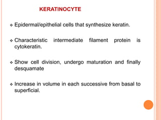
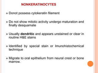
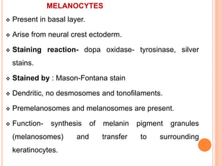
This document provides an overview of epithelium, including its definition, development, characteristics, classification, functions, cell polarity, membrane specializations, glands, and structure of the oral epithelium. Some key points include:
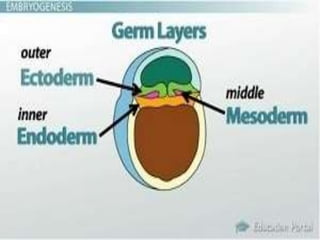
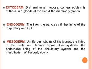
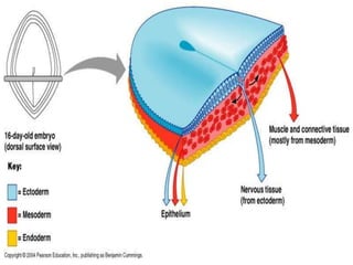
- Epithelium covers body surfaces and lines cavities, and is composed of cells attached to a basement membrane. It develops from ectoderm, mesoderm, or endoderm.
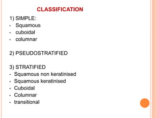
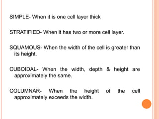
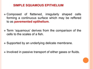
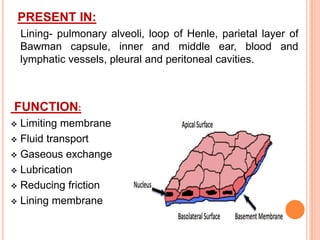
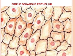
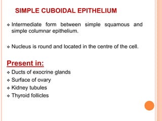
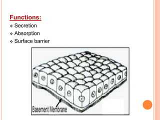
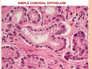
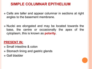
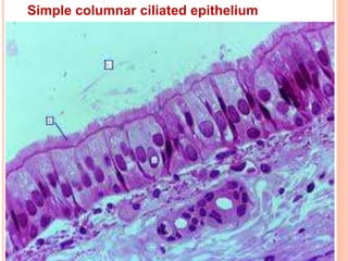
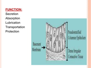
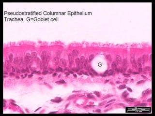
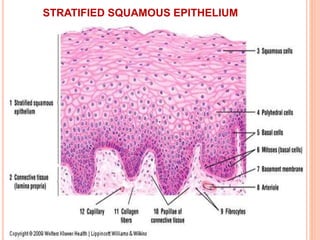
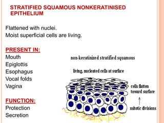
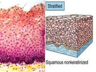
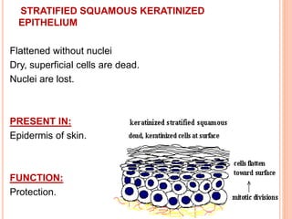
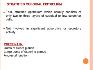
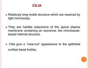
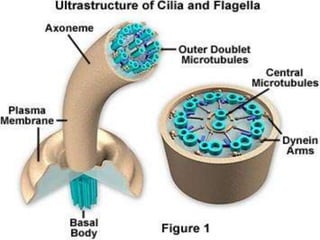
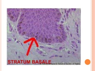
- Epithelia are classified based on number of cell layers as simple (1 cell layer), pseudostratified (cells appear in multiple layers but are all attached to the basement membrane), or stratified (multiple cell layers).
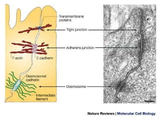
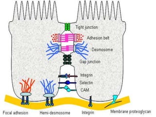
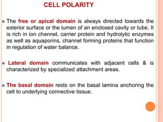
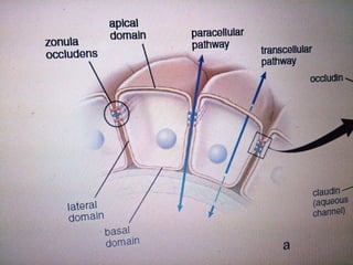
- Epithelial cells exhibit polarity with specialized domains, and form
epithelium covers body surfaces, lines body cavities and constitute glands.so it is important to know about epithelium in detail to deal with tissue of different type and origin.
. Definition of Tissues Biological tissue is a collection of interconnected cells that perform a similar function within an organism. In other words, it is a group of cells working together mainly inside an organ.
3. Classification of Tissues Human body is composed of 4 basic types of tissue: •Epithelial tissue •Connective tissue •Muscular tissue •Nervous tissue
4. Origin of Tissue A fertilized egg divides to produce 3 primary germ cell layers. These layers differentiate to form the tissues of the body.
5. Epithelial Tissue Epithelial cells cover or line all body surfaces, cavities and tubes. So, These are called covering epithelia. Epithelial cells form the functional units of secretory glands. So, These are called glandular epithelia.
detail notes on connective tissue..
Connective tissue (CT) is one of the four basic types of animal tissue, along with epithelial tissue, muscle tissue, and nervous tissue. It develops from the mesoderm. Connective tissue is found in between other tissues everywhere in the body, including the nervous system. In the central nervous system, the three outer membranes (the meninges) that envelop the brain and spinal cord are composed of connective tissue.
All connective tissue consists of three main components: fibers (elastic and collagenous fibers), ground substance and cells. Not all authorities include blood or lymph as connective tissue because they lack the fiber component. All are immersed in the body water.
a brief ppt description about cartilage which may be usefull for teaching for first year mbbs, bds and paramedical students, hope it is helpfull to everyone
oral mucous membranes-1 /orthodontic courses by Indian dental academy Indian dental academy
The Indian Dental Academy is the Leader in continuing dental education , training dentists in all aspects of dentistry and
offering a wide range of dental certified courses in different formats.for more details please visit
www.indiandentalacademy.com
RED CELL MEMBRANE: PAST, PRESENT, AND FUTURE / certified fixed orthodontic co...Indian dental academy
The Indian Dental Academy is the Leader in continuing dental education , training dentists in all aspects of dentistry and
offering a wide range of dental certified courses in different formats.for more details please visit
www.indiandentalacademy.com
This presentation has a short introduction about the different types of tissues (epithelium, connective, muscular, nervous), but focuses mainly on epithelial tissues - its characteristics, functions, and types.
This also contains the different surface modifications of epithelial tissues - apical, lateral and basal.
Lastly, glands are also discussed here. Endocrine and exocrine glands are differentiated based on characteristics and functions.
Epithelial tissue, also known as the epithelium, is one of the four tissues found in the human body. It exists in various parts of the body, such as our digestive system, outer surfaces of organs and blood vessels throughout the body, as well as the inner surfaces of cavities in many internal organs.
Histology
Junqueira’s Basic Histology Text and Atlas, 15th Ed
Epithelial tissue is a type of tissue that covers the surfaces of organs, lines body cavities and forms glands. This PDF provides a comprehensive overview of epithelial tissue, including its types, structure, function, and location in the body. The document covers the different types of epithelial cells, their shapes, and layers, as well as their functions in different organs and tissues. The PDF also delves into the unique features of epithelial tissue, such as its polarity, cell junctions, and the basement membrane. Additionally, Whether you are a student, medical professional, or someone interested in learning about the human body, this PDF provides a detailed understanding of epithelial tissue and its importance in the body.
Ozempic: Preoperative Management of Patients on GLP-1 Receptor Agonists Saeid Safari
Preoperative Management of Patients on GLP-1 Receptor Agonists like Ozempic and Semiglutide
ASA GUIDELINE
NYSORA Guideline
2 Case Reports of Gastric Ultrasound
New Directions in Targeted Therapeutic Approaches for Older Adults With Mantl...i3 Health
i3 Health is pleased to make the speaker slides from this activity available for use as a non-accredited self-study or teaching resource.
This slide deck presented by Dr. Kami Maddocks, Professor-Clinical in the Division of Hematology and
Associate Division Director for Ambulatory Operations
The Ohio State University Comprehensive Cancer Center, will provide insight into new directions in targeted therapeutic approaches for older adults with mantle cell lymphoma.
STATEMENT OF NEED
Mantle cell lymphoma (MCL) is a rare, aggressive B-cell non-Hodgkin lymphoma (NHL) accounting for 5% to 7% of all lymphomas. Its prognosis ranges from indolent disease that does not require treatment for years to very aggressive disease, which is associated with poor survival (Silkenstedt et al, 2021). Typically, MCL is diagnosed at advanced stage and in older patients who cannot tolerate intensive therapy (NCCN, 2022). Although recent advances have slightly increased remission rates, recurrence and relapse remain very common, leading to a median overall survival between 3 and 6 years (LLS, 2021). Though there are several effective options, progress is still needed towards establishing an accepted frontline approach for MCL (Castellino et al, 2022). Treatment selection and management of MCL are complicated by the heterogeneity of prognosis, advanced age and comorbidities of patients, and lack of an established standard approach for treatment, making it vital that clinicians be familiar with the latest research and advances in this area. In this activity chaired by Michael Wang, MD, Professor in the Department of Lymphoma & Myeloma at MD Anderson Cancer Center, expert faculty will discuss prognostic factors informing treatment, the promising results of recent trials in new therapeutic approaches, and the implications of treatment resistance in therapeutic selection for MCL.
Target Audience
Hematology/oncology fellows, attending faculty, and other health care professionals involved in the treatment of patients with mantle cell lymphoma (MCL).
Learning Objectives
1.) Identify clinical and biological prognostic factors that can guide treatment decision making for older adults with MCL
2.) Evaluate emerging data on targeted therapeutic approaches for treatment-naive and relapsed/refractory MCL and their applicability to older adults
3.) Assess mechanisms of resistance to targeted therapies for MCL and their implications for treatment selection
Recomendações da OMS sobre cuidados maternos e neonatais para uma experiência pós-natal positiva.
Em consonância com os ODS – Objetivos do Desenvolvimento Sustentável e a Estratégia Global para a Saúde das Mulheres, Crianças e Adolescentes, e aplicando uma abordagem baseada nos direitos humanos, os esforços de cuidados pós-natais devem expandir-se para além da cobertura e da simples sobrevivência, de modo a incluir cuidados de qualidade.
Estas diretrizes visam melhorar a qualidade dos cuidados pós-natais essenciais e de rotina prestados às mulheres e aos recém-nascidos, com o objetivo final de melhorar a saúde e o bem-estar materno e neonatal.
Uma “experiência pós-natal positiva” é um resultado importante para todas as mulheres que dão à luz e para os seus recém-nascidos, estabelecendo as bases para a melhoria da saúde e do bem-estar a curto e longo prazo. Uma experiência pós-natal positiva é definida como aquela em que as mulheres, pessoas que gestam, os recém-nascidos, os casais, os pais, os cuidadores e as famílias recebem informação consistente, garantia e apoio de profissionais de saúde motivados; e onde um sistema de saúde flexível e com recursos reconheça as necessidades das mulheres e dos bebês e respeite o seu contexto cultural.
Estas diretrizes consolidadas apresentam algumas recomendações novas e já bem fundamentadas sobre cuidados pós-natais de rotina para mulheres e neonatos que recebem cuidados no pós-parto em unidades de saúde ou na comunidade, independentemente dos recursos disponíveis.
É fornecido um conjunto abrangente de recomendações para cuidados durante o período puerperal, com ênfase nos cuidados essenciais que todas as mulheres e recém-nascidos devem receber, e com a devida atenção à qualidade dos cuidados; isto é, a entrega e a experiência do cuidado recebido. Estas diretrizes atualizam e ampliam as recomendações da OMS de 2014 sobre cuidados pós-natais da mãe e do recém-nascido e complementam as atuais diretrizes da OMS sobre a gestão de complicações pós-natais.
O estabelecimento da amamentação e o manejo das principais intercorrências é contemplada.
Recomendamos muito.
Vamos discutir essas recomendações no nosso curso de pós-graduação em Aleitamento no Instituto Ciclos.
Esta publicação só está disponível em inglês até o momento.
Prof. Marcus Renato de Carvalho
www.agostodourado.com
TEST BANK for Operations Management, 14th Edition by William J. Stevenson, Ve...kevinkariuki227
TEST BANK for Operations Management, 14th Edition by William J. Stevenson, Verified Chapters 1 - 19, Complete Newest Version.pdf
Flu Vaccine Alert in Bangalore Karnatakaaddon Scans
As flu season approaches, health officials in Bangalore, Karnataka, are urging residents to get their flu vaccinations. The seasonal flu, while common, can lead to severe health complications, particularly for vulnerable populations such as young children, the elderly, and those with underlying health conditions.
Dr. Vidisha Kumari, a leading epidemiologist in Bangalore, emphasizes the importance of getting vaccinated. "The flu vaccine is our best defense against the influenza virus. It not only protects individuals but also helps prevent the spread of the virus in our communities," he says.
This year, the flu season is expected to coincide with a potential increase in other respiratory illnesses. The Karnataka Health Department has launched an awareness campaign highlighting the significance of flu vaccinations. They have set up multiple vaccination centers across Bangalore, making it convenient for residents to receive their shots.
To encourage widespread vaccination, the government is also collaborating with local schools, workplaces, and community centers to facilitate vaccination drives. Special attention is being given to ensuring that the vaccine is accessible to all, including marginalized communities who may have limited access to healthcare.
Residents are reminded that the flu vaccine is safe and effective. Common side effects are mild and may include soreness at the injection site, mild fever, or muscle aches. These side effects are generally short-lived and far less severe than the flu itself.
Healthcare providers are also stressing the importance of continuing COVID-19 precautions. Wearing masks, practicing good hand hygiene, and maintaining social distancing are still crucial, especially in crowded places.
Protect yourself and your loved ones by getting vaccinated. Together, we can help keep Bangalore healthy and safe this flu season. For more information on vaccination centers and schedules, residents can visit the Karnataka Health Department’s official website or follow their social media pages.
Stay informed, stay safe, and get your flu shot today!
Tom Selleck Health: A Comprehensive Look at the Iconic Actor’s Wellness Journeygreendigital
Tom Selleck, an enduring figure in Hollywood. has captivated audiences for decades with his rugged charm, iconic moustache. and memorable roles in television and film. From his breakout role as Thomas Magnum in Magnum P.I. to his current portrayal of Frank Reagan in Blue Bloods. Selleck's career has spanned over 50 years. But beyond his professional achievements. fans have often been curious about Tom Selleck Health. especially as he has aged in the public eye.
Follow us on: Pinterest
Introduction
Many have been interested in Tom Selleck health. not only because of his enduring presence on screen but also because of the challenges. and lifestyle choices he has faced and made over the years. This article delves into the various aspects of Tom Selleck health. exploring his fitness regimen, diet, mental health. and the challenges he has encountered as he ages. We'll look at how he maintains his well-being. the health issues he has faced, and his approach to ageing .
Early Life and Career
Childhood and Athletic Beginnings
Tom Selleck was born on January 29, 1945, in Detroit, Michigan, and grew up in Sherman Oaks, California. From an early age, he was involved in sports, particularly basketball. which played a significant role in his physical development. His athletic pursuits continued into college. where he attended the University of Southern California (USC) on a basketball scholarship. This early involvement in sports laid a strong foundation for his physical health and disciplined lifestyle.
Transition to Acting
Selleck's transition from an athlete to an actor came with its physical demands. His first significant role in "Magnum P.I." required him to perform various stunts and maintain a fit appearance. This role, which he played from 1980 to 1988. necessitated a rigorous fitness routine to meet the show's demands. setting the stage for his long-term commitment to health and wellness.
Fitness Regimen
Workout Routine
Tom Selleck health and fitness regimen has evolved. adapting to his changing roles and age. During his "Magnum, P.I." days. Selleck's workouts were intense and focused on building and maintaining muscle mass. His routine included weightlifting, cardiovascular exercises. and specific training for the stunts he performed on the show.
Selleck adjusted his fitness routine as he aged to suit his body's needs. Today, his workouts focus on maintaining flexibility, strength, and cardiovascular health. He incorporates low-impact exercises such as swimming, walking, and light weightlifting. This balanced approach helps him stay fit without putting undue strain on his joints and muscles.
Importance of Flexibility and Mobility
In recent years, Selleck has emphasized the importance of flexibility and mobility in his fitness regimen. Understanding the natural decline in muscle mass and joint flexibility with age. he includes stretching and yoga in his routine. These practices help prevent injuries, improve posture, and maintain mobilit
Lung Cancer: Artificial Intelligence, Synergetics, Complex System Analysis, S...Oleg Kshivets
RESULTS: Overall life span (LS) was 2252.1±1742.5 days and cumulative 5-year survival (5YS) reached 73.2%, 10 years – 64.8%, 20 years – 42.5%. 513 LCP lived more than 5 years (LS=3124.6±1525.6 days), 148 LCP – more than 10 years (LS=5054.4±1504.1 days).199 LCP died because of LC (LS=562.7±374.5 days). 5YS of LCP after bi/lobectomies was significantly superior in comparison with LCP after pneumonectomies (78.1% vs.63.7%, P=0.00001 by log-rank test). AT significantly improved 5YS (66.3% vs. 34.8%) (P=0.00000 by log-rank test) only for LCP with N1-2. Cox modeling displayed that 5YS of LCP significantly depended on: phase transition (PT) early-invasive LC in terms of synergetics, PT N0—N12, cell ratio factors (ratio between cancer cells- CC and blood cells subpopulations), G1-3, histology, glucose, AT, blood cell circuit, prothrombin index, heparin tolerance, recalcification time (P=0.000-0.038). Neural networks, genetic algorithm selection and bootstrap simulation revealed relationships between 5YS and PT early-invasive LC (rank=1), PT N0—N12 (rank=2), thrombocytes/CC (3), erythrocytes/CC (4), eosinophils/CC (5), healthy cells/CC (6), lymphocytes/CC (7), segmented neutrophils/CC (8), stick neutrophils/CC (9), monocytes/CC (10); leucocytes/CC (11). Correct prediction of 5YS was 100% by neural networks computing (area under ROC curve=1.0; error=0.0).
CONCLUSIONS: 5YS of LCP after radical procedures significantly depended on: 1) PT early-invasive cancer; 2) PT N0--N12; 3) cell ratio factors; 4) blood cell circuit; 5) biochemical factors; 6) hemostasis system; 7) AT; 8) LC characteristics; 9) LC cell dynamics; 10) surgery type: lobectomy/pneumonectomy; 11) anthropometric data. Optimal diagnosis and treatment strategies for LC are: 1) screening and early detection of LC; 2) availability of experienced thoracic surgeons because of complexity of radical procedures; 3) aggressive en block surgery and adequate lymph node dissection for completeness; 4) precise prediction; 5) adjuvant chemoimmunoradiotherapy for LCP with unfavorable prognosis.
HOT NEW PRODUCT! BIG SALES FAST SHIPPING NOW FROM CHINA!! EU KU DB BK substit...GL Anaacs
Contact us if you are interested:
Email / Skype : kefaya1771@gmail.com
Threema: PXHY5PDH
New BATCH Ku !!! MUCH IN DEMAND FAST SALE EVERY BATCH HAPPY GOOD EFFECT BIG BATCH !
Contact me on Threema or skype to start big business!!
Hot-sale products:
NEW HOT EUTYLONE WHITE CRYSTAL!!
5cl-adba precursor (semi finished )
5cl-adba raw materials
ADBB precursor (semi finished )
ADBB raw materials
APVP powder
5fadb/4f-adb
Jwh018 / Jwh210
Eutylone crystal
Protonitazene (hydrochloride) CAS: 119276-01-6
Flubrotizolam CAS: 57801-95-3
Metonitazene CAS: 14680-51-4
Payment terms: Western Union,MoneyGram,Bitcoin or USDT.
Deliver Time: Usually 7-15days
Shipping method: FedEx, TNT, DHL,UPS etc.Our deliveries are 100% safe, fast, reliable and discreet.
Samples will be sent for your evaluation!If you are interested in, please contact me, let's talk details.
We specializes in exporting high quality Research chemical, medical intermediate, Pharmaceutical chemicals and so on. Products are exported to USA, Canada, France, Korea, Japan,Russia, Southeast Asia and other countries.
NVBDCP.pptx Nation vector borne disease control programSapna Thakur
NVBDCP was launched in 2003-2004 . Vector-Borne Disease: Disease that results from an infection transmitted to humans and other animals by blood-feeding arthropods, such as mosquitoes, ticks, and fleas. Examples of vector-borne diseases include Dengue fever, West Nile Virus, Lyme disease, and malaria.
Prix Galien International 2024 Forum ProgramLevi Shapiro
June 20, 2024, Prix Galien International and Jerusalem Ethics Forum in ROME. Detailed agenda including panels:
- ADVANCES IN CARDIOLOGY: A NEW PARADIGM IS COMING
- WOMEN’S HEALTH: FERTILITY PRESERVATION
- WHAT’S NEW IN THE TREATMENT OF INFECTIOUS,
ONCOLOGICAL AND INFLAMMATORY SKIN DISEASES?
- ARTIFICIAL INTELLIGENCE AND ETHICS
- GENE THERAPY
- BEYOND BORDERS: GLOBAL INITIATIVES FOR DEMOCRATIZING LIFE SCIENCE TECHNOLOGIES AND PROMOTING ACCESS TO HEALTHCARE
- ETHICAL CHALLENGES IN LIFE SCIENCES
- Prix Galien International Awards Ceremony
Pulmonary Thromboembolism - etilogy, types, medical- Surgical and nursing man...VarunMahajani
Disruption of blood supply to lung alveoli due to blockage of one or more pulmonary blood vessels is called as Pulmonary thromboembolism. In this presentation we will discuss its causes, types and its management in depth.
Report Back from SGO 2024: What’s the Latest in Cervical Cancer?bkling
Are you curious about what’s new in cervical cancer research or unsure what the findings mean? Join Dr. Emily Ko, a gynecologic oncologist at Penn Medicine, to learn about the latest updates from the Society of Gynecologic Oncology (SGO) 2024 Annual Meeting on Women’s Cancer. Dr. Ko will discuss what the research presented at the conference means for you and answer your questions about the new developments.
2. CONTENTS
Definition
Development
Characteristics
Classification
Functions
Cell polarity
Membrane specialization of epithelia
Glands
Epithelial cell renewal
Structure of the oral epithelium
3. DEFINITION
Epithelium is an avascular tissue composed of cells that
cover the exterior body surfaces and line internal closed
cavities (including the vascular system) and body tubes
that communicate with the exterior (the alimentary,
respiratory, and genitourinary tracts). Epithelium also
forms the secretory portion (parenchyma) of glands and
their ducts. In addition, specialized epithelial cells function
as receptors for the special senses (smell, taste, hearing,
and vision).
4. DEVELOPMENT
EPITHELIUM IS DERIVED FROM 3 GEREM LAYERS
ECTODERM
MESODERM
ENDODERM
ALTHOUGH MOST OF THE EPITHELIAARE DERIVED
FROM ECTODERM AND ENDODEREM
5.
6. ECTODERM: Oral and nasal mucosa, cornea, epidermis
of the skin & glands of the skin & the mammary glands.
ENDODERM: The liver, the pancreas & the lining of the
respiratory and GIT.
MESODERM: Uriniferous tubules of the kidney, the lining
of the male and female reproductive systems, the
endothelial lining of the circulatory system and the
mesothelium of the body cavity.
7.
8. CHARACTERISTICS
They are closely apposed and adhere to one another by
specialized cell junctions.
They exhibit functional and morphologic polarity through
a free surface or apical domain, a lateral domain, and a
basal domain.
Their basal surface is attached to an underlying basement
membrane.
9.
10.
11. EPITHELIAL CELLS that lack a free surface are seen
in:
interstitial cells of Leydig in the testis
the lutein cells of the ovary
the parenchyma of the adrenal gland
anterior lobe of the pituitary gland
Epithelioreticular cells of the thymus
certain types of injury, Infections and tumors
12. EPITHELIUM creates a selective barrier between the
external environment and the underlying connective
tissue seen in
Blood
lymph
14. SIMPLE- When it is one cell layer thick
STRATIFIED- When it has two or more cell layer.
SQUAMOUS- When the width of the cell is greater than
its height.
CUBOIDAL- When the width, depth & height are
approximately the same.
COLUMNAR- When the height of the cell
approximately exceeds the width.
15.
16. SIMPLE SQUAMOUS EPITHELIUM
Composed of flattened, irregularly shaped cells
forming a continuous surface which may be reffered
to as pavemented epithelium.
Term ‘squamous’ derives from the comparison of the
cells to the scales of a fish.
Supported by an underlying delicate membrane.
Involved in passive transport of either gases or fluids.
17. PRESENT IN:
Lining- pulmonary alveoli, loop of Henle, parietal layer of
Bawman capsule, inner and middle ear, blood and
lymphatic vessels, pleural and peritoneal cavities.
FUNCTION:
Limiting membrane
Fluid transport
Gaseous exchange
Lubrication
Reducing friction
Lining membrane
19. SIMPLE CUBOIDAL EPITHELIUM
Intermediate form between simple squamous and
simple columnar epithelium.
Nucleus is round and located in the centre of the cell.
Present in:
Ducts of exocrine glands
Surface of ovary
Kidney tubules
Thyroid follicles
22. SIMPLE COLUMNAR EPITHELIUM
Cells are taller and appear columnar in sections at right
angles to the basement membrane.
Nuclei are elongated and may be located towards the
base, the centre or occasionally the apex of the
cytoplasm, this is known as polarity.
PRESENT IN:
Small intestine & colon
Stomach lining and gastric glands
Gall bladder
25. SIMPLE COLUMNAR CILIATED EPITHELIUM
Described as a special entity because of the presence of
surface specialisation called cilia.
Each cilia consists of a finger like projection of the
plasma membrane.
Not common in humans except in the female
reproductive tract.
27. GOBLET CELL
These are modified columnar epithelial cells which
synthesize and secrete mucin.
Scattered among the cells of many epithelial linings
particularly respiratory and GIT.
Mucus content stain for glycoprotein using PAS.
The part of the cell where secretory vesicle
accumulates bulges and compresses the
neighboring cells.
28.
29. PSEUDOSTRATIFIED COLUMNAR EPITHELIUM
The term pseudostratified is derived from the appearance
of this epithelium in section which conveys the erroneous
impression that there is more than one layer of cells.
True simple epithelium since all the cells rest on the
basement membrane.
Nuclei are disposed at different levels thus creating the
illusion of cellular stratification.
30. Exhibit polarity with nuclei confined to the basal two-third
of the epithelium, cilia are never present on stratified
epithelium
PRESENT IN:
Trachea & bronchial tree
Ductus deferens
Auditory tube and tympanic cavity
Nasal cavity & lacrimal sac
Male urethra
Large excretory ducts
33. STRATIFIED SQUAMOUS EPITHELIUM
It consists of a variable number of cells layer which
exhibit transition from a cuboidal basal layer to a
flattened surface.
Basal layer divide continuously.
Well adapted to withstand abrasion since loss of
surface cells does not compromise the underlying
tissue.
Nuclei become progressively condensed (pyknotic)
and flattened, before ultimately disintegrating.
39. STRATIFIED CUBOIDAL EPITHELIUM
Thin, stratified epithelium which usually consists of
only two or three layers of cuboidal or low columnar
cells.
Not involved in significant absorptive or secretory
activity
PRESENT IN:
Ducts of sweat glands
Large ducts of exocrine glands
Anorectal junction
42. STRATIFIED COLUMNAR EPITHELIUM
PRESENT IN:
Conjunctiva of eye
Some large excretory ducts
Portions of male urethra
FUNCTION:
Secretion
Absorption
Protection
43.
44. TRANSITIONAL EPITHELIUM
Form of stratified epithelium.
Highly specialized to accommodate a great degree of
stretch.
So named because it has some features which are
intermediate (transitional) between stratified cuboidal and
stratified squamous epithelia.
In relaxed state (contracted) state, transitional epithelium
appear to be about 4-5 layers thick. basal cells are
roughly cuboidal , the intermediate cells are polygonal, and
the surface cells are large and rounded and may contain 2
nuclei.
45. In the stretched state, it appears 2-3 cells thick (although
the actual number of layers remains constant) and the
intermediate and surface layers are extremely flattened.
PRESENT IN:
Urethra
Ureters
Bladders
Renal calyces
FUNCTION:
Protection
Distensible
46.
47. FUNCTIONS
1) Protection of underlying tissue of the body from
abrasion and injury
2) Transcellular transport of molecules across
epithelial layers
3) Secretion of mucus, hormones, enzymes and so
forth from various glands.
48. 4) Absorption of materials from a lumen
5) Control of movement of materials between body
compartments via selective permeability of
intracellular junctions between epithelial cells.
6) Detection of sensations via taste buds, retina of the
eye and specialized hair cells in the ear.
49. CELL POLARITY
The free or apical domain is always directed towards the
exterior surface or the lumen of an enclosed cavity or tube. It
is rich in ion channel, carrier protein and hydrolytic enzymes
as well as aquaporins, channel forming proteins that function
in regulation of water balance.
Lateral domain communicates with adjacent cells & is
characterized by specialized attachment areas.
The basal domain rests on the basal lamina anchoring the
cell to underlying connective tissue.
50.
51. MEMBRANE SPECIALIZATION OF EPITHELIA
The intercellular, luminal and basal surface of epithelial
cells exhibit a variety of specialization.
1) INTERCELLULAR SURFACE:
The apposed surface of epithelial cells are lined by
several different types of membrane and cytoskeletal
specialization. Cell junctions are:
a) Occluding or tight junctions:
Located immediately behind the luminal surface of
simple columnar epithelium
Intercellular spaces are oblitereted
Transmembrane adhesive protein- occludin, claudin,
junctional adhesive molecule
52. Each tight junction forms a continuous
circumferential band or zonules around the cell and
are thus known as zonula occludens.
FUNCTIONS:
Seal adjacent cells together
Involved in cell signaling
Defines apical and basolateral domain of
plasma membrane.
Tightness of the junction is related to the claudins
present
53.
54.
55. b) Adhering junctions:
Tightly binds the constituent cells of the epithelium
together and acts as an anchorage
Cytoskeleton of all the cells are effectively linked into a
single functional unit
Apoptosis, loss of cell polarity, unregulated cell
proliferation are absent.
Important in cellular signaling
Intercellular space- 20nm
56.
57. CELL TO CELL ADHESIVE JUNCTION:
Transmembrane protein – cadherin
Cytoplasmic adaptar protein- catenin
Zona adherens- e-cadherin
A&B catenins, nectins
actin filaments
Macula adherens- A desmosome also known as macula
adherens is a cell structure specialized for cell-to-
cell adhesion.
Desmoglein & desmocollin
Desmoplakin, Plakoglobin,
Plakophollin
Intermediate filaments
58.
59.
60. CELL TO CELL MATRIX JUNCTION:
Focal adhesion which anchor actin filaments of the
cytoskeleton into the basement membrane
Integrin, A-actinin, vinculin, talin, actin filaments, remodelling
of actin filaments.
Hemidesmosomes which anchor the intermediate filaments
of the cytoskeleton into the basement membrane.
Hemidesmosomes are asymmetrical and are found in
epithelial cells connecting the basal face of the cell to basal
lamina. Similar in form to desmosomes when visualized by
electron microscopy
Integrin, A6B4, BP230, Plectin, intermediate filaments links
the cells to the basal lamina.
62. c) Gap junction:
Circular intercellular contacts areas containing hundreds
of tiny pores which permit passage of small molecules
between adjacent cells.
Intercellular space- 2-3 nm
Transmembrane protein- connexin (form aqueous
channels)
Function-:-
Creates a (nexus) adjacent cell conduct between two
adjacent cells for passage of small ions and
informational micromolecules.
63.
64. 2) LUMINAL SURFACE:
Luminal surface of epithelial cells may incorporate
3 main types of specialization:
a)cilia
b)microvilli
c)stereocilli
65. CILIA
Relatively long motile structure which are resolved by
light microscopy
They are hairlike extensions of the apical plasma
membrane containing an axoneme, the microtubule-
based internal structure.
Cilia give a “crew-cut” appearance to the epithelial
surface basal bodies.
66.
67. MOTILE CILIA
Active movement due to the presence of microtubule
associated proteins; rapid forward movement with slow
recovery stroke (half cone trajectory).
most commonly found on epithelia which function in
transporting secretions .
present on sperm cells as flagella; provides a forward
movement to the sperm cell.
Motile cilia are capable of moving fluid and particles
along epithelial surfaces eg. Tracheobronchial tree and
oviduct.
68. PRIMARY CILIA
found in almost all cells in the body .
transmit signals from extracellular space into the
cell.
No active movement; passively bend due to flow
of fluid.
Function: chemosensors
osmosensors
mechanosensors.
69. NODAL CILIA
Structure similar to primary cilia except they have
an ability for active transport, active rotational
movement
Found in the embryo during gastrulation on the
bilaminar disc near the area of primitive node.
Essential in developing left-right asymmetry of
internal organs.
70. MICROVILLI
Microvilli are fingerlike cytoplasmic projections on the
apical surface of most epithelial cells .
In intestinal absorptive cell this surface structure
was originally called the striated border; in the
kidney tubule cells, it is called the brush border.
Can not be individually resolved with the microscope
Internal structure contain a core of actin filament that
are cross linked by several actin binding protein
Increase absorptive capacity
71.
72. STREOCILIA
Stereocilia are unusually long, immotile microvilli.
Found only singly or in small number in odd sites
such as the male reproductive tracts.
Contains ezrin and A-actinin.
Treadmilling effect- structure renewal process
74. 3) BASAL SURFACE
The interface between all epithelia and underlying
supporting structures is marked by a noncellular
structure known as the basement membrane.
It provides structural support for epithelia and
constitute a selective barrier to the passage of
material between epithelium and supporting tissue.
75. Hemidesmosomes provides a mean of anchorage
of the cells via its cytoskeleton to the basement
membrane and underlying supporting tissue
Consists of 3 zones:
lamina lucida
lamina densa
lamina fibroreticularis or sublamina densa
76. LAMINA DENSA
The lamina densa is a component of the basement
membrane zone between the epidermis and dermis of
the skin, and is an electron-dense zone between
the lamina lucida and dermis.
Synthesized by the basal cells of the epidermis
Electron dense matrix 50nm thick between the epithelium
and the adjacent connective tissue
Exhibit a network of fine, 3-4nm filaments composed of
laminins, a type iv collagen molecule (chicken-wire) and
proteoglycans and glycoprotein.
77.
78. LAMINA LUCIDA
The lamina lucida is a component of the basement
membrane which is found between the epithelium and
underlying connective tissue.
Clear zone 40nm thick that attach the cells to the
basal lamina
Contain- collagen type xvii, integrins, laminin v
Anchoring fibrils consists of collagen type vii attach
basal lamina to connective tissue.
79. GLANDS
Typically glands are classified into:
1)Exocrine glands discharge their secretory product via a
duct onto an epithelial surface. Cells of which are
composed of highly specialized epithelial cells, the
internal structure of the cells reflecting the nature of the
secretory product and the mode of secretion.
Morphology: a)simple: single, unbranched duct.
b)compound: branched duct system.
2)Endocrine glands are ductless. Secrete their product
into the connective tissue where they enter the blood
stream to reach the target cells. The products of
endocrine glands are called hormone.
80.
81.
82. In some epithelia, individual cells secrete a
substance that does not reach the blood stream but
rather affects other cells within the same epithelia.
Such secretory activity is referred to as paracrine.
The secretory material reaches the target cells by
diffusion through the extracellular space or
immediately subjacent connective tissue.
83.
84. Cells of exocrine glands exhibit different
mechanism of secretion:
Merocrine secretion- involves the process of
exocytosis and is the most common form of
secretion, protein are usually the major secretory
product
Apocrine secretion- involves discharge of free,
unbroken, membrane bound vesicles containing
secretory product. This is an unusual mode of
secretion and appears to lipid secretory products in
the breasts and some sweat glands
85. Holocrine secretion- involves discharge of whole
secretory cells with subsequent disintegration of the
cells to release the secretory product. Occurs
principally on the sebaceous glands.
86.
87. EPITHELIAL CELL RENEWAL
The stratified squamous epithelium of skin is replaced in
approximately 28 days.
Cells in the stratum basale undergo mitosis to provide
for cell renewal.
As these cells differentiate they are pushed toward the
surface by new cells in the basal layer.
Ultimately, the cells become keratinized and slough off.
88. Thus a steady state is maintained within the
epithelium, with new cells normally replacing
exfoliated cells at the same rate.
Cells arising by division in the basal layer may
remain in the progenitor cell population or undergo a
process of maturation as they move to surface.
89. EPITHELIAL METAPLASIA
Epithelial metaplasia is a reversible conversion of
one mature epithelial cell type to another mature
epithelial cell type.
Metaplasia is generally an adaptive response to
stress, chronic inflammation, or other abnormal
stimuli.
91. TUMOURS ARISING FROM EPITHELIA
A tumour can arise from any tissue if there is uncontrolled
growth of cells.
A malignant tumour arising from an epithelia is a
carcinoma.
If it arises from squamous epithelium it is a squamous cell
carcinoma
If tumour arising from glandular epithelium it is called
adenoma.
Diagnosis can be made by Immuno histochemical
technique.
92. STRUCTURE OF THE ORAL EPITHELIUM
Stratified squamous variety.
May be keratinized (ortho or parakeratinized) or
nonkeratinized depending on location.
Keratinized: gingiva and hard palate (masticatory
mocosa). In many gingival epithelium is parakeratinized.
Non keratinized: cheeks, faucial and sublingual tissue.
Both keratinized and nonkeratinized contains 2 groups of
cells- keratinocytes and nonkeratinocytes.
93. TURNOVER TIME OF THE EPITHELIUM
Turnover time- time taken for a cell to divide and
pass through the entire epithelium.
E.g. – * skin - 52 to 75 days .
* gut - 4 to 14 days.
* gingiva - 41 to 57 days.
* cheek - 25 days.
Nonkeratinised buccal epithelium turns over faster
than keratinized gingival epithelium.
95. STRATUM BASALE
Single layer of cuboidal cells
Made up of cells that synthesize DNA and undergo
mitosis thus providing new cells
Basal cells and parabasal cells are referred to as
stratum germinativum but only basal cells can
divide.
Basal cells synthesize proteins
96. Hemidesmosomes are found in basal layer.
Lateral borders of the adjacent cells are closely
apposed and connected by desmosomes.
The basal cells contain tonofilaments and are
attached to the attachment plaque
Desmosomes consists of 2 principal proteins:
transmembranous protein and proteins within the
cells and related to attachment plaque.
98. STRATUM SPINOSUM
Irregular polyhedral cells larger than basal cells.
In light microscopy, it appears these are joined by
“intercellular bridges”
Tonofilaments seems to course from cell to cell
across the bridge.
Electron microscopy revels- intercellular bridges are
desmosomes and tonofibrils are bundles of
tonofilament.
99. Desmosome attachment plaques contain the
polypeptides desmoplakin and plakoglobin.
Intercellular space contains glycoprotein,
glycosaminoglycan and fibronectin.
Prickle cell layer- shrinks away from each other
remaining in contact at the desmosomes.
Most active layer in protein synthesis.
101. STRATUM GRANULOSUM
Flatter and wider cells larger than spinous cells
Contains basophilic keratohyalin granules
Nucleus show degeration and pyknosis.
Tonofilaments are more dense in quantity and are often
seen associated with keratohyalin granules.
Cell surface are more regular and more closely
attached to adjacent cell surface.
102. Lamellar granules:
keratinosome or odland body-
membrane coating acts as permeability barrier.
Involucrin (keratolin)- protein present at
the upper half.
Membrane coating granules are glycoprotein.
104. STRATUM CORNEUM
keratinized squamae which are larger and flatter than
granular cells.
Nuclei and organelles have disappeared.
Acidophilic and histologically amorphous layer.
Keratohyalin granules have disappeared.
Cells are composed of densely packed filaments coated
by basic protein of keratohyaline granules, filaggrin.
106. Orthokeratinized epithelium:
do not contain nuclei.
Parakeratinized epithelium:
the stratum corneum retains pyknotic nuclei.
Incomplete removal of the organelles from the
cells of the granular layer occur so that the nuclei
remain as shrunken pyknotic structure, and
remnants of other organelles also may be
present in the keratinized layer
110. Basal cells are similar.
Cells of stratum intermedium are larger than
spinosum and are attached by desmosomes and
other junction.
More closely attached than spinous cells.
No Stratum Granulosum
111. No Stratum Corneum.
Stratum Superficiale – nucleated cells
Less number of tonofilaments
Lack keratohyaline granules.
112. Have higher rate of mitosis than keratinized
epithelium.
Parakeratosis –physiologic
normally keratinizing tissue becomes
parakeratinized.
Keratosis- Pathologic
keratinization occurs in anormally nonkeratinized
tissue.
113. KERATINOCYTE
Epidermal/epithelial cells that synthesize keratin.
Characteristic intermediate filament protein is
cytokeratin.
Show cell division, undergo maturation and finally
desquamate
Increase in volume in each successive from basal to
superficial.
114. NONKERATINOCYTES
Donot possess cytokeratin filament
Do not show mitotic activity undergo maturation and
finally desquamate
Usually dendritic and appears unstained or clear in
routine H&E stains
Identified by special stain or Imunohistochemical
technique
Migrate to oral epithelium from neural crest or bone
marrow.
115. MELANOCYTES
Present in basal layer.
Arise from neural crest ectoderm.
Staining reaction- dopa oxidase- tyrosinase, silver
stains.
Stained by : Mason-Fontana stain
Dendritic, no desmosomes and tonofilaments.
Premelanosomes and melanosomes are present.
Function- synthesis of melanin pigment granules
(melanosomes) and transfer to surrounding
keratinocytes.
116.
117. LANGERHANS CELL
Present in suprabasal layer.
Arise from bone marrow.
Dendritic or clear cells with no desmosomes or tonofilaments.
Characteristic langerhans granule- Birbeck granules
Staining reactions- cell surface antigen markers
Stains by: gold chloride, ATPase & immunofluorescent
markers.
Function-
antigen trapping and processing.
120. MERKEL CELLS
Present in basal layer.
Arise from division of epithelial cell.
Staining reaction- PAS positive.
Seen in masticatory mucosa but are absent in lining
mucosa
Non-dendritic with less desmosomes and
tonofilaments.
sensory and respond to touch.
Characteristic electron-dense vesicles and associated
nerve axon.
123. REFERENCES
Michael H. Ross and Wojciech Pawlina;
Histology A Text & Atlas; 6th edition; p.105-146
Kumar GS , Orban’s Oral Histology and
Embryology, 12th Ed,2009,Elsevier,New Delhi,
p.210-226.
Nanci A , Ten Cate’s Oral Histology Development
structure and function, 7th Ed,2008,Mosby,New
Delhi,p.320-336.
124. Singh.I,Histology of Human Histology Colour Atlas,5th Ed
, Jaypee brothers , 2009,New Delhi, p.45-53.
Wheaters, functional histology, a text and colour atlas,
4th edition, page 80-96
BKB Berkovitz, oral anatomy, histology and embryology,
3rd edition, page 220-224
Leslic P. Gartner, colour textbook of histology, 3rd
edition, page 85-109