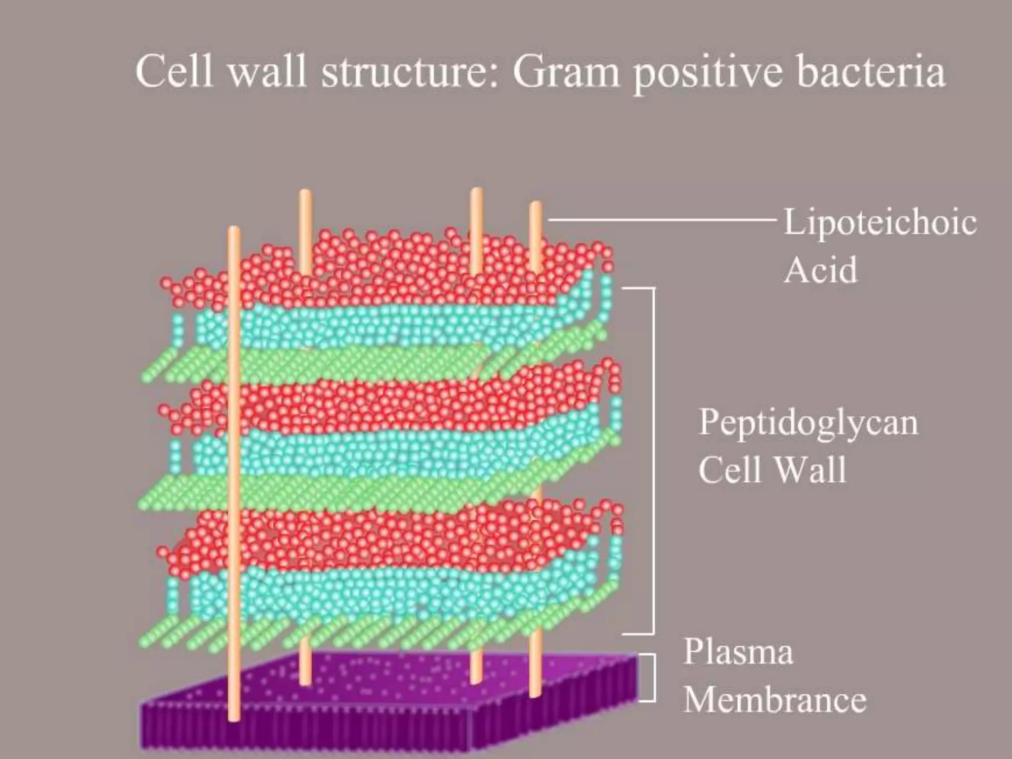
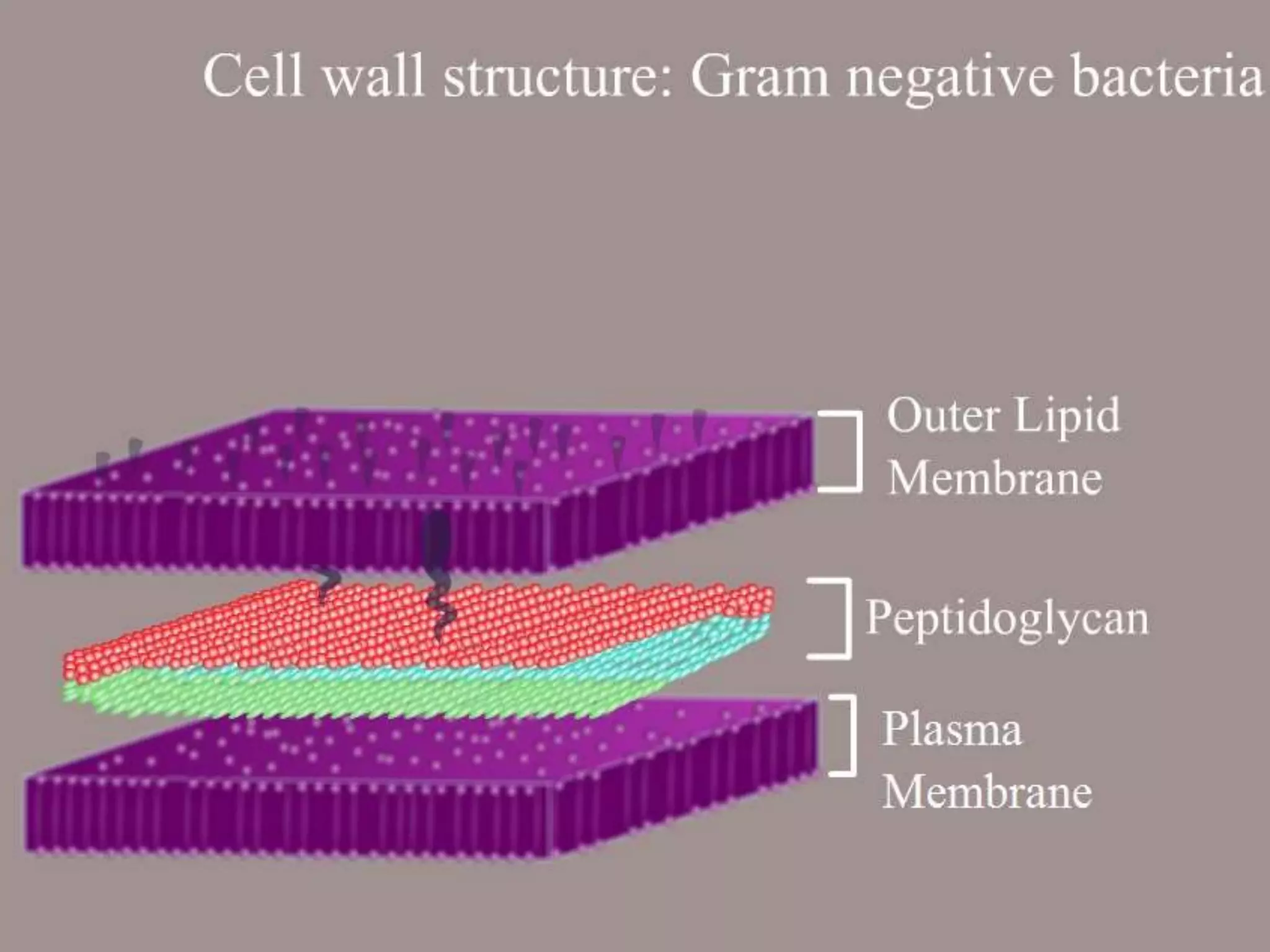
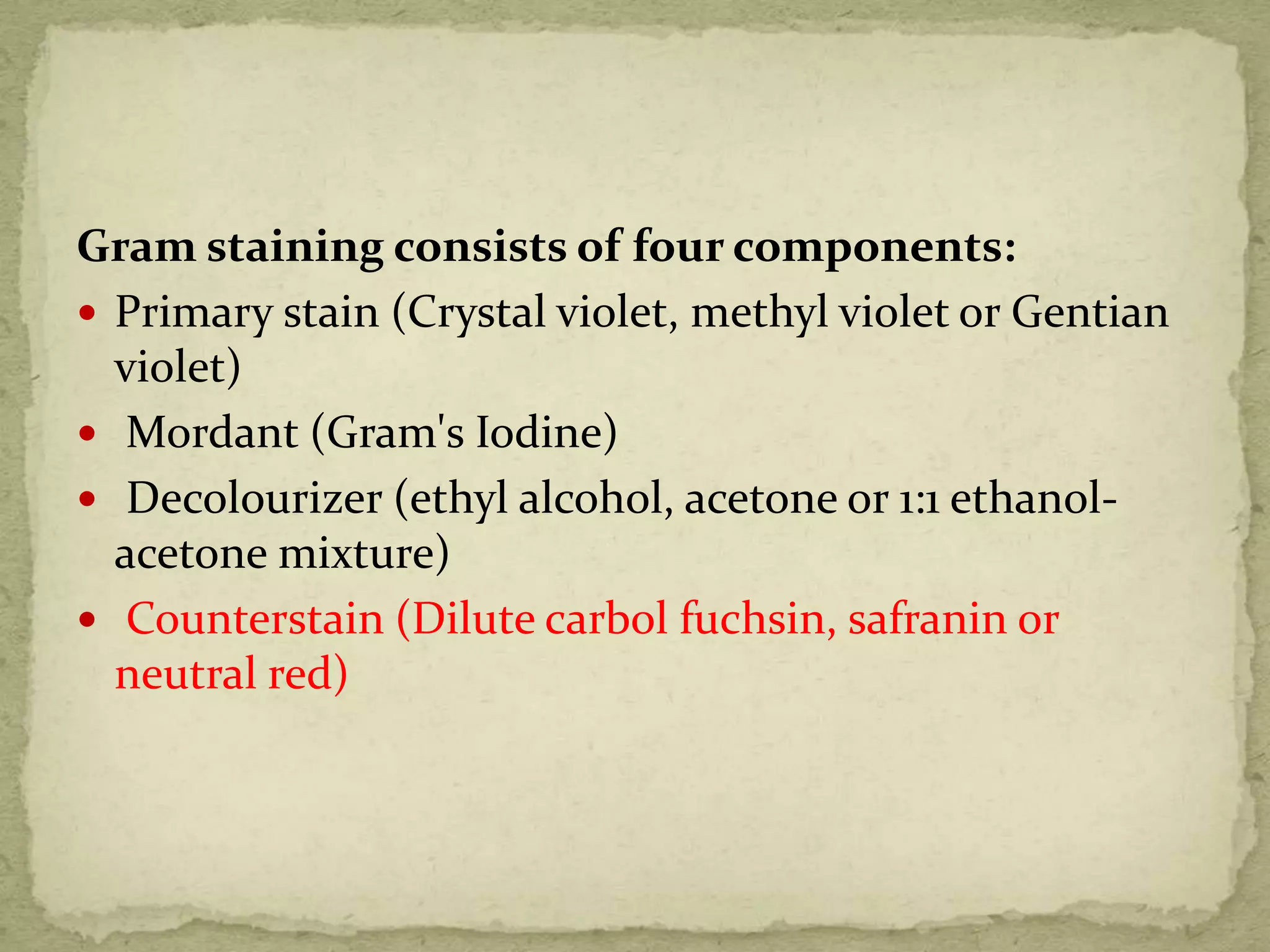
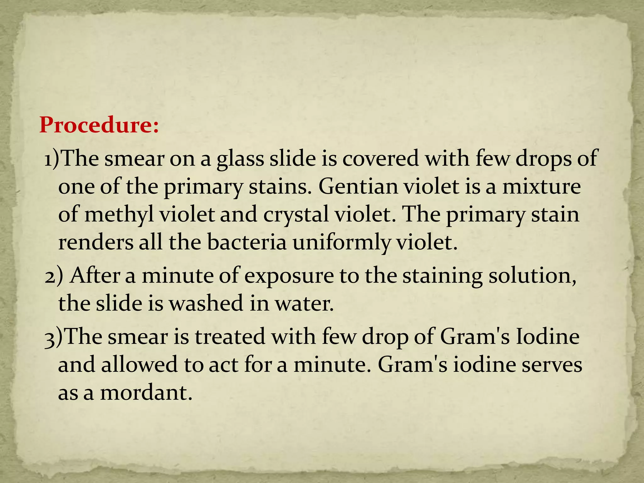
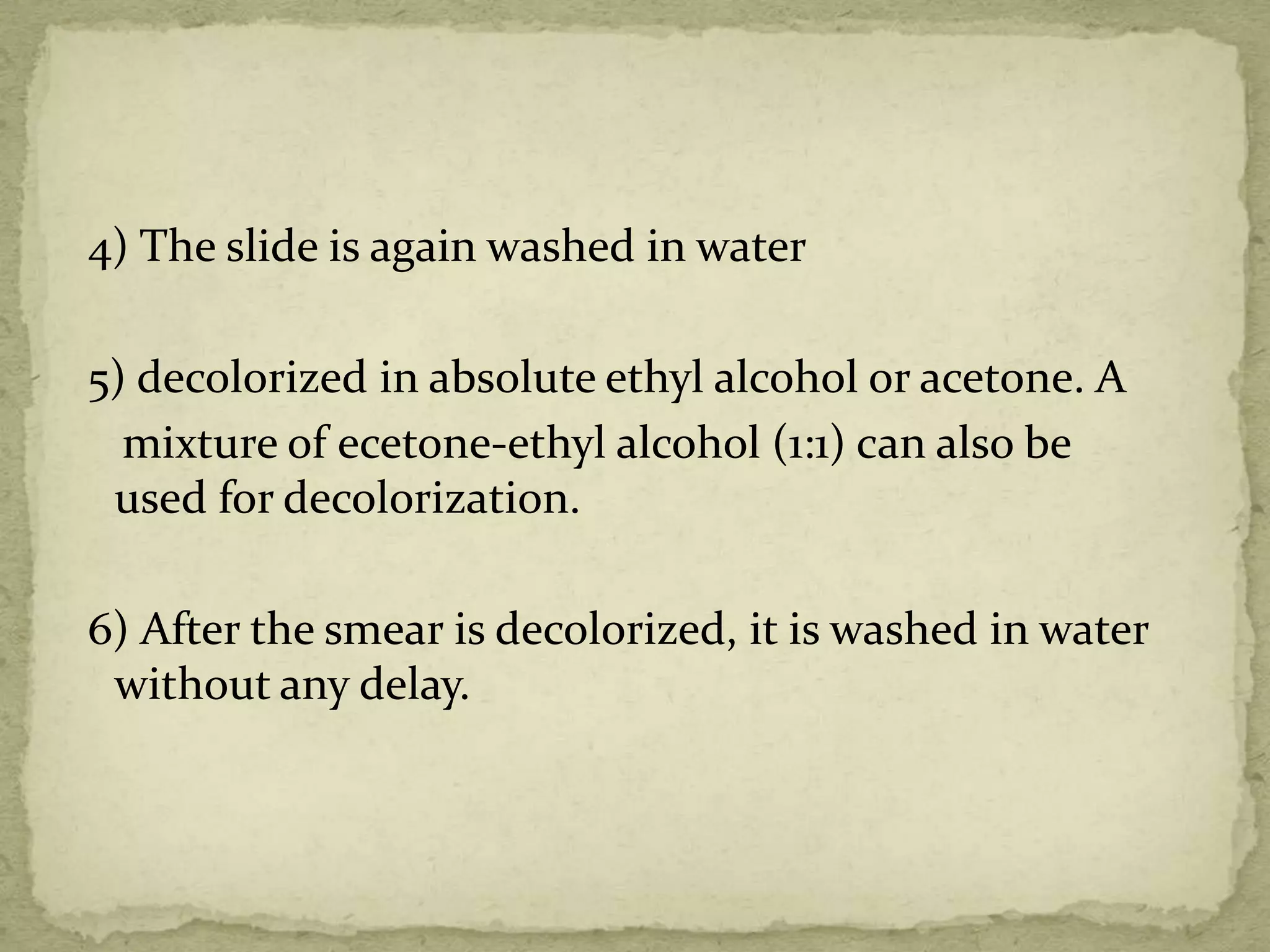
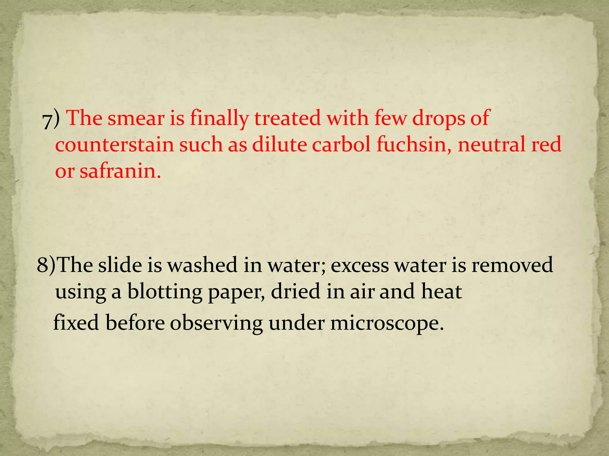
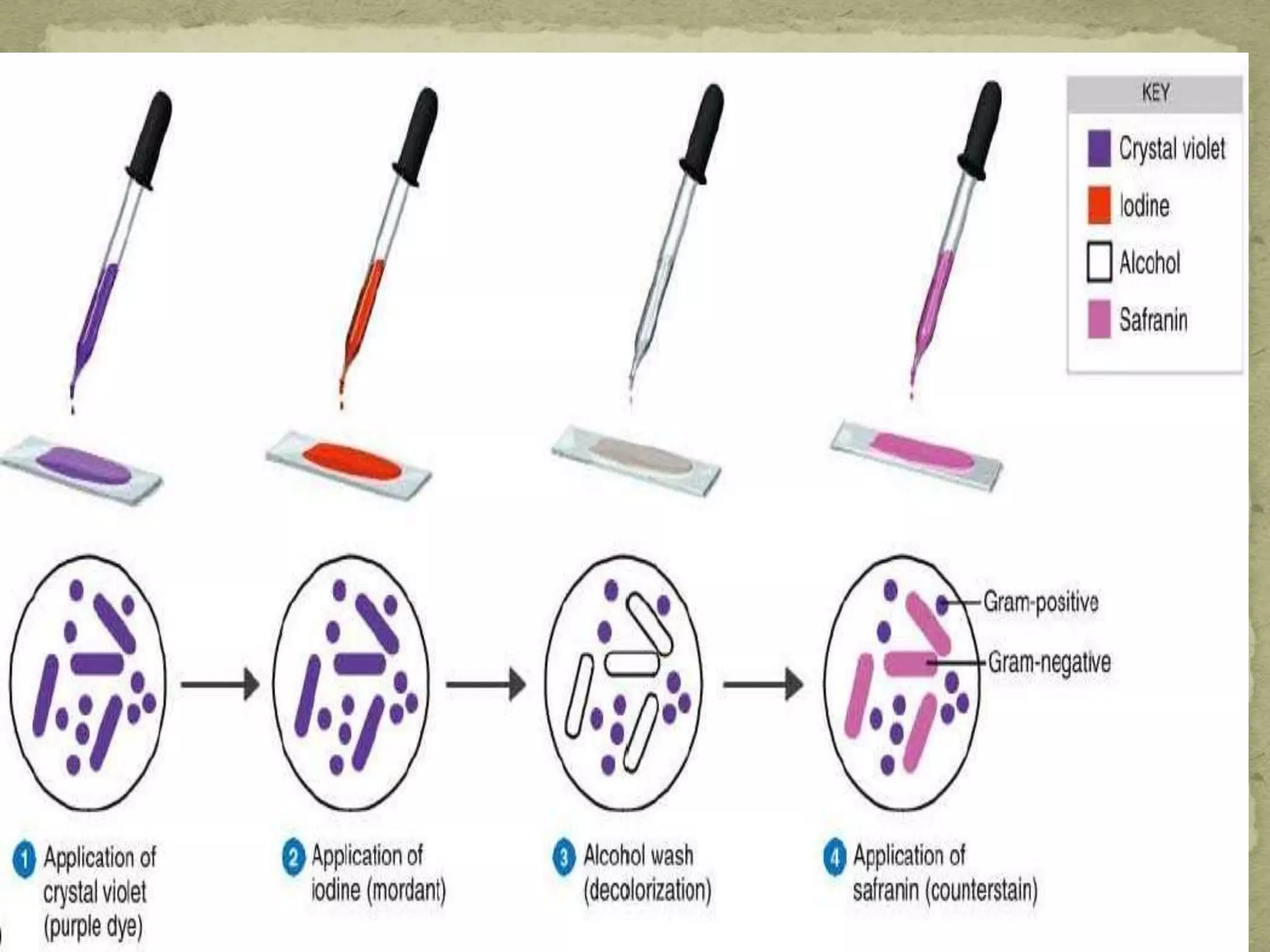
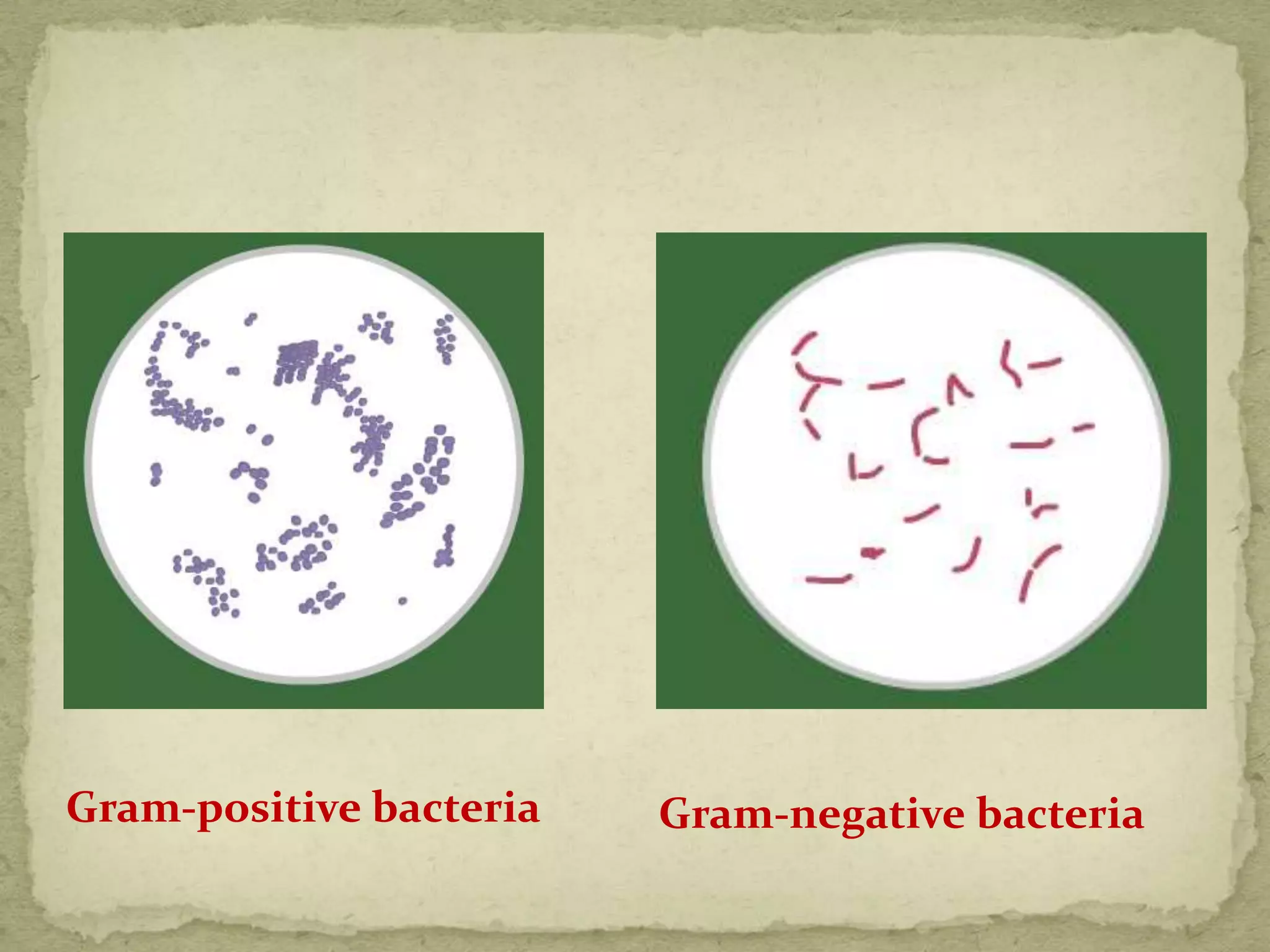
SlideShare a Scribd company logo
MADE BY:
DR. KALPAJYOTIBHATTACHARJEE
 DEFINATIONS
 EXAMPLES OF COUNTERSTAIN
 USES OF COUNTERSTAIN
 H&E STAIN
 VERHOEFF’S HAEMATOXYLIN
 ORANGE G
 GRAM’S STAIN
 ZIEHL-NEELSEN STAIN
 GIMENEZ STAIN
 NEUTRAL RED
 IHC COUNTERSTAIN
STAINS :
Are substances used to impart color to tissues or
cells, to facilitate microscopic study &
identification.
STAINING :
It is the artificial coloration of a substance to
facilitate examination of tissues ,microorganisms,
or other cells under the microscope.
COUNTERSTAIN :
A stain applied to render the effects of another
stain more discernable.
1) EOSIN counterstain to haemotoxyllin in H&E stain.
2) Malachite green counterstain to the fuschine stain in
Gimenez staining technique.
3) In Gram’s staining- safranin counterstain is applied
which stains all cells even following the
identification of gram –ve bacteria as well.
4) PAS- Orange G
5) Ziehl- Neelson technique- Methylene blue
6) Neutral Red.
7) Verhoeff’s stain
8) IHC counterstain- chromogenic
fluorescent
 Used to differentiate one component or cellular
structure from another or to differentiate an entity
from another in a specimen.
 It does so by coloring a portion of a specimen that
remained uncolored following the 1st staining using a
dye of different color.
 Most widely used stain.
 It clearly demonstrates an enormous number of
different tissue structures
 Haematoxylin :
Stains cell nuclei blue – black
Good intra nuclear detail
 Eosin :
Stains cell cytoplasm & most connective tissue fibers.
HAEMATOXYLIN
 Extracted from : Heartwood (logwood) of the tree
Haematoxylon campechianum that originated in the
Mexican State of Campeche.
 It is extracted from logwood with hot water & then
precipitated out from the aqueous solution using urea.
 Haematoxylin itself is not a stain
 Its major oxidation product is hematein- natural dye
responsible for the color properties.
 Hematein is produced in two ways
a)Natural oxidation- exposure to light & air.
slow process- 3-4 months.
E.g- Ehrlich’s and Delafield’s hematoxylin.
oxidation
b)Chemical oxidation- using sodium nitrate (Mayer’s
hematoxylin) or mercuric oxide (Harris heatoxyllin).
 ready for use immediately after preparation
 shorter life than natural oxidation one.
 Mordents used- salts of aluminum, iron and tungsten.
TYPES OF HEMATOXYLIN:
 Alum hematoxylins
 Iron hematoxylins
 Tungsten hematoxylins
 Molybdenum hematoxylins
 Lead hematoxylins
 Hematoxylin without mordant
Counter stain
EOSIN-
 Most suitable stain to combine with an alum
hematoxylin.
 It is a xanthene dye
 Variants:
a) Eosin B- (eosin bluish, erythrosin B) dibromodinitro
derivative of flurorescein
b) Eosin Y- (eosin yellowish, eosin water- soluble)
tetrabromo derivative of fluorescein.
c) Ethyl Eosin- (Eosin S, eosin alcohol soluble)
• Eosin Y is most widely used, satisfactorily soluble
in alcohol- “water and alcohol soluble”.
• As a counterstain- 0.5 - 1% aqueous solution water
– soluble solution eosin is generally preferred.
• Differentiation of eosin staining occurs in the
subsequent tap water wash, and a little
differentiation occurs during the dehydration
through the alcohol.
• Ethyl eosin and eosin B are rarely used. E.g.- Harris
stain for negri bodies.
USES OF EOSIN:
 Its ability with proper differentiation, to distinguish
between the cytoplasm of different types of cell, and
between the different types of connective tissue fibers
and matrices, by staining them differing shades of red
and pink.
 Counterstain to haemotoxylin in H&E staining-
cytoplasm stains pink-orange & nuclei stained darkly
blue-purple.
 Stains RBC intensely red.
Method :
1) Remove paraffin wax with xylene, 5 minutes.
2) Treat with absolute alcohol, two changes, 1 minute
each.
3) Wash in water.
4) Stain in haematoxylin, 10 minutes.
5) Wash in water.
6) If regressive stain is used, differentiate in acid /
alcohol until only nuclei remain blue.
7) Wash in water.
8) Blue in running water - 10 minutes.
9) Rinse in water.
10) Stain in 1 % aqueous eosin for 10-15 seconds.
11) Wash in running water for 30 seconds.
12) Dehydrate in 3changes absolute alcohol for 1 minute.
13)Clear in 2changes xylene.
14)Mount in suitable synthetic resin.
Results :
 Nuclei
 RNA rich cytoplasm :Blue
 calcium
 Muscle, fibrin , keratin : Bright red
 Collagen : Pink
 Red blood cells : Orange/ red
 Useful stain to demonstrate elastic fibers.
PURPOSE:
 This stain is useful in demonstrating atrophy of
elastic tissue in cases of emphysema, and the
thinning and loss of elastic fibers in
arteriosclerosis, and other vascular diseases.
 With increasing age, changes such as splitting and
fragmentation occur, these changes are most
obvious in the skin which becomes wrinkled and
rather 'loose-fitting'
PRINCIPLE:
 The tissue is stained with a regressive hematoxylin,
consisting of ferric chloride and iodine.
 The differentiating is accomplished by using excess
mordant (ferric chloride) to break the tissue-mordant dye
complex.
 The dye will be attracted to the larger amount of mordant
in the differentiating solution and will be removed from
the tissue.
 The elastic tissue has the strongest affinity of the iron
hematoxylin complex and will retain the dye longer than
the other tissue elements.
 CONTROL: Artery or skin.
Reagent :
 5 % haematoxylin in absolute alcohol : 20 ml
 10% ferric chloride : 8 ml
 Verhoeff’s iodine : 8 ml
Method :
1) Bring sections to water.
2) Stain in Verhoeff’s iron haematoxylin for 15-30 mins
until sections are jet-black.
3) Differentiate in 2% ferric chloride.
4) Wash in water, then in 95% alcohol to remove iodine
coloration.
5) Wash in water for 5 mins
6) counterstain in Van Gieson’s stain for 3 mins.
7) Dehydrate rapidly, clear and mount in synthetic
resin.
Results :
 Elastic fibers : Black
 Collagen : Red
 Muscle : Yellow
 Nuclei : Black
Result: Elastic fibres - black; collagen - red;
cytoplasm and muscle - yellow
 The universal stain for cytological preparation is the
Papanicolaou stain.
 Pap staining is used to differentiate cells in smear
preparations of various bodily secretions.
REAGENTS:
 Harris’s hematoxylin
 Orange G6 Counterstain (OG5 OG8)
 EA 50(Eosin Azure) Counterstain
ORANGE G:
 The main use of Orange G is in the OG-
6 Papanicolaou stain, to stain keratin
 It is also a major component of the Alexander test for
pollen staining.
 It is often combined with other yellow dyes and used to
stain erythrocytes in the trichrome methods.
EOSIN AZURE:
 EA (Eosin Azure) counterstain, comprising three
dyes; the number denotes the proportion of the
dyes, e.g. EA-36, EA-50, EA-65.
a) Eosin Y
b) Light Green SF
c) Bismarck brown Y.
 The Gram staining method is named
after the Danish bacteriologist Hans
Christian Gram (1853 –1938) who
originally devised it in 1882.
 It is a differential staining method of
differentiating bacterial species into
two large groups (Gram-positive and
Gram-negative) based on the chemical
and physical properties of their cell
walls.
Principle:
 Gram staining is a differential staining technique that
differentiates bacteria into two groups: gram-positives and
gram-negatives.
 The procedure is based on the ability of microorganisms to
retain color of the stains used during the gram stain
reaction.
 Gram-negative bacteria are decolorized by the
alcohol, losing the color of the primary stain, purple.
 Gram-positive bacteria are not decolorized by alcohol and
will remain as purple.
 After decolorization step, a counterstain is used to impart a
pink color to the decolorized gram-negative organisms.
 Gram-positive bacteria have a thick mesh-like cell wall
which is made up of peptidoglycan (50-90% of cell wall),
which stains purple.
 Peptidoglycan is mainly a polysaccharide composed of
two subunits called N-acetyl glucosamine and N-acetyl
muramic acid.
 As adjacent layers of peptidoglycan are formed, they are
cross linked by short chains of peptides by means of a
transpeptidase enzyme, resulting in the shape and rigidity
of the cell wall.
 The thick peptidoglycan layer of Gram-positive organisms
allows these organisms to retain the crystal violet-iodine
complex and stains the cells as purple.
 Lipoteichoic acid (LTA) is another major constituent of the
cell wall of Gram-positive bacteria which is embedded in
the peptidoglycan layer. It acts as regulator of autolytic
wall enzymes.
 Gram-negative bacteria have a thinner layer of
peptidoglycan (10% of the cell wall) and lose the
crystal violet-iodine complex during decolorization
with the alcohol rinse, but retain the counter stain
Safranin, thus appearing reddish or pink.
 They also have an additional outer membrane which
contains lipids, which is separated from the cell wall
by means of periplasmic space.
Gram staining consists of four components:
 Primary stain (Crystal violet, methyl violet or Gentian
violet)
 Mordant (Gram's Iodine)
 Decolourizer (ethyl alcohol, acetone or 1:1 ethanol-
acetone mixture)
 Counterstain (Dilute carbol fuchsin, safranin or
neutral red)
Procedure:
1)The smear on a glass slide is covered with few drops of
one of the primary stains. Gentian violet is a mixture
of methyl violet and crystal violet. The primary stain
renders all the bacteria uniformly violet.
2) After a minute of exposure to the staining solution,
the slide is washed in water.
3)The smear is treated with few drop of Gram's Iodine
and allowed to act for a minute. Gram's iodine serves
as a mordant.
4) The slide is again washed in water
5) decolorized in absolute ethyl alcohol or acetone. A
mixture of ecetone-ethyl alcohol (1:1) can also be
used for decolorization.
6) After the smear is decolorized, it is washed in water
without any delay.
7) The smear is finally treated with few drops of
counterstain such as dilute carbol fuchsin, neutral red
or safranin.
8)The slide is washed in water; excess water is removed
using a blotting paper, dried in air and heat
fixed before observing under microscope.
Gram-positive bacteria Gram-negative bacteria
Safranin (also Safranin O or basic red 2)
 a biological stain used in histology and cytology
Use:
a) as a counterstain in some staining protocols,
colouring all cell nuclei red.
b) This is the classic counterstain in both Gram stains,
and endospore staining.
c) It can also be used for the detection
of cartilage, mucin and mast cell granules
Carbol fuchsin:
 Mixture of phenol and basic fuchsin.
USES:
 in bacterial staining procedures.
 Component of Ziehi- Neelson Stain.
 Used as a dye to detect acid fast bacteria because it is
more soluble in the cell wall lipids than in the acid
alcohol.
 Topical antiseptic.
 The Ziehl–Neelsen stain, also known as the acid-fast
stain
PURPOSE:
Used in the demonstration of acid-fast bacteria
belonging to the genus 'mycobacterium', which
include the causative agent for tuberculosis.
PRINCIPLE:
The lipoid capsule of the acid-fast organism takes up
carbol fuchsin and resists decolorization with a dilute
acid rinse. The lipoid capsule of the mycobacteria is of
such high molecular weight that it is waxy at room
temperature and successful penetration by the
aqueous based staining solutions (such as Gram's) is
prevented.
 CONTROL: Any tissue containing acid-fast organisms
 FIXATIVE: 10% formalin
Reagents
 Carbol fuschin (basic dye)
 Mordant (heat)
 20% sulphuric acid (decolorizer)
 Methylene blue (counter stain) or Malachite green
Procedure
 Fix the smear of the specimen over the glass slide,
either by heating or alcohol fixation.
 Pour carbol fuschin over smear and heat gently until
fumes appear. Do not overheat and allow it to stand for
5 minutes, then wash it off with water.
 Pour 20% sulphuric acid, wait for one minute and keep
on repeating this step until the slide appears light pink
in color. Wash off with water.
 Pour methylene blue, wait for two minutes, again wash
with water
 Allow it to air dry and examine under oil immersion
lens.
Result
 Acid fast bacilli - pink, straight or slightly curved rods,
at times having beaded appearance.
 Background - blue due to methylene blue.
Methylene blue
 heterocyclic aromatic chemical compound
 molecular formula C16H18 N3SCl
 At room temperature- solid, odourless, dark green
powder that yields a blue solution when dissolved in
water.
USES:
 redox indicator in analytical chemistry- indicates the
presence or absence of oxygen.
 Intravital and supravital staining of nerve fibers
 Antidote to potassium cyanide poisonong
 Treatment of methemoglobinemia
 placebo
 Gimenez staining technique uses biological
stains to detect and
identify bacterial infections in tissue samples.
 Basic fuchsin stain in aqueous solution
with phenol and ethanol colours many bacteria
(both gram positive and Gram negative) red, magenta,
or pink.
 A malachite green counterstain gives a blue-green
background cast to the surrounding tissue.
Mechanism
 Gimenez stain is made up of basic fuchsin,ethanol and
phenol,sodium Di-hydrogen phosphate and Di-
sodium hydrogen phosphate.
 Gimenez stain has a property to stain both type of
bacteria that is Gram positive as well as Gram negative
bacteria.
 Basic fuchsin stains negatively charged cytoplasm and
stains cytoplasm in red colour.
 Where as when we treat smear with Malachite green
and as we know malachite green is a basic dye it has a
strong affinity towards host cell material as compared
to cytoplasm and so it stains host cell material that is
background of cell in bluish green colour.
METHOD
1. Sections to water.
2. Filter on carbol fuchsin solution for 1 minute.
3. Wash well in water.
4. Stain with 1% malachite green for up to 45 seconds.
5. Wash well in water.
6. Repeat step 4 until sections appear blue/green.
7. Wash, blot dry, and dry in air.
8. Mount sections in DPX
RESULTS
 Bacteria- red/magenta
 Background- blue/green
Malachite green:
 Malachite green is an organic compound that is used
as a dyestuff
 First prepared by Fischer in 1877
USES:
 used as a dye
 parasiticide and antibacterial.
 used in endospore staining
 MG has frequently been used to catch thieves
 is a dye used for staining in histology.
 It stains lysosomes red.
 It is used as a general stain in histology, as
a counterstain in combination with other dyes, and for
many staining methods.
PRINCIPLE :
 The neutral red (NR) assay procedure is a cell
survival/viability assay based on the ability of viable cells to
incorporate and bind neutral red within lysosomes.
 It is generally performed on adherent cells.
 NR is a weak cationic dye that readily penetrates the cell
membrane and accumulates intracellularly in lysosomes
(lysosomal pH < cytoplasmic pH), where it binds with
anionic sites to the lysosomal matrix .
 It is thus possible to distinguish between viable, damaged,
or dead cells, which is the basis of the assay.
USES:
 used as a general stain in histology, as a counterstain in
combination with other dyes, and for many staining
methods
 Stains Golgi apparatus in cells and Nissl
granules in neurons.
 Added to some growth media for bacterial and cell
cultures.
 acts as a pH indicator, changing from red to yellow
between pH 6.8 and 8.0.
 as a chloride salt
 After immunohistochemical staining the target
antigen, a second stain is often applied to provide
contast that helps the primary stain stand out.
 Mayers Hematoxylin is one of the most common
dyes used in diagnostic histology, as well as a common
nuclear counterstain in IHC.
 Nuclear fast red, also called Kernechtrot dye, is also
a nuclear stain. Differences between this dye and
hematoxylin, though, are that Nuclear fast red dyes
nucleic acids, results in red nuclear staining and only
takes 5 minutes to stain, instead of an hour with
hematoxylin.
 Methyl green is also a nucleic acid dye that rapidly
stains the nuclei green.
 Methyl green
 From blue to pink – Although not so often used as a
counterstain, another common blue dye is toluidine
blue.
 This is a strong basic dye that stains nuclei a deep blue
colour; however, it will also stain polysaccharadies a
pink/red colour.
 This colour shift is called metachromasia, a term used
when a dye stains a tissue component a different colour
to the dye solution.
 Eosin can also be used as a counterstain when an
antibody localized to the nucleus is used.
 Eosin is an anionic dye and works best in acidic
conditions.
 As nearly all proteins contain these two amino acids,
eosin is bound by the majority of structures in any
tissue.
 A ‘golden standard’ for fluorescence: DAPI.
 DAPI (4', 6-diamidino-2-phenylindole)
and Hoechst are common nuclear dyes used for
fluorescent IHC because they intercalate into the DNA
to give a strong blue color under UV excitation.
 Propidium iodide is another nucleic acid dye that is
frequently used to dye the nucleus red.
References :
 Culling.C.F.A ,Allison .R.T & Barr.W.T,Cellular
Pathology Technique; Butterworths; Fourth ed,
Page no.211 -312.
 Bancroft.J.D & Gamble .M, Theory & practice of
histological techniques; Elsevier,China; Sixth ed,
Page no 121-183.
COUNTERSTAIN ppt

More Related Content

What's hot

Special stain in histopathology
Special stain in histopathologySpecial stain in histopathology
Special stain in histopathology
aghara mahesh
 
DECALCIFICATION
DECALCIFICATIONDECALCIFICATION
DECALCIFICATION
Shravya Kishore
 
Embedding
EmbeddingEmbedding
Hematoxylin and Eosin Staining
Hematoxylin and Eosin StainingHematoxylin and Eosin Staining
Hematoxylin and Eosin Staining
Dr. Varughese George
 
Pas staining
Pas staining Pas staining
Cytotechniques
Cytotechniques  Cytotechniques
Cytotechniques
drtousif
 
Fixation of tissues
Fixation of tissuesFixation of tissues
Fixation of tissues
SUNIL KUMAR PEDDANA
 
Museum techniques
Museum techniquesMuseum techniques
Museum techniques
Sizan Thapa
 
Nucleic acid stains
Nucleic acid stainsNucleic acid stains
Nucleic acid stains
Sizan Thapa
 
Histopathology - CRYOSTAT
Histopathology - CRYOSTATHistopathology - CRYOSTAT
Histopathology - CRYOSTAT
SHRUTHI VASAN
 
Fixatives
Fixatives Fixatives
Fixatives
Aseem Jain
 
Decalcification
Decalcification  Decalcification
Decalcification
Layal Fahad
 
Hematological stains
Hematological stainsHematological stains
Hematological stains
Akash Dhiman
 
Mounting media
Mounting mediaMounting media
Mounting media
MAHMOUD IBRAHIM
 
Special stains in histopathology
Special stains in histopathologySpecial stains in histopathology
Special stains in histopathologyEkta Jajodia
 
Fixatives used in histopathology
Fixatives used in histopathologyFixatives used in histopathology
Fixatives used in histopathologyHitendra Prajapati
 
Hematoxylin and Eosin staining for UGs
Hematoxylin and Eosin staining for UGsHematoxylin and Eosin staining for UGs
Hematoxylin and Eosin staining for UGs
Dr. Varughese George
 
Anticoagulants used in haematology
Anticoagulants used in haematologyAnticoagulants used in haematology
Anticoagulants used in haematology
SUNIL KUMAR PEDDANA
 
H & e staining part 2
H & e staining part 2H & e staining part 2
H & e staining part 2
DrShrikant Sonune
 

What's hot (20)

Special stain in histopathology
Special stain in histopathologySpecial stain in histopathology
Special stain in histopathology
 
DECALCIFICATION
DECALCIFICATIONDECALCIFICATION
DECALCIFICATION
 
Embedding
EmbeddingEmbedding
Embedding
 
Hematoxylin and Eosin Staining
Hematoxylin and Eosin StainingHematoxylin and Eosin Staining
Hematoxylin and Eosin Staining
 
Pas staining
Pas staining Pas staining
Pas staining
 
Cytotechniques
Cytotechniques  Cytotechniques
Cytotechniques
 
Fixation of tissues
Fixation of tissuesFixation of tissues
Fixation of tissues
 
Museum techniques
Museum techniquesMuseum techniques
Museum techniques
 
Nucleic acid stains
Nucleic acid stainsNucleic acid stains
Nucleic acid stains
 
Histopathology - CRYOSTAT
Histopathology - CRYOSTATHistopathology - CRYOSTAT
Histopathology - CRYOSTAT
 
Fixatives
Fixatives Fixatives
Fixatives
 
Decalcification
Decalcification  Decalcification
Decalcification
 
Hematological stains
Hematological stainsHematological stains
Hematological stains
 
Mounting media
Mounting mediaMounting media
Mounting media
 
Special stains in histopathology
Special stains in histopathologySpecial stains in histopathology
Special stains in histopathology
 
Fixatives used in histopathology
Fixatives used in histopathologyFixatives used in histopathology
Fixatives used in histopathology
 
stains
stainsstains
stains
 
Hematoxylin and Eosin staining for UGs
Hematoxylin and Eosin staining for UGsHematoxylin and Eosin staining for UGs
Hematoxylin and Eosin staining for UGs
 
Anticoagulants used in haematology
Anticoagulants used in haematologyAnticoagulants used in haematology
Anticoagulants used in haematology
 
H & e staining part 2
H & e staining part 2H & e staining part 2
H & e staining part 2
 

Viewers also liked

Strain counterstrain powerpoint
Strain counterstrain powerpointStrain counterstrain powerpoint
Strain counterstrain powerpointsarathprabha
 
Staining Techniques in Microbiology
Staining Techniques in MicrobiologyStaining Techniques in Microbiology
Staining Techniques in Microbiology
Guddeti Prashanth Kumar
 
SIP slides on CYTO - gynae
SIP slides on CYTO - gynaeSIP slides on CYTO - gynae
SIP slides on CYTO - gynaeguestdcd535
 
FINE NEEDLE ASPIRATION CYTOLOGY / FNAC
FINE NEEDLE ASPIRATION CYTOLOGY / FNACFINE NEEDLE ASPIRATION CYTOLOGY / FNAC
FINE NEEDLE ASPIRATION CYTOLOGY / FNAC
K BHATTACHARJEE
 
Exfoliative cytology
Exfoliative cytology Exfoliative cytology
Exfoliative cytology
Atifa Ambreen
 
Cell lab
Cell labCell lab
Cell lab
Bryon Spicci
 
Detection of Abnormal Cervical Cytology by Papanicolaou Stained (PAP) Smears ...
Detection of Abnormal Cervical Cytology by Papanicolaou Stained (PAP) Smears ...Detection of Abnormal Cervical Cytology by Papanicolaou Stained (PAP) Smears ...
Detection of Abnormal Cervical Cytology by Papanicolaou Stained (PAP) Smears ...
SSR Institute of International Journal of Life Sciences
 
Reflective essay biomedical techniques
Reflective essay biomedical techniquesReflective essay biomedical techniques
Reflective essay biomedical techniquesjosearzon99
 
Training on Tuberculosis & Its Treatment
Training on Tuberculosis & Its TreatmentTraining on Tuberculosis & Its Treatment
Training on Tuberculosis & Its TreatmentDr. ANSHU GOKARN
 
hematoxylin and eosin stains
hematoxylin and eosin stainshematoxylin and eosin stains
hematoxylin and eosin stains
Upama Sishan
 
B.sc. (micro) i em unit 4.1 staining techniques
B.sc. (micro) i em unit 4.1 staining techniquesB.sc. (micro) i em unit 4.1 staining techniques
B.sc. (micro) i em unit 4.1 staining techniques
Rai University
 
Povidone iodine
Povidone iodinePovidone iodine
Povidone iodine
Vohinh Ngo
 

Viewers also liked (17)

Strain counterstrain powerpoint
Strain counterstrain powerpointStrain counterstrain powerpoint
Strain counterstrain powerpoint
 
Pap smear
Pap smear Pap smear
Pap smear
 
Staining Techniques in Microbiology
Staining Techniques in MicrobiologyStaining Techniques in Microbiology
Staining Techniques in Microbiology
 
SIP slides on CYTO - gynae
SIP slides on CYTO - gynaeSIP slides on CYTO - gynae
SIP slides on CYTO - gynae
 
Routine H &amp; E
Routine H &amp; ERoutine H &amp; E
Routine H &amp; E
 
FINE NEEDLE ASPIRATION CYTOLOGY / FNAC
FINE NEEDLE ASPIRATION CYTOLOGY / FNACFINE NEEDLE ASPIRATION CYTOLOGY / FNAC
FINE NEEDLE ASPIRATION CYTOLOGY / FNAC
 
Exfoliative cytology
Exfoliative cytology Exfoliative cytology
Exfoliative cytology
 
Cell lab
Cell labCell lab
Cell lab
 
Detection of Abnormal Cervical Cytology by Papanicolaou Stained (PAP) Smears ...
Detection of Abnormal Cervical Cytology by Papanicolaou Stained (PAP) Smears ...Detection of Abnormal Cervical Cytology by Papanicolaou Stained (PAP) Smears ...
Detection of Abnormal Cervical Cytology by Papanicolaou Stained (PAP) Smears ...
 
Reflective essay biomedical techniques
Reflective essay biomedical techniquesReflective essay biomedical techniques
Reflective essay biomedical techniques
 
Training on Tuberculosis & Its Treatment
Training on Tuberculosis & Its TreatmentTraining on Tuberculosis & Its Treatment
Training on Tuberculosis & Its Treatment
 
Fnac
FnacFnac
Fnac
 
hematoxylin and eosin stains
hematoxylin and eosin stainshematoxylin and eosin stains
hematoxylin and eosin stains
 
B.sc. (micro) i em unit 4.1 staining techniques
B.sc. (micro) i em unit 4.1 staining techniquesB.sc. (micro) i em unit 4.1 staining techniques
B.sc. (micro) i em unit 4.1 staining techniques
 
Povidone iodine
Povidone iodinePovidone iodine
Povidone iodine
 
Pradip vitamin
Pradip vitaminPradip vitamin
Pradip vitamin
 
Fnac
FnacFnac
Fnac
 

Similar to COUNTERSTAIN ppt

Histopatholgy staining by suchit kumar
Histopatholgy staining by suchit kumarHistopatholgy staining by suchit kumar
Histopatholgy staining by suchit kumar
suchitkumar24
 
Staining ( rouine and special in cytology) rajiv kumar
Staining ( rouine and special in cytology) rajiv kumarStaining ( rouine and special in cytology) rajiv kumar
Staining ( rouine and special in cytology) rajiv kumar
rajusehrawat
 
5. Staining techniques (Microbiology)
5. Staining techniques (Microbiology)5. Staining techniques (Microbiology)
5. Staining techniques (Microbiology)
Jay Khaniya
 
Gram staining
Gram stainingGram staining
Gram staining
Suprakash Das
 
Romanowsky stain
Romanowsky stainRomanowsky stain
Romanowsky stain
sandeep singh
 
Special Staining in Histopathology.pptx
Special Staining in Histopathology.pptxSpecial Staining in Histopathology.pptx
Special Staining in Histopathology.pptx
Bhatyasir1234
 
Hematoxylin and eosin staining Dr.Abhinav Golla MD Pathology
Hematoxylin and eosin staining Dr.Abhinav Golla MD PathologyHematoxylin and eosin staining Dr.Abhinav Golla MD Pathology
Hematoxylin and eosin staining Dr.Abhinav Golla MD Pathology
Aadhya Medicure Pathlabs Vijayanagar colony.hyderabad
 
staining of blood smears.pptx
staining of blood smears.pptxstaining of blood smears.pptx
staining of blood smears.pptx
abdiasis omar mohamed
 
CYTOLOGIC STAINING TECHNIQUES.ppt
CYTOLOGIC STAINING TECHNIQUES.pptCYTOLOGIC STAINING TECHNIQUES.ppt
CYTOLOGIC STAINING TECHNIQUES.ppt
MEDTECH LAB
 
HISTOPATH Staining.pptx
HISTOPATH Staining.pptxHISTOPATH Staining.pptx
HISTOPATH Staining.pptx
WynlorAbarca1
 
Staining of microorganisms
Staining of microorganismsStaining of microorganisms
Staining of microorganisms
Dr.Dinesh Jain
 
COURSE MLSC 417 Staining in Bacteriology.ppt
COURSE MLSC 417 Staining in Bacteriology.pptCOURSE MLSC 417 Staining in Bacteriology.ppt
COURSE MLSC 417 Staining in Bacteriology.ppt
petry2
 
COURSE MLSC 417 Staining in Bacteriology (1).ppt
COURSE MLSC 417 Staining in Bacteriology (1).pptCOURSE MLSC 417 Staining in Bacteriology (1).ppt
COURSE MLSC 417 Staining in Bacteriology (1).ppt
RahulVishwakarma722529
 
Hema I Chapter 6_staining.ppt
Hema I Chapter 6_staining.pptHema I Chapter 6_staining.ppt
Hema I Chapter 6_staining.ppt
chandreshmishra13
 
Hema -staining smears.ppt
Hema -staining smears.pptHema -staining smears.ppt
Hema -staining smears.ppt
ssuser75fd45
 
Hema I Chapter 6_staining.ppt
Hema I Chapter 6_staining.pptHema I Chapter 6_staining.ppt
Hema I Chapter 6_staining.ppt
deribew genetu
 
Staining techniques
Staining techniquesStaining techniques
Staining techniques
Maninderjitkaur4
 
romanowskystains-180521083448 (1).pptx
romanowskystains-180521083448 (1).pptxromanowskystains-180521083448 (1).pptx
romanowskystains-180521083448 (1).pptx
rohitshrivastava97
 

Similar to COUNTERSTAIN ppt (20)

Histopatholgy staining by suchit kumar
Histopatholgy staining by suchit kumarHistopatholgy staining by suchit kumar
Histopatholgy staining by suchit kumar
 
Staining ( rouine and special in cytology) rajiv kumar
Staining ( rouine and special in cytology) rajiv kumarStaining ( rouine and special in cytology) rajiv kumar
Staining ( rouine and special in cytology) rajiv kumar
 
5. Staining techniques (Microbiology)
5. Staining techniques (Microbiology)5. Staining techniques (Microbiology)
5. Staining techniques (Microbiology)
 
Stains
StainsStains
Stains
 
Gram staining
Gram stainingGram staining
Gram staining
 
Romanowsky stain
Romanowsky stainRomanowsky stain
Romanowsky stain
 
Special Staining in Histopathology.pptx
Special Staining in Histopathology.pptxSpecial Staining in Histopathology.pptx
Special Staining in Histopathology.pptx
 
Hematoxylin and eosin staining Dr.Abhinav Golla MD Pathology
Hematoxylin and eosin staining Dr.Abhinav Golla MD PathologyHematoxylin and eosin staining Dr.Abhinav Golla MD Pathology
Hematoxylin and eosin staining Dr.Abhinav Golla MD Pathology
 
staining of blood smears.pptx
staining of blood smears.pptxstaining of blood smears.pptx
staining of blood smears.pptx
 
DOUBLE STAINING
DOUBLE STAININGDOUBLE STAINING
DOUBLE STAINING
 
CYTOLOGIC STAINING TECHNIQUES.ppt
CYTOLOGIC STAINING TECHNIQUES.pptCYTOLOGIC STAINING TECHNIQUES.ppt
CYTOLOGIC STAINING TECHNIQUES.ppt
 
HISTOPATH Staining.pptx
HISTOPATH Staining.pptxHISTOPATH Staining.pptx
HISTOPATH Staining.pptx
 
Staining of microorganisms
Staining of microorganismsStaining of microorganisms
Staining of microorganisms
 
COURSE MLSC 417 Staining in Bacteriology.ppt
COURSE MLSC 417 Staining in Bacteriology.pptCOURSE MLSC 417 Staining in Bacteriology.ppt
COURSE MLSC 417 Staining in Bacteriology.ppt
 
COURSE MLSC 417 Staining in Bacteriology (1).ppt
COURSE MLSC 417 Staining in Bacteriology (1).pptCOURSE MLSC 417 Staining in Bacteriology (1).ppt
COURSE MLSC 417 Staining in Bacteriology (1).ppt
 
Hema I Chapter 6_staining.ppt
Hema I Chapter 6_staining.pptHema I Chapter 6_staining.ppt
Hema I Chapter 6_staining.ppt
 
Hema -staining smears.ppt
Hema -staining smears.pptHema -staining smears.ppt
Hema -staining smears.ppt
 
Hema I Chapter 6_staining.ppt
Hema I Chapter 6_staining.pptHema I Chapter 6_staining.ppt
Hema I Chapter 6_staining.ppt
 
Staining techniques
Staining techniquesStaining techniques
Staining techniques
 
romanowskystains-180521083448 (1).pptx
romanowskystains-180521083448 (1).pptxromanowskystains-180521083448 (1).pptx
romanowskystains-180521083448 (1).pptx
 

More from K BHATTACHARJEE

Fibroosseous lesions
Fibroosseous lesionsFibroosseous lesions
Fibroosseous lesions
K BHATTACHARJEE
 
Bone tumors
Bone tumors   Bone tumors
Bone tumors
K BHATTACHARJEE
 
Blood disorders ppt
Blood disorders pptBlood disorders ppt
Blood disorders ppt
K BHATTACHARJEE
 
PULP AND PERIAPICAL LESIONS OF THE TOOTH ppt
PULP AND PERIAPICAL LESIONS OF THE TOOTH pptPULP AND PERIAPICAL LESIONS OF THE TOOTH ppt
PULP AND PERIAPICAL LESIONS OF THE TOOTH ppt
K BHATTACHARJEE
 
FORENSIC ODONTOLOGY ppt
FORENSIC ODONTOLOGY pptFORENSIC ODONTOLOGY ppt
FORENSIC ODONTOLOGY ppt
K BHATTACHARJEE
 
Immunohistochemistry METHODS
Immunohistochemistry  METHODSImmunohistochemistry  METHODS
Immunohistochemistry METHODS
K BHATTACHARJEE
 
ORAL SUBMUCOUS FIBROSIS- PPT
ORAL SUBMUCOUS FIBROSIS- PPTORAL SUBMUCOUS FIBROSIS- PPT
ORAL SUBMUCOUS FIBROSIS- PPT
K BHATTACHARJEE
 
Salivary gland tumors
Salivary gland tumorsSalivary gland tumors
Salivary gland tumors
K BHATTACHARJEE
 
Inflamation
InflamationInflamation
Inflamation
K BHATTACHARJEE
 
Osteology of facial skeleton
Osteology of facial skeletonOsteology of facial skeleton
Osteology of facial skeleton
K BHATTACHARJEE
 
Epithelium
EpitheliumEpithelium
Epithelium
K BHATTACHARJEE
 
Development of face, palate and jaw
Development of face, palate and jawDevelopment of face, palate and jaw
Development of face, palate and jaw
K BHATTACHARJEE
 

More from K BHATTACHARJEE (12)

Fibroosseous lesions
Fibroosseous lesionsFibroosseous lesions
Fibroosseous lesions
 
Bone tumors
Bone tumors   Bone tumors
Bone tumors
 
Blood disorders ppt
Blood disorders pptBlood disorders ppt
Blood disorders ppt
 
PULP AND PERIAPICAL LESIONS OF THE TOOTH ppt
PULP AND PERIAPICAL LESIONS OF THE TOOTH pptPULP AND PERIAPICAL LESIONS OF THE TOOTH ppt
PULP AND PERIAPICAL LESIONS OF THE TOOTH ppt
 
FORENSIC ODONTOLOGY ppt
FORENSIC ODONTOLOGY pptFORENSIC ODONTOLOGY ppt
FORENSIC ODONTOLOGY ppt
 
Immunohistochemistry METHODS
Immunohistochemistry  METHODSImmunohistochemistry  METHODS
Immunohistochemistry METHODS
 
ORAL SUBMUCOUS FIBROSIS- PPT
ORAL SUBMUCOUS FIBROSIS- PPTORAL SUBMUCOUS FIBROSIS- PPT
ORAL SUBMUCOUS FIBROSIS- PPT
 
Salivary gland tumors
Salivary gland tumorsSalivary gland tumors
Salivary gland tumors
 
Inflamation
InflamationInflamation
Inflamation
 
Osteology of facial skeleton
Osteology of facial skeletonOsteology of facial skeleton
Osteology of facial skeleton
 
Epithelium
EpitheliumEpithelium
Epithelium
 
Development of face, palate and jaw
Development of face, palate and jawDevelopment of face, palate and jaw
Development of face, palate and jaw
 

Recently uploaded

Charaka Samhita Sutra Sthana 9 Chapter khuddakachatuspadadhyaya
Charaka Samhita Sutra Sthana 9 Chapter khuddakachatuspadadhyayaCharaka Samhita Sutra Sthana 9 Chapter khuddakachatuspadadhyaya
Charaka Samhita Sutra Sthana 9 Chapter khuddakachatuspadadhyaya
Dr KHALID B.M
 
Evaluation of antidepressant activity of clitoris ternatea in animals
Evaluation of antidepressant activity of clitoris ternatea in animalsEvaluation of antidepressant activity of clitoris ternatea in animals
Evaluation of antidepressant activity of clitoris ternatea in animals
Shweta
 
micro teaching on communication m.sc nursing.pdf
micro teaching on communication m.sc nursing.pdfmicro teaching on communication m.sc nursing.pdf
micro teaching on communication m.sc nursing.pdf
Anurag Sharma
 
Surat @ℂall @Girls ꧁❤8527049040❤꧂@ℂall @Girls Service Vip Top Model Safe
Surat @ℂall @Girls ꧁❤8527049040❤꧂@ℂall @Girls Service Vip Top Model SafeSurat @ℂall @Girls ꧁❤8527049040❤꧂@ℂall @Girls Service Vip Top Model Safe
Surat @ℂall @Girls ꧁❤8527049040❤꧂@ℂall @Girls Service Vip Top Model Safe
Savita Shen $i11
 
The POPPY STUDY (Preconception to post-partum cardiovascular function in prim...
The POPPY STUDY (Preconception to post-partum cardiovascular function in prim...The POPPY STUDY (Preconception to post-partum cardiovascular function in prim...
The POPPY STUDY (Preconception to post-partum cardiovascular function in prim...
Catherine Liao
 
24 Upakrama.pptx class ppt useful in all
24 Upakrama.pptx class ppt useful in all24 Upakrama.pptx class ppt useful in all
24 Upakrama.pptx class ppt useful in all
DrSathishMS1
 
Ozempic: Preoperative Management of Patients on GLP-1 Receptor Agonists
Ozempic: Preoperative Management of Patients on GLP-1 Receptor Agonists  Ozempic: Preoperative Management of Patients on GLP-1 Receptor Agonists
Ozempic: Preoperative Management of Patients on GLP-1 Receptor Agonists
Saeid Safari
 
Are There Any Natural Remedies To Treat Syphilis.pdf
Are There Any Natural Remedies To Treat Syphilis.pdfAre There Any Natural Remedies To Treat Syphilis.pdf
Are There Any Natural Remedies To Treat Syphilis.pdf
Little Cross Family Clinic
 
New Directions in Targeted Therapeutic Approaches for Older Adults With Mantl...
New Directions in Targeted Therapeutic Approaches for Older Adults With Mantl...New Directions in Targeted Therapeutic Approaches for Older Adults With Mantl...
New Directions in Targeted Therapeutic Approaches for Older Adults With Mantl...
i3 Health
 
HOT NEW PRODUCT! BIG SALES FAST SHIPPING NOW FROM CHINA!! EU KU DB BK substit...
HOT NEW PRODUCT! BIG SALES FAST SHIPPING NOW FROM CHINA!! EU KU DB BK substit...HOT NEW PRODUCT! BIG SALES FAST SHIPPING NOW FROM CHINA!! EU KU DB BK substit...
HOT NEW PRODUCT! BIG SALES FAST SHIPPING NOW FROM CHINA!! EU KU DB BK substit...
GL Anaacs
 
Hemodialysis: Chapter 3, Dialysis Water Unit - Dr.Gawad
Hemodialysis: Chapter 3, Dialysis Water Unit - Dr.GawadHemodialysis: Chapter 3, Dialysis Water Unit - Dr.Gawad
Hemodialysis: Chapter 3, Dialysis Water Unit - Dr.Gawad
NephroTube - Dr.Gawad
 
Flu Vaccine Alert in Bangalore Karnataka
Flu Vaccine Alert in Bangalore KarnatakaFlu Vaccine Alert in Bangalore Karnataka
Flu Vaccine Alert in Bangalore Karnataka
addon Scans
 
POST OPERATIVE OLIGURIA and its management
POST OPERATIVE OLIGURIA and its managementPOST OPERATIVE OLIGURIA and its management
POST OPERATIVE OLIGURIA and its management
touseefaziz1
 
The Normal Electrocardiogram - Part I of II
The Normal Electrocardiogram - Part I of IIThe Normal Electrocardiogram - Part I of II
The Normal Electrocardiogram - Part I of II
MedicoseAcademics
 
Cervical & Brachial Plexus By Dr. RIG.pptx
Cervical & Brachial Plexus By Dr. RIG.pptxCervical & Brachial Plexus By Dr. RIG.pptx
Cervical & Brachial Plexus By Dr. RIG.pptx
Dr. Rabia Inam Gandapore
 
Lung Cancer: Artificial Intelligence, Synergetics, Complex System Analysis, S...
Lung Cancer: Artificial Intelligence, Synergetics, Complex System Analysis, S...Lung Cancer: Artificial Intelligence, Synergetics, Complex System Analysis, S...
Lung Cancer: Artificial Intelligence, Synergetics, Complex System Analysis, S...
Oleg Kshivets
 
Pulmonary Thromboembolism - etilogy, types, medical- Surgical and nursing man...
Pulmonary Thromboembolism - etilogy, types, medical- Surgical and nursing man...Pulmonary Thromboembolism - etilogy, types, medical- Surgical and nursing man...
Pulmonary Thromboembolism - etilogy, types, medical- Surgical and nursing man...
VarunMahajani
 
Physiology of Special Chemical Sensation of Taste
Physiology of Special Chemical Sensation of TastePhysiology of Special Chemical Sensation of Taste
Physiology of Special Chemical Sensation of Taste
MedicoseAcademics
 
Report Back from SGO 2024: What’s the Latest in Cervical Cancer?
Report Back from SGO 2024: What’s the Latest in Cervical Cancer?Report Back from SGO 2024: What’s the Latest in Cervical Cancer?
Report Back from SGO 2024: What’s the Latest in Cervical Cancer?
bkling
 
Couples presenting to the infertility clinic- Do they really have infertility...
Couples presenting to the infertility clinic- Do they really have infertility...Couples presenting to the infertility clinic- Do they really have infertility...
Couples presenting to the infertility clinic- Do they really have infertility...
Sujoy Dasgupta
 

Recently uploaded (20)

Charaka Samhita Sutra Sthana 9 Chapter khuddakachatuspadadhyaya
Charaka Samhita Sutra Sthana 9 Chapter khuddakachatuspadadhyayaCharaka Samhita Sutra Sthana 9 Chapter khuddakachatuspadadhyaya
Charaka Samhita Sutra Sthana 9 Chapter khuddakachatuspadadhyaya
 
Evaluation of antidepressant activity of clitoris ternatea in animals
Evaluation of antidepressant activity of clitoris ternatea in animalsEvaluation of antidepressant activity of clitoris ternatea in animals
Evaluation of antidepressant activity of clitoris ternatea in animals
 
micro teaching on communication m.sc nursing.pdf
micro teaching on communication m.sc nursing.pdfmicro teaching on communication m.sc nursing.pdf
micro teaching on communication m.sc nursing.pdf
 
Surat @ℂall @Girls ꧁❤8527049040❤꧂@ℂall @Girls Service Vip Top Model Safe
Surat @ℂall @Girls ꧁❤8527049040❤꧂@ℂall @Girls Service Vip Top Model SafeSurat @ℂall @Girls ꧁❤8527049040❤꧂@ℂall @Girls Service Vip Top Model Safe
Surat @ℂall @Girls ꧁❤8527049040❤꧂@ℂall @Girls Service Vip Top Model Safe
 
The POPPY STUDY (Preconception to post-partum cardiovascular function in prim...
The POPPY STUDY (Preconception to post-partum cardiovascular function in prim...The POPPY STUDY (Preconception to post-partum cardiovascular function in prim...
The POPPY STUDY (Preconception to post-partum cardiovascular function in prim...
 
24 Upakrama.pptx class ppt useful in all
24 Upakrama.pptx class ppt useful in all24 Upakrama.pptx class ppt useful in all
24 Upakrama.pptx class ppt useful in all
 
Ozempic: Preoperative Management of Patients on GLP-1 Receptor Agonists
Ozempic: Preoperative Management of Patients on GLP-1 Receptor Agonists  Ozempic: Preoperative Management of Patients on GLP-1 Receptor Agonists
Ozempic: Preoperative Management of Patients on GLP-1 Receptor Agonists
 
Are There Any Natural Remedies To Treat Syphilis.pdf
Are There Any Natural Remedies To Treat Syphilis.pdfAre There Any Natural Remedies To Treat Syphilis.pdf
Are There Any Natural Remedies To Treat Syphilis.pdf
 
New Directions in Targeted Therapeutic Approaches for Older Adults With Mantl...
New Directions in Targeted Therapeutic Approaches for Older Adults With Mantl...New Directions in Targeted Therapeutic Approaches for Older Adults With Mantl...
New Directions in Targeted Therapeutic Approaches for Older Adults With Mantl...
 
HOT NEW PRODUCT! BIG SALES FAST SHIPPING NOW FROM CHINA!! EU KU DB BK substit...
HOT NEW PRODUCT! BIG SALES FAST SHIPPING NOW FROM CHINA!! EU KU DB BK substit...HOT NEW PRODUCT! BIG SALES FAST SHIPPING NOW FROM CHINA!! EU KU DB BK substit...
HOT NEW PRODUCT! BIG SALES FAST SHIPPING NOW FROM CHINA!! EU KU DB BK substit...
 
Hemodialysis: Chapter 3, Dialysis Water Unit - Dr.Gawad
Hemodialysis: Chapter 3, Dialysis Water Unit - Dr.GawadHemodialysis: Chapter 3, Dialysis Water Unit - Dr.Gawad
Hemodialysis: Chapter 3, Dialysis Water Unit - Dr.Gawad
 
Flu Vaccine Alert in Bangalore Karnataka
Flu Vaccine Alert in Bangalore KarnatakaFlu Vaccine Alert in Bangalore Karnataka
Flu Vaccine Alert in Bangalore Karnataka
 
POST OPERATIVE OLIGURIA and its management
POST OPERATIVE OLIGURIA and its managementPOST OPERATIVE OLIGURIA and its management
POST OPERATIVE OLIGURIA and its management
 
The Normal Electrocardiogram - Part I of II
The Normal Electrocardiogram - Part I of IIThe Normal Electrocardiogram - Part I of II
The Normal Electrocardiogram - Part I of II
 
Cervical & Brachial Plexus By Dr. RIG.pptx
Cervical & Brachial Plexus By Dr. RIG.pptxCervical & Brachial Plexus By Dr. RIG.pptx
Cervical & Brachial Plexus By Dr. RIG.pptx
 
Lung Cancer: Artificial Intelligence, Synergetics, Complex System Analysis, S...
Lung Cancer: Artificial Intelligence, Synergetics, Complex System Analysis, S...Lung Cancer: Artificial Intelligence, Synergetics, Complex System Analysis, S...
Lung Cancer: Artificial Intelligence, Synergetics, Complex System Analysis, S...
 
Pulmonary Thromboembolism - etilogy, types, medical- Surgical and nursing man...
Pulmonary Thromboembolism - etilogy, types, medical- Surgical and nursing man...Pulmonary Thromboembolism - etilogy, types, medical- Surgical and nursing man...
Pulmonary Thromboembolism - etilogy, types, medical- Surgical and nursing man...
 
Physiology of Special Chemical Sensation of Taste
Physiology of Special Chemical Sensation of TastePhysiology of Special Chemical Sensation of Taste
Physiology of Special Chemical Sensation of Taste
 
Report Back from SGO 2024: What’s the Latest in Cervical Cancer?
Report Back from SGO 2024: What’s the Latest in Cervical Cancer?Report Back from SGO 2024: What’s the Latest in Cervical Cancer?
Report Back from SGO 2024: What’s the Latest in Cervical Cancer?
 
Couples presenting to the infertility clinic- Do they really have infertility...
Couples presenting to the infertility clinic- Do they really have infertility...Couples presenting to the infertility clinic- Do they really have infertility...
Couples presenting to the infertility clinic- Do they really have infertility...
 

COUNTERSTAIN ppt

  • 2.  DEFINATIONS  EXAMPLES OF COUNTERSTAIN  USES OF COUNTERSTAIN  H&E STAIN  VERHOEFF’S HAEMATOXYLIN  ORANGE G  GRAM’S STAIN  ZIEHL-NEELSEN STAIN  GIMENEZ STAIN  NEUTRAL RED  IHC COUNTERSTAIN
  • 3. STAINS : Are substances used to impart color to tissues or cells, to facilitate microscopic study & identification.
  • 4. STAINING : It is the artificial coloration of a substance to facilitate examination of tissues ,microorganisms, or other cells under the microscope.
  • 5. COUNTERSTAIN : A stain applied to render the effects of another stain more discernable.
  • 6. 1) EOSIN counterstain to haemotoxyllin in H&E stain. 2) Malachite green counterstain to the fuschine stain in Gimenez staining technique. 3) In Gram’s staining- safranin counterstain is applied which stains all cells even following the identification of gram –ve bacteria as well. 4) PAS- Orange G
  • 7. 5) Ziehl- Neelson technique- Methylene blue 6) Neutral Red. 7) Verhoeff’s stain 8) IHC counterstain- chromogenic fluorescent
  • 8.  Used to differentiate one component or cellular structure from another or to differentiate an entity from another in a specimen.  It does so by coloring a portion of a specimen that remained uncolored following the 1st staining using a dye of different color.
  • 9.  Most widely used stain.  It clearly demonstrates an enormous number of different tissue structures  Haematoxylin : Stains cell nuclei blue – black Good intra nuclear detail  Eosin : Stains cell cytoplasm & most connective tissue fibers.
  • 10. HAEMATOXYLIN  Extracted from : Heartwood (logwood) of the tree Haematoxylon campechianum that originated in the Mexican State of Campeche.
  • 11.  It is extracted from logwood with hot water & then precipitated out from the aqueous solution using urea.  Haematoxylin itself is not a stain  Its major oxidation product is hematein- natural dye responsible for the color properties.  Hematein is produced in two ways a)Natural oxidation- exposure to light & air. slow process- 3-4 months. E.g- Ehrlich’s and Delafield’s hematoxylin. oxidation
  • 12. b)Chemical oxidation- using sodium nitrate (Mayer’s hematoxylin) or mercuric oxide (Harris heatoxyllin).  ready for use immediately after preparation  shorter life than natural oxidation one.  Mordents used- salts of aluminum, iron and tungsten.
  • 13. TYPES OF HEMATOXYLIN:  Alum hematoxylins  Iron hematoxylins  Tungsten hematoxylins  Molybdenum hematoxylins  Lead hematoxylins  Hematoxylin without mordant
  • 14. Counter stain EOSIN-  Most suitable stain to combine with an alum hematoxylin.  It is a xanthene dye  Variants: a) Eosin B- (eosin bluish, erythrosin B) dibromodinitro derivative of flurorescein b) Eosin Y- (eosin yellowish, eosin water- soluble) tetrabromo derivative of fluorescein. c) Ethyl Eosin- (Eosin S, eosin alcohol soluble)
  • 15. • Eosin Y is most widely used, satisfactorily soluble in alcohol- “water and alcohol soluble”. • As a counterstain- 0.5 - 1% aqueous solution water – soluble solution eosin is generally preferred. • Differentiation of eosin staining occurs in the subsequent tap water wash, and a little differentiation occurs during the dehydration through the alcohol. • Ethyl eosin and eosin B are rarely used. E.g.- Harris stain for negri bodies.
  • 16. USES OF EOSIN:  Its ability with proper differentiation, to distinguish between the cytoplasm of different types of cell, and between the different types of connective tissue fibers and matrices, by staining them differing shades of red and pink.  Counterstain to haemotoxylin in H&E staining- cytoplasm stains pink-orange & nuclei stained darkly blue-purple.  Stains RBC intensely red.
  • 17. Method : 1) Remove paraffin wax with xylene, 5 minutes. 2) Treat with absolute alcohol, two changes, 1 minute each. 3) Wash in water. 4) Stain in haematoxylin, 10 minutes.
  • 18. 5) Wash in water. 6) If regressive stain is used, differentiate in acid / alcohol until only nuclei remain blue. 7) Wash in water. 8) Blue in running water - 10 minutes.
  • 19. 9) Rinse in water. 10) Stain in 1 % aqueous eosin for 10-15 seconds. 11) Wash in running water for 30 seconds. 12) Dehydrate in 3changes absolute alcohol for 1 minute. 13)Clear in 2changes xylene. 14)Mount in suitable synthetic resin.
  • 20. Results :  Nuclei  RNA rich cytoplasm :Blue  calcium  Muscle, fibrin , keratin : Bright red  Collagen : Pink  Red blood cells : Orange/ red
  • 21.
  • 22.  Useful stain to demonstrate elastic fibers. PURPOSE:  This stain is useful in demonstrating atrophy of elastic tissue in cases of emphysema, and the thinning and loss of elastic fibers in arteriosclerosis, and other vascular diseases.  With increasing age, changes such as splitting and fragmentation occur, these changes are most obvious in the skin which becomes wrinkled and rather 'loose-fitting'
  • 23. PRINCIPLE:  The tissue is stained with a regressive hematoxylin, consisting of ferric chloride and iodine.  The differentiating is accomplished by using excess mordant (ferric chloride) to break the tissue-mordant dye complex.  The dye will be attracted to the larger amount of mordant in the differentiating solution and will be removed from the tissue.  The elastic tissue has the strongest affinity of the iron hematoxylin complex and will retain the dye longer than the other tissue elements.
  • 24.  CONTROL: Artery or skin. Reagent :  5 % haematoxylin in absolute alcohol : 20 ml  10% ferric chloride : 8 ml  Verhoeff’s iodine : 8 ml
  • 25. Method : 1) Bring sections to water. 2) Stain in Verhoeff’s iron haematoxylin for 15-30 mins until sections are jet-black. 3) Differentiate in 2% ferric chloride. 4) Wash in water, then in 95% alcohol to remove iodine coloration.
  • 26. 5) Wash in water for 5 mins 6) counterstain in Van Gieson’s stain for 3 mins. 7) Dehydrate rapidly, clear and mount in synthetic resin. Results :  Elastic fibers : Black  Collagen : Red  Muscle : Yellow  Nuclei : Black
  • 27. Result: Elastic fibres - black; collagen - red; cytoplasm and muscle - yellow
  • 28.  The universal stain for cytological preparation is the Papanicolaou stain.  Pap staining is used to differentiate cells in smear preparations of various bodily secretions. REAGENTS:  Harris’s hematoxylin  Orange G6 Counterstain (OG5 OG8)  EA 50(Eosin Azure) Counterstain
  • 29. ORANGE G:  The main use of Orange G is in the OG- 6 Papanicolaou stain, to stain keratin  It is also a major component of the Alexander test for pollen staining.  It is often combined with other yellow dyes and used to stain erythrocytes in the trichrome methods.
  • 30.
  • 31. EOSIN AZURE:  EA (Eosin Azure) counterstain, comprising three dyes; the number denotes the proportion of the dyes, e.g. EA-36, EA-50, EA-65. a) Eosin Y b) Light Green SF c) Bismarck brown Y.
  • 32.  The Gram staining method is named after the Danish bacteriologist Hans Christian Gram (1853 –1938) who originally devised it in 1882.  It is a differential staining method of differentiating bacterial species into two large groups (Gram-positive and Gram-negative) based on the chemical and physical properties of their cell walls.
  • 33. Principle:  Gram staining is a differential staining technique that differentiates bacteria into two groups: gram-positives and gram-negatives.  The procedure is based on the ability of microorganisms to retain color of the stains used during the gram stain reaction.  Gram-negative bacteria are decolorized by the alcohol, losing the color of the primary stain, purple.  Gram-positive bacteria are not decolorized by alcohol and will remain as purple.  After decolorization step, a counterstain is used to impart a pink color to the decolorized gram-negative organisms.
  • 34.  Gram-positive bacteria have a thick mesh-like cell wall which is made up of peptidoglycan (50-90% of cell wall), which stains purple.  Peptidoglycan is mainly a polysaccharide composed of two subunits called N-acetyl glucosamine and N-acetyl muramic acid.  As adjacent layers of peptidoglycan are formed, they are cross linked by short chains of peptides by means of a transpeptidase enzyme, resulting in the shape and rigidity of the cell wall.  The thick peptidoglycan layer of Gram-positive organisms allows these organisms to retain the crystal violet-iodine complex and stains the cells as purple.  Lipoteichoic acid (LTA) is another major constituent of the cell wall of Gram-positive bacteria which is embedded in the peptidoglycan layer. It acts as regulator of autolytic wall enzymes.
  • 35.
  • 36.  Gram-negative bacteria have a thinner layer of peptidoglycan (10% of the cell wall) and lose the crystal violet-iodine complex during decolorization with the alcohol rinse, but retain the counter stain Safranin, thus appearing reddish or pink.  They also have an additional outer membrane which contains lipids, which is separated from the cell wall by means of periplasmic space.
  • 37.
  • 38. Gram staining consists of four components:  Primary stain (Crystal violet, methyl violet or Gentian violet)  Mordant (Gram's Iodine)  Decolourizer (ethyl alcohol, acetone or 1:1 ethanol- acetone mixture)  Counterstain (Dilute carbol fuchsin, safranin or neutral red)
  • 39. Procedure: 1)The smear on a glass slide is covered with few drops of one of the primary stains. Gentian violet is a mixture of methyl violet and crystal violet. The primary stain renders all the bacteria uniformly violet. 2) After a minute of exposure to the staining solution, the slide is washed in water. 3)The smear is treated with few drop of Gram's Iodine and allowed to act for a minute. Gram's iodine serves as a mordant.
  • 40. 4) The slide is again washed in water 5) decolorized in absolute ethyl alcohol or acetone. A mixture of ecetone-ethyl alcohol (1:1) can also be used for decolorization. 6) After the smear is decolorized, it is washed in water without any delay.
  • 41. 7) The smear is finally treated with few drops of counterstain such as dilute carbol fuchsin, neutral red or safranin. 8)The slide is washed in water; excess water is removed using a blotting paper, dried in air and heat fixed before observing under microscope.
  • 42.
  • 44. Safranin (also Safranin O or basic red 2)  a biological stain used in histology and cytology Use: a) as a counterstain in some staining protocols, colouring all cell nuclei red. b) This is the classic counterstain in both Gram stains, and endospore staining. c) It can also be used for the detection of cartilage, mucin and mast cell granules
  • 45. Carbol fuchsin:  Mixture of phenol and basic fuchsin. USES:  in bacterial staining procedures.  Component of Ziehi- Neelson Stain.  Used as a dye to detect acid fast bacteria because it is more soluble in the cell wall lipids than in the acid alcohol.  Topical antiseptic.
  • 46.  The Ziehl–Neelsen stain, also known as the acid-fast stain PURPOSE: Used in the demonstration of acid-fast bacteria belonging to the genus 'mycobacterium', which include the causative agent for tuberculosis.
  • 47. PRINCIPLE: The lipoid capsule of the acid-fast organism takes up carbol fuchsin and resists decolorization with a dilute acid rinse. The lipoid capsule of the mycobacteria is of such high molecular weight that it is waxy at room temperature and successful penetration by the aqueous based staining solutions (such as Gram's) is prevented.
  • 48.  CONTROL: Any tissue containing acid-fast organisms  FIXATIVE: 10% formalin
  • 49. Reagents  Carbol fuschin (basic dye)  Mordant (heat)  20% sulphuric acid (decolorizer)  Methylene blue (counter stain) or Malachite green
  • 50. Procedure  Fix the smear of the specimen over the glass slide, either by heating or alcohol fixation.  Pour carbol fuschin over smear and heat gently until fumes appear. Do not overheat and allow it to stand for 5 minutes, then wash it off with water.  Pour 20% sulphuric acid, wait for one minute and keep on repeating this step until the slide appears light pink in color. Wash off with water.
  • 51.  Pour methylene blue, wait for two minutes, again wash with water  Allow it to air dry and examine under oil immersion lens. Result  Acid fast bacilli - pink, straight or slightly curved rods, at times having beaded appearance.  Background - blue due to methylene blue.
  • 52.
  • 53. Methylene blue  heterocyclic aromatic chemical compound  molecular formula C16H18 N3SCl  At room temperature- solid, odourless, dark green powder that yields a blue solution when dissolved in water.
  • 54. USES:  redox indicator in analytical chemistry- indicates the presence or absence of oxygen.  Intravital and supravital staining of nerve fibers  Antidote to potassium cyanide poisonong  Treatment of methemoglobinemia  placebo
  • 55.  Gimenez staining technique uses biological stains to detect and identify bacterial infections in tissue samples.  Basic fuchsin stain in aqueous solution with phenol and ethanol colours many bacteria (both gram positive and Gram negative) red, magenta, or pink.  A malachite green counterstain gives a blue-green background cast to the surrounding tissue.
  • 56. Mechanism  Gimenez stain is made up of basic fuchsin,ethanol and phenol,sodium Di-hydrogen phosphate and Di- sodium hydrogen phosphate.  Gimenez stain has a property to stain both type of bacteria that is Gram positive as well as Gram negative bacteria.  Basic fuchsin stains negatively charged cytoplasm and stains cytoplasm in red colour.  Where as when we treat smear with Malachite green and as we know malachite green is a basic dye it has a strong affinity towards host cell material as compared to cytoplasm and so it stains host cell material that is background of cell in bluish green colour.
  • 57. METHOD 1. Sections to water. 2. Filter on carbol fuchsin solution for 1 minute. 3. Wash well in water. 4. Stain with 1% malachite green for up to 45 seconds. 5. Wash well in water. 6. Repeat step 4 until sections appear blue/green. 7. Wash, blot dry, and dry in air. 8. Mount sections in DPX
  • 58. RESULTS  Bacteria- red/magenta  Background- blue/green
  • 59.
  • 60. Malachite green:  Malachite green is an organic compound that is used as a dyestuff  First prepared by Fischer in 1877 USES:  used as a dye  parasiticide and antibacterial.  used in endospore staining  MG has frequently been used to catch thieves
  • 61.  is a dye used for staining in histology.  It stains lysosomes red.  It is used as a general stain in histology, as a counterstain in combination with other dyes, and for many staining methods.
  • 62. PRINCIPLE :  The neutral red (NR) assay procedure is a cell survival/viability assay based on the ability of viable cells to incorporate and bind neutral red within lysosomes.  It is generally performed on adherent cells.  NR is a weak cationic dye that readily penetrates the cell membrane and accumulates intracellularly in lysosomes (lysosomal pH < cytoplasmic pH), where it binds with anionic sites to the lysosomal matrix .  It is thus possible to distinguish between viable, damaged, or dead cells, which is the basis of the assay.
  • 63. USES:  used as a general stain in histology, as a counterstain in combination with other dyes, and for many staining methods  Stains Golgi apparatus in cells and Nissl granules in neurons.  Added to some growth media for bacterial and cell cultures.  acts as a pH indicator, changing from red to yellow between pH 6.8 and 8.0.  as a chloride salt
  • 64.
  • 65.  After immunohistochemical staining the target antigen, a second stain is often applied to provide contast that helps the primary stain stand out.
  • 66.  Mayers Hematoxylin is one of the most common dyes used in diagnostic histology, as well as a common nuclear counterstain in IHC.  Nuclear fast red, also called Kernechtrot dye, is also a nuclear stain. Differences between this dye and hematoxylin, though, are that Nuclear fast red dyes nucleic acids, results in red nuclear staining and only takes 5 minutes to stain, instead of an hour with hematoxylin.  Methyl green is also a nucleic acid dye that rapidly stains the nuclei green.
  • 67.
  • 69.  From blue to pink – Although not so often used as a counterstain, another common blue dye is toluidine blue.  This is a strong basic dye that stains nuclei a deep blue colour; however, it will also stain polysaccharadies a pink/red colour.  This colour shift is called metachromasia, a term used when a dye stains a tissue component a different colour to the dye solution.
  • 70.  Eosin can also be used as a counterstain when an antibody localized to the nucleus is used.  Eosin is an anionic dye and works best in acidic conditions.  As nearly all proteins contain these two amino acids, eosin is bound by the majority of structures in any tissue.
  • 71.  A ‘golden standard’ for fluorescence: DAPI.  DAPI (4', 6-diamidino-2-phenylindole) and Hoechst are common nuclear dyes used for fluorescent IHC because they intercalate into the DNA to give a strong blue color under UV excitation.  Propidium iodide is another nucleic acid dye that is frequently used to dye the nucleus red.
  • 72. References :  Culling.C.F.A ,Allison .R.T & Barr.W.T,Cellular Pathology Technique; Butterworths; Fourth ed, Page no.211 -312.  Bancroft.J.D & Gamble .M, Theory & practice of histological techniques; Elsevier,China; Sixth ed, Page no 121-183.