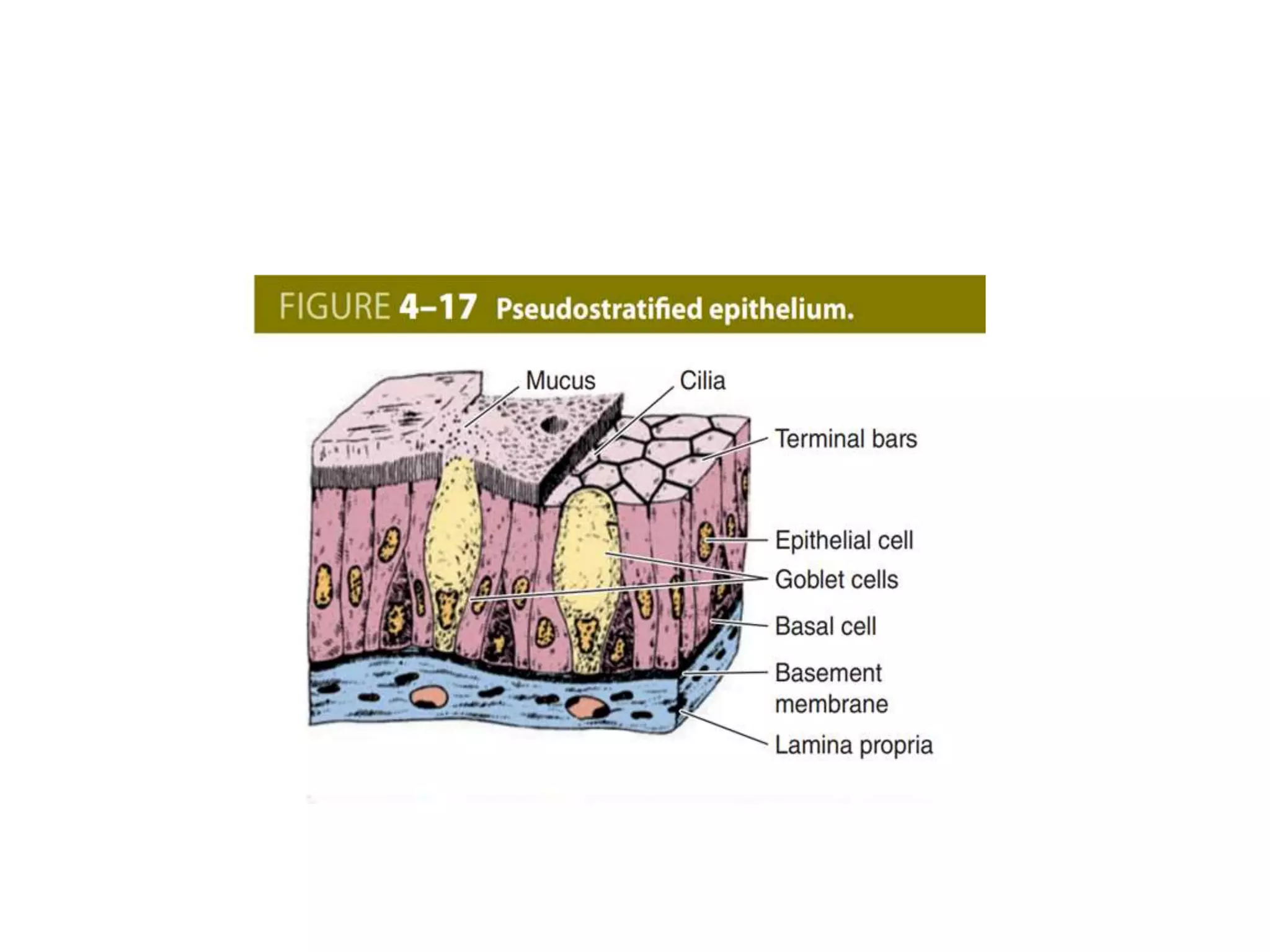
Epithelial tissue lines body surfaces and forms glands. It has several key functions, including protection, absorption, and secretion. Epithelial cells are closely packed and polarized. They communicate through junctions and microstructures. There are several types of epithelia defined by their cell shape, number of layers, and location in the body. Epithelial tissues contain secretory cells that produce molecules for exocrine and endocrine glands.
introduction to tissues-human body is made up of 4 basic tissues- connective tissues, epithelium tissue, nervous tissue, muscular tissue--biological tissues is a collection of interconnected cells that perform a similar function and an embryological origin with similar structure , types of tissues , microscopic diagram, diagrams of tissues, epithelial tissue and types, connective tissue , its components and types
Types of tissues with characteristic's and distribution regions
Epithelium cellstissues histology
1. Chapter 4 Tissues and Histology • Tissues - collections of similar cells and the substances surrounding them • Tissue classification based on structure of cells, composition of noncellular extracellular matrix, and cell function • Major types of adult tissues – Epithelial – Connective – Muscle – Nervous • Histology: Microscopic Study of Tissues – Biopsy: removal of tissues for diagnostic purposes – Autopsy: examination of organs of a dead body to determine cause of death
epithelium covers body surfaces, lines body cavities and constitute glands.so it is important to know about epithelium in detail to deal with tissue of different type and origin.
i. The glycogen synthase can add glucose units only in
alpha-1, 4 linkage. A branching enzyme is needed to
create the alpha-1, 6 linkages.
ii. When the chain is lengthened to 11–12 glucose
residues, the branching enzyme will transfer a block
of 6 to 8 glucose residues from this chain to another
site on the growing molecule. The enzyme amylo-
[1, 4]→[1, 6]-transglucosidase (branching enzyme)
forms this alpha-1, 6 linkage. (Fig. 10.4).
iii. To this newly created branch, further glucose units can
be added in alpha-1, 4 linkage by glycogen synthase.i. The glycogen synthase can add glucose units only in
alpha-1, 4 linkage. A branching enzyme is needed to
create the alpha-1, 6 linkages.
ii. When the chain is lengthened to 11–12 glucose
residues, the branching enzyme will transfer a block
of 6 to 8 glucose residues from this chain to another
site on the growing molecule. The enzyme amylo-
[1, 4]→[1, 6]-transglucosidase (branching enzyme)
forms this alpha-1, 6 linkage. (Fig. 10.4).
iii. To this newly created branch, further glucose units can
be added in alpha-1, 4 linkage by glycogen synthase.Vi. The glycogen synthase can add glucose units only in
alpha-1, 4 linkage. A branching enzyme is needed to
create the alpha-1, 6 linkages.
ii. When the chain is lengthened to 11–12 glucose
residues, the branching enzyme will transfer a block
of
Biology Class 11 Chapter 8
FOR FURTHER DETAILS YOU CAN WATCH THE RELATED VIDEO AT THE GIVEN LINK
https://www.youtube.com/channel/UCxo06Nj-QWo_7SNvMyDnJCQ?view_as=subscriber
Prix Galien International 2024 Forum ProgramLevi Shapiro
June 20, 2024, Prix Galien International and Jerusalem Ethics Forum in ROME. Detailed agenda including panels:
- ADVANCES IN CARDIOLOGY: A NEW PARADIGM IS COMING
- WOMEN’S HEALTH: FERTILITY PRESERVATION
- WHAT’S NEW IN THE TREATMENT OF INFECTIOUS,
ONCOLOGICAL AND INFLAMMATORY SKIN DISEASES?
- ARTIFICIAL INTELLIGENCE AND ETHICS
- GENE THERAPY
- BEYOND BORDERS: GLOBAL INITIATIVES FOR DEMOCRATIZING LIFE SCIENCE TECHNOLOGIES AND PROMOTING ACCESS TO HEALTHCARE
- ETHICAL CHALLENGES IN LIFE SCIENCES
- Prix Galien International Awards Ceremony
These simplified slides by Dr. Sidra Arshad present an overview of the non-respiratory functions of the respiratory tract.
Learning objectives:
1. Enlist the non-respiratory functions of the respiratory tract
2. Briefly explain how these functions are carried out
3. Discuss the significance of dead space
4. Differentiate between minute ventilation and alveolar ventilation
5. Describe the cough and sneeze reflexes
Study Resources:
1. Chapter 39, Guyton and Hall Textbook of Medical Physiology, 14th edition
2. Chapter 34, Ganong’s Review of Medical Physiology, 26th edition
3. Chapter 17, Human Physiology by Lauralee Sherwood, 9th edition
4. Non-respiratory functions of the lungs https://academic.oup.com/bjaed/article/13/3/98/278874
TEST BANK for Operations Management, 14th Edition by William J. Stevenson, Ve...kevinkariuki227
TEST BANK for Operations Management, 14th Edition by William J. Stevenson, Verified Chapters 1 - 19, Complete Newest Version.pdf
Title: Sense of Taste
Presenter: Dr. Faiza, Assistant Professor of Physiology
Qualifications:
MBBS (Best Graduate, AIMC Lahore)
FCPS Physiology
ICMT, CHPE, DHPE (STMU)
MPH (GC University, Faisalabad)
MBA (Virtual University of Pakistan)
Learning Objectives:
Describe the structure and function of taste buds.
Describe the relationship between the taste threshold and taste index of common substances.
Explain the chemical basis and signal transduction of taste perception for each type of primary taste sensation.
Recognize different abnormalities of taste perception and their causes.
Key Topics:
Significance of Taste Sensation:
Differentiation between pleasant and harmful food
Influence on behavior
Selection of food based on metabolic needs
Receptors of Taste:
Taste buds on the tongue
Influence of sense of smell, texture of food, and pain stimulation (e.g., by pepper)
Primary and Secondary Taste Sensations:
Primary taste sensations: Sweet, Sour, Salty, Bitter, Umami
Chemical basis and signal transduction mechanisms for each taste
Taste Threshold and Index:
Taste threshold values for Sweet (sucrose), Salty (NaCl), Sour (HCl), and Bitter (Quinine)
Taste index relationship: Inversely proportional to taste threshold
Taste Blindness:
Inability to taste certain substances, particularly thiourea compounds
Example: Phenylthiocarbamide
Structure and Function of Taste Buds:
Composition: Epithelial cells, Sustentacular/Supporting cells, Taste cells, Basal cells
Features: Taste pores, Taste hairs/microvilli, and Taste nerve fibers
Location of Taste Buds:
Found in papillae of the tongue (Fungiform, Circumvallate, Foliate)
Also present on the palate, tonsillar pillars, epiglottis, and proximal esophagus
Mechanism of Taste Stimulation:
Interaction of taste substances with receptors on microvilli
Signal transduction pathways for Umami, Sweet, Bitter, Sour, and Salty tastes
Taste Sensitivity and Adaptation:
Decrease in sensitivity with age
Rapid adaptation of taste sensation
Role of Saliva in Taste:
Dissolution of tastants to reach receptors
Washing away the stimulus
Taste Preferences and Aversions:
Mechanisms behind taste preference and aversion
Influence of receptors and neural pathways
Impact of Sensory Nerve Damage:
Degeneration of taste buds if the sensory nerve fiber is cut
Abnormalities of Taste Detection:
Conditions: Ageusia, Hypogeusia, Dysgeusia (parageusia)
Causes: Nerve damage, neurological disorders, infections, poor oral hygiene, adverse drug effects, deficiencies, aging, tobacco use, altered neurotransmitter levels
Neurotransmitters and Taste Threshold:
Effects of serotonin (5-HT) and norepinephrine (NE) on taste sensitivity
Supertasters:
25% of the population with heightened sensitivity to taste, especially bitterness
Increased number of fungiform papillae
Flu Vaccine Alert in Bangalore Karnatakaaddon Scans
As flu season approaches, health officials in Bangalore, Karnataka, are urging residents to get their flu vaccinations. The seasonal flu, while common, can lead to severe health complications, particularly for vulnerable populations such as young children, the elderly, and those with underlying health conditions.
Dr. Vidisha Kumari, a leading epidemiologist in Bangalore, emphasizes the importance of getting vaccinated. "The flu vaccine is our best defense against the influenza virus. It not only protects individuals but also helps prevent the spread of the virus in our communities," he says.
This year, the flu season is expected to coincide with a potential increase in other respiratory illnesses. The Karnataka Health Department has launched an awareness campaign highlighting the significance of flu vaccinations. They have set up multiple vaccination centers across Bangalore, making it convenient for residents to receive their shots.
To encourage widespread vaccination, the government is also collaborating with local schools, workplaces, and community centers to facilitate vaccination drives. Special attention is being given to ensuring that the vaccine is accessible to all, including marginalized communities who may have limited access to healthcare.
Residents are reminded that the flu vaccine is safe and effective. Common side effects are mild and may include soreness at the injection site, mild fever, or muscle aches. These side effects are generally short-lived and far less severe than the flu itself.
Healthcare providers are also stressing the importance of continuing COVID-19 precautions. Wearing masks, practicing good hand hygiene, and maintaining social distancing are still crucial, especially in crowded places.
Protect yourself and your loved ones by getting vaccinated. Together, we can help keep Bangalore healthy and safe this flu season. For more information on vaccination centers and schedules, residents can visit the Karnataka Health Department’s official website or follow their social media pages.
Stay informed, stay safe, and get your flu shot today!
micro teaching on communication m.sc nursing.pdfAnurag Sharma
Microteaching is a unique model of practice teaching. It is a viable instrument for the. desired change in the teaching behavior or the behavior potential which, in specified types of real. classroom situations, tends to facilitate the achievement of specified types of objectives.
Report Back from SGO 2024: What’s the Latest in Cervical Cancer?bkling
Are you curious about what’s new in cervical cancer research or unsure what the findings mean? Join Dr. Emily Ko, a gynecologic oncologist at Penn Medicine, to learn about the latest updates from the Society of Gynecologic Oncology (SGO) 2024 Annual Meeting on Women’s Cancer. Dr. Ko will discuss what the research presented at the conference means for you and answer your questions about the new developments.
Factory Supply Best Quality Pmk Oil CAS 28578–16–7 PMK Powder in Stockrebeccabio
Factory Supply Best Quality Pmk Oil CAS 28578–16–7 PMK Powder in Stock
Telegram: bmksupplier
signal: +85264872720
threema: TUD4A6YC
You can contact me on Telegram or Threema
Communicate promptly and reply
Free of customs clearance, Double Clearance 100% pass delivery to USA, Canada, Spain, Germany, Netherland, Poland, Italy, Sweden, UK, Czech Republic, Australia, Mexico, Russia, Ukraine, Kazakhstan.Door to door service
Hot Selling Organic intermediates
- Video recording of this lecture in English language: https://youtu.be/lK81BzxMqdo
- Video recording of this lecture in Arabic language: https://youtu.be/Ve4P0COk9OI
- Link to download the book free: https://nephrotube.blogspot.com/p/nephrotube-nephrology-books.html
- Link to NephroTube website: www.NephroTube.com
- Link to NephroTube social media accounts: https://nephrotube.blogspot.com/p/join-nephrotube-on-social-media.html
Couples presenting to the infertility clinic- Do they really have infertility...Sujoy Dasgupta
Dr Sujoy Dasgupta presented the study on "Couples presenting to the infertility clinic- Do they really have infertility? – The unexplored stories of non-consummation" in the 13th Congress of the Asia Pacific Initiative on Reproduction (ASPIRE 2024) at Manila on 24 May, 2024.
The prostate is an exocrine gland of the male mammalian reproductive system
It is a walnut-sized gland that forms part of the male reproductive system and is located in front of the rectum and just below the urinary bladder
Function is to store and secrete a clear, slightly alkaline fluid that constitutes 10-30% of the volume of the seminal fluid that along with the spermatozoa, constitutes semen
A healthy human prostate measures (4cm-vertical, by 3cm-horizontal, 2cm ant-post ).
It surrounds the urethra just below the urinary bladder. It has anterior, median, posterior and two lateral lobes
It’s work is regulated by androgens which are responsible for male sex characteristics
Generalised disease of the prostate due to hormonal derangement which leads to non malignant enlargement of the gland (increase in the number of epithelial cells and stromal tissue)to cause compression of the urethra leading to symptoms (LUTS
Lung Cancer: Artificial Intelligence, Synergetics, Complex System Analysis, S...Oleg Kshivets
RESULTS: Overall life span (LS) was 2252.1±1742.5 days and cumulative 5-year survival (5YS) reached 73.2%, 10 years – 64.8%, 20 years – 42.5%. 513 LCP lived more than 5 years (LS=3124.6±1525.6 days), 148 LCP – more than 10 years (LS=5054.4±1504.1 days).199 LCP died because of LC (LS=562.7±374.5 days). 5YS of LCP after bi/lobectomies was significantly superior in comparison with LCP after pneumonectomies (78.1% vs.63.7%, P=0.00001 by log-rank test). AT significantly improved 5YS (66.3% vs. 34.8%) (P=0.00000 by log-rank test) only for LCP with N1-2. Cox modeling displayed that 5YS of LCP significantly depended on: phase transition (PT) early-invasive LC in terms of synergetics, PT N0—N12, cell ratio factors (ratio between cancer cells- CC and blood cells subpopulations), G1-3, histology, glucose, AT, blood cell circuit, prothrombin index, heparin tolerance, recalcification time (P=0.000-0.038). Neural networks, genetic algorithm selection and bootstrap simulation revealed relationships between 5YS and PT early-invasive LC (rank=1), PT N0—N12 (rank=2), thrombocytes/CC (3), erythrocytes/CC (4), eosinophils/CC (5), healthy cells/CC (6), lymphocytes/CC (7), segmented neutrophils/CC (8), stick neutrophils/CC (9), monocytes/CC (10); leucocytes/CC (11). Correct prediction of 5YS was 100% by neural networks computing (area under ROC curve=1.0; error=0.0).
CONCLUSIONS: 5YS of LCP after radical procedures significantly depended on: 1) PT early-invasive cancer; 2) PT N0--N12; 3) cell ratio factors; 4) blood cell circuit; 5) biochemical factors; 6) hemostasis system; 7) AT; 8) LC characteristics; 9) LC cell dynamics; 10) surgery type: lobectomy/pneumonectomy; 11) anthropometric data. Optimal diagnosis and treatment strategies for LC are: 1) screening and early detection of LC; 2) availability of experienced thoracic surgeons because of complexity of radical procedures; 3) aggressive en block surgery and adequate lymph node dissection for completeness; 4) precise prediction; 5) adjuvant chemoimmunoradiotherapy for LCP with unfavorable prognosis.
MANAGEMENT OF ATRIOVENTRICULAR CONDUCTION BLOCK.pdfJim Jacob Roy
Cardiac conduction defects can occur due to various causes.
Atrioventricular conduction blocks ( AV blocks ) are classified into 3 types.
This document describes the acute management of AV block.
2. Introduction
• the organs of the human body are composed of only
four basic tissue types:
• Epithelial
• Connective
• Muscular
• Nervous tissues
3. • Epithelial tissues are composed of closely aggregated
polyhedral cells adhering strongly to one another and
to a thin layer of ECM, forming cellular sheets that
line the cavities of organs and cover the body surface.
• Epithelia (Gr. epi, upon + thele, nipple) line all
external and internal surfaces of the body and all
substances that enter or leave an organ must cross this
type of tissue.
4. • The principal functions of epithelial tissues include
the following:
- Covering, lining, and protecting surfaces (eg,
epidermis)
- Absorption (eg, the intestinal lining)
- Secretion (eg, parenchymal cells of glands)
5. Characteristic features of epithelial cells
• Most epithelia are adjacent to connective tissue
containing blood vessels from which the epithelial
cells receive nutrients and O2.
• The connective tissue that underlies the epithelia
lining the organs of the digestive, respiratory, and
urinary systems is called the lamina propria.
• The area of contact between the two tissues may be
increased by small evaginations called papillae
projecting from the connective tissue into the
epithelium.
6. • Epithelial cells generally show polarity.
• The region of the cell contacting the ECM and
connective tissue is called the basal pole and the
opposite end, usually facing a space, is the apical
pole, with the two poles differing significantly in both
structure and function.
7.
8.
9. • Basement Membrane
• The basal surface of all epithelia rests on a thin
extracellular, felt-like sheet of macromolecules
referred to as the basement membrane , a
semipermeable filter for substances reaching
epithelial cells from below.
• Nearest the epithelial cells is the basal lamina, a thin,
electron-dense, sheet-like layer of fine fibrils, and
beneath this layer is a more diffuse and fibrous
reticular lamina.
10. • basal lamina characteristically include the following:
Type IV collagen, Laminin, integrin, Nidogen and
perlecan.
• The more diffuse meshwork of the reticular lamina
contains type III collagen and is bound to the basal
lamina by anchoring fibrils of type VII collagen, both
of which are produced by cells of the connective
tissue
11. • Intercellular Adhesion & Other Junctions
• Tight or occluding junctions form a seal between
adjacent cells.
• Adherent or anchoring junctions are sites of strong
cell adhesion.
• Gap junctions are channels for communication
between adjacent cells.
12.
13. • Zonula occludens (Tight junctions):
• The most apical junction
• form bands completely encircling each cell
• close off the space between the cells
• The seal between the membranes is due primarily to
direct interactions between the transmembrane
protein claudin on each cell
• The number of these fusions sites is inversely
correlated with the leakiness of the epithelium
14.
15. • Prevent the flow of material between the base and the
apex of the cell in either directions.
• It contribute to the formation of two separate
functional units , apical and basal.
• Composed of several proteins:-
- Junctional associated molecule (JAM)-
- Occludins
- Claudins
16. • Zonula adherens
• Also encircles the cell
• Immediately below the zonula occludens
• Adhesion is mediated by transmembrane
glycoproteins of each cell, the cadherins. which lose
their adhesive properties in the absence of Ca2+.
• Inside the cell, cadherins bind the protein catenin
which is linked to actin filaments
17. • Desmosome:
• does not form a belt around the cell
• Disk like structure that faces mirror image on
adjacent cell (20-30 nm gap).
• Circular plaque called attachment plaque made up of
12 proteins of anchoring proteins.
• Intermediate cytokeratin filaments are inserted to the
plaque.
18.
19. • Hemidesmosome:
• connection between cell and BL
• Take the shape of half desmosome.
• The attachment plaque contain mainly integrin not
cadherin.
20. • gap junctions:
• Passageway between two adjacent cells
• Let small molecules move directly between
neighboring cells
• Cells are connected by hollow cylinders of protein
• Each junction is composed of 6 connexin form
hexameric complexes called connexons proteins that
span the membrane and form one half of the junction
• Permit Cell to cell exchange of low MW molecules
such as: ions, cAMP, small hormones.
• Not present in skeletal muscle.
21. Specializations of cell surface
• Microvilli:
• Non-branching fingerlike projections of cell 1 μm
high, 0.08 μm wide,at the free surface of the cell.
• Increase of cell surface
• Covered by plasma membrane
• The glycocalyx is thicker contain glycoproteins and
enzymes that allow final stage digestion
• Glycocalyx and microvilli seen under light
microscope called striated or brush border.
22. • It’s a cluster of actin filaments that crossed linked to
each other proteins such as fimbrin and villin and to
plasma membran by proteins such as myosin I.
• Hundred of microvilli can be seen in absorptive cells.
23.
24. • Stereocilia
• Branched non – motile microvilli of cells of
epididymis and ductus deference
• Its function to increase the surface area
• Cilia
• Non-branching motile projections
• Transport of extracellular mass
• May be numerous
• 270 per cell in trachea
25. • 5-10 μm tall ,0.2 μm wide
• Attached to basal bodies ( analogus to the centrioles)
at the apical area
• Flagella
• very long cillia 100 μm , only in spermatozoa
26. Type of Epithelia
• Covering epithelia
Number of cell layer
• Simple
• Stratified
Shape of the cell at the surface layer
• Squamous
• Cuboidal
• Columnar
• Glandular epithelia
27. • Simple Squamous Epithelium
• single layer of flat cells with disc-shaped nuclei
• Special types
- Endothelium (inner covering) slick lining of hollow
organs
- Mesothelium (middle covering)Lines peritoneal,
pleural, and pericardial cavities
• Covers visceral organs of those cavities
28.
29. • Simple Cuboidal Epithelium
• single layer of cube-like cells with large, spherical
central nuclei
• Function
- secretion and absorption
• Location
- kidney tubules
- secretory portions of small glands, ovary & thyroid
follicles
30.
31. • Simple Columnar Epithelium
• Single layer of column-shaped (rectangular) cells
with oval nuclei
• Some bear cilia at their apical surface
• May contain goblet cells
• Function: Absorption; secretion of mucus, enzymes,
and other substances
• Ciliated type propels mucus or reproductive cells by
ciliary action
32.
33. • Non-ciliated form
- Lines digestive tract, gallbladder, ducts of some
glands
• Ciliated form
- Lines small bronchi, uterine tubes, uterus
34. • Pseudostratified Columnar Epithelium
• All cells originate at basement membrane
• Only tall cells reach the apical surface
• May contain goblet cells and bear cilia
• Nuclei lie at varying heights within cells
• Gives false impression of stratification
• Function: secretion of mucus; propulsion of mucus by
cilia
35.
36. • Non-ciliated type
- Ducts of male reproductive tubes
- Ducts of large glands
• Ciliated variety
- Lines trachea and most of upper respiratory tract
37. • Stratified Squamous Epithelium
• Many layers of cells – squamous in shape
• Deeper layers of cells appear cuboidal or columnar
• Thickest epithelial tissue – adapted for protection
• Specific types
Keratinized – contain the protective protein keratin
• Surface cells are dead and full of keratin
38.
39. Non-keratinized – forms moist lining of body
openings
• Function: Protects underlying tissues in areas subject
to abrasion
• Location
- Keratinized – forms epidermis
- Non-keratinized – forms lining of esophagus, mouth,
and vagina
40. • Stratified Cuboidal Epithelium
• generally two layers of cube-shaped cells
• Location:
- Forms largest ducts of sweat glands
- Forms ducts of mammary glands and salivary
glands
41. • Stratified Columnar Epithelium
• several layers; basal cells usually cuboidal;
superficial cells elongated
• Function: protection and secretion
• Location:
- Rare tissue type
- Found in male urethra and vas deferens, largest
ducts of salivary glands, nasopharynx
42. • Transitional Epithelium
• Basal cells usually cuboidal or columnar
• Superficial cells dome-shaped or squamous
• Function: stretches and permits distension of urinary
bladder
• Location: Lines ureters, urinary bladder and part of
urethra
43.
44. • Secretory Epithelia & Glands
• Epithelial cells that function mainly to produce and
secrete various macromolecules may occur in
epithelia with other major functions or comprise
specialized organs called glands.
• Secretory cells may synthesize, store, and release
proteins (eg, in the pancreas), lipids (eg, adrenal,
sebaceous glands), or complexes of carbohydrates
and proteins (eg, salivary glands). Epithelia of
mammary glands secrete all three substances.
45. • Exocrine glands remain connected with the surface
epithelium, the connection forming the tubular ducts
lined with epithelium that deliver the secreted
material where it is used.
• Endocrine glands lose the connection to their original
epithelium and therefore lack ducts.
46. • glands can be simple (ducts not branched) or
compound (ducts with two or more branches).
• Secretory portions can be tubular (either short or long
and coiled) or acinar (rounded and saclike); either
type of secretory unit may be branched, even if the
duct is not branched.
• Compound glands can have branching ducts and can
have multiple tubular, acinar, or tubuloacinar
secretory portions.