Downloaded 256 times


![ Muscle hypertrophy
Weight training (repeated intense workouts): increases diameter and
strength of “fast” muscle fibers by increasing production of
Mitochondria
Actin and myosin protein
Myofilaments containing these contractile proteins
The myofibril organelles these myofilaments form
Fibers enlarge (hypertrophy) as number and size of myofibrils
increase
[Muscle fibers (=muscle cells) don’t increase in number but increase
in diameter producing large muscles]
Endurance training (aerobic): doesn’t produce hypertrophy
Muscle atrophy: loss of tone and mass from lack of
stimulation
Muscle becomes smaller and weaker
Note on terminology: in general, increased size is hypertrophy; increased number
of cells is hyperplasia](https://image.slidesharecdn.com/musclehistologyby-dr-150302034017-conversion-gate01/75/Muscle-histology-by-dr-armaan-singh-22-2048.jpg)
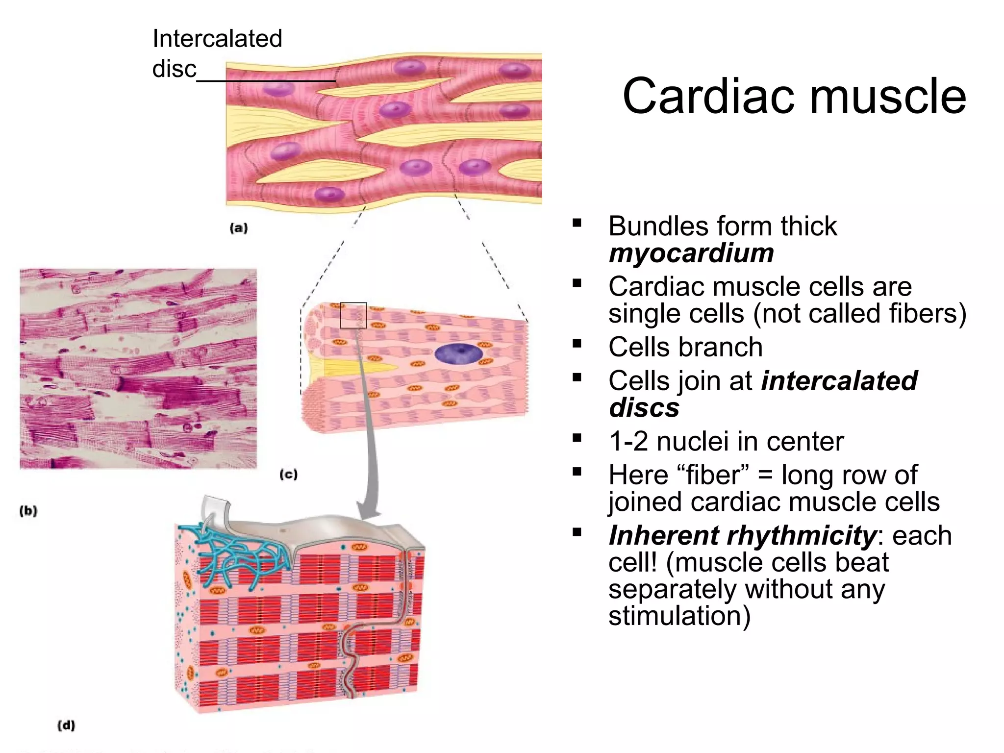


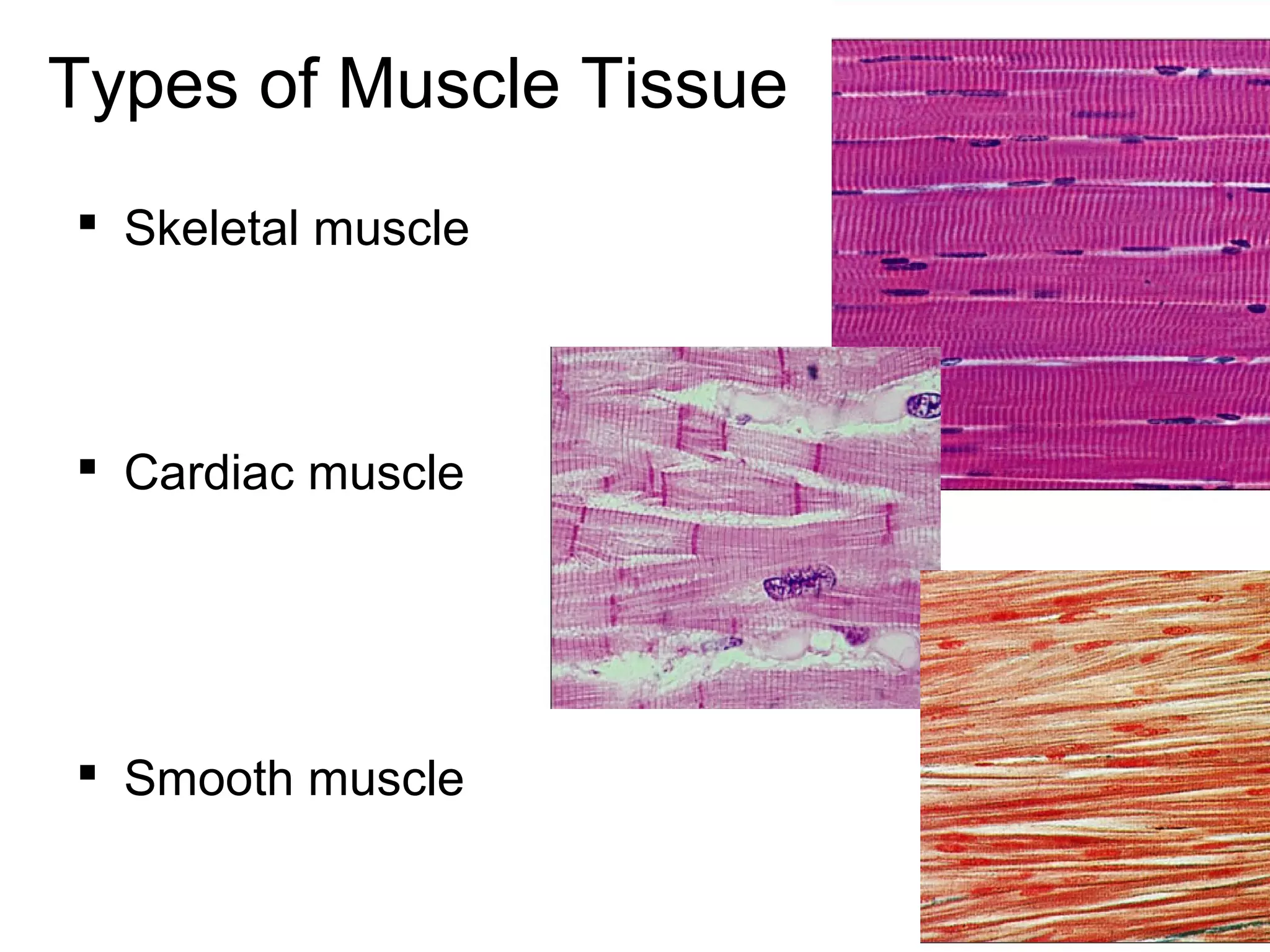
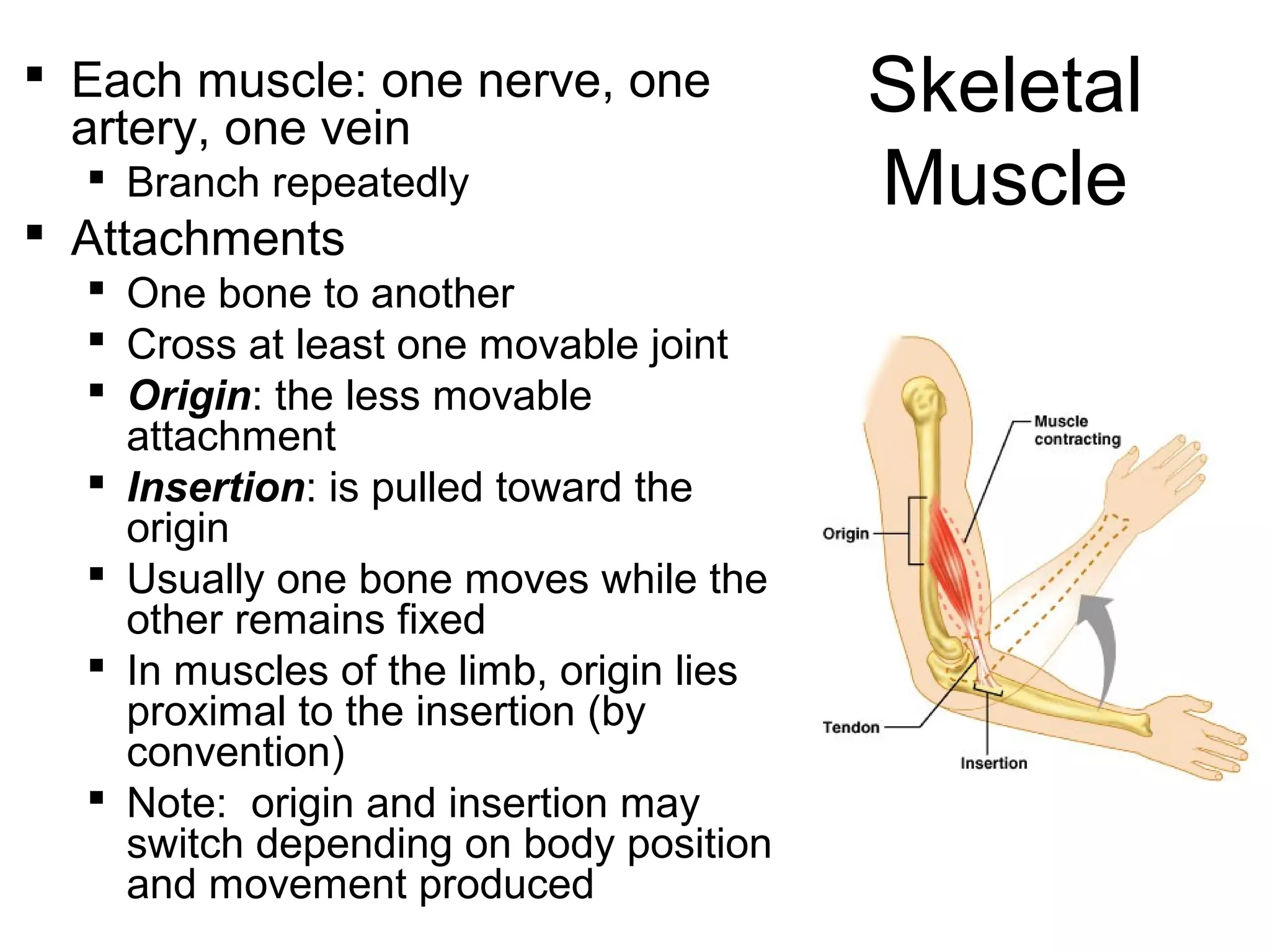
Skeletal muscle tissue functions include movement, posture maintenance, joint stabilization, and heat generation. The main types of muscle tissue are skeletal, cardiac, and smooth muscle. Skeletal muscle is striated and voluntary, attaching to bones and moving the skeleton. Cardiac muscle is only found in the heart walls and has involuntary, rhythmic contractions. Smooth muscle lacks striations and controls involuntary functions like digestion and blood flow. All muscle tissues contain contractile filaments that slide past each other to cause shortening, but the tissues differ in organization, fiber type, and control.


