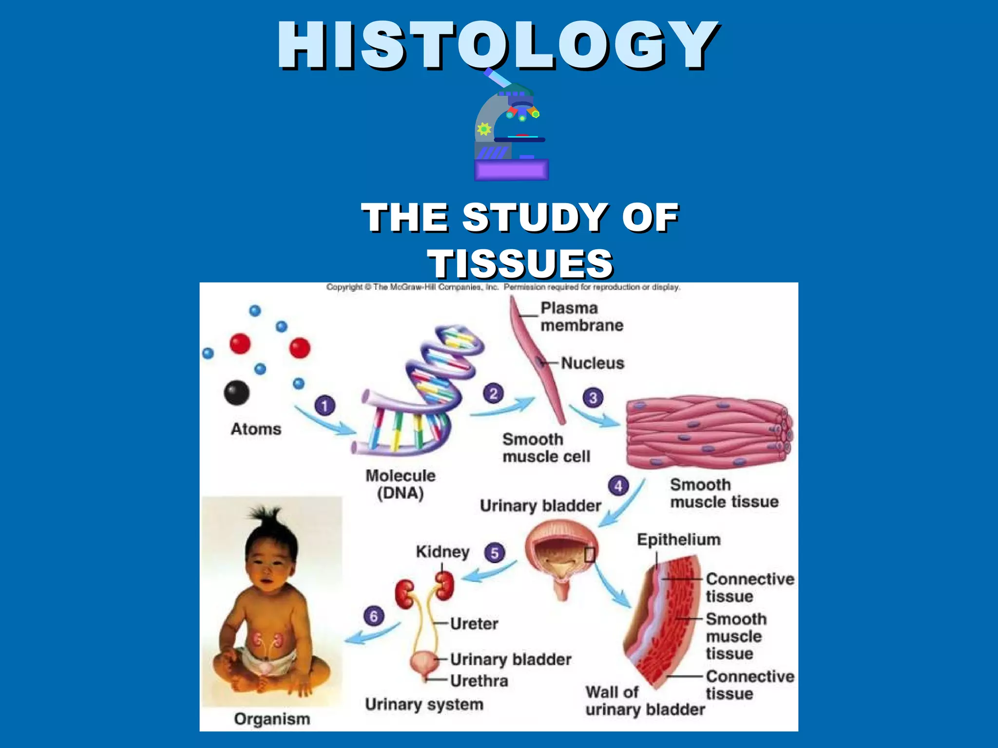
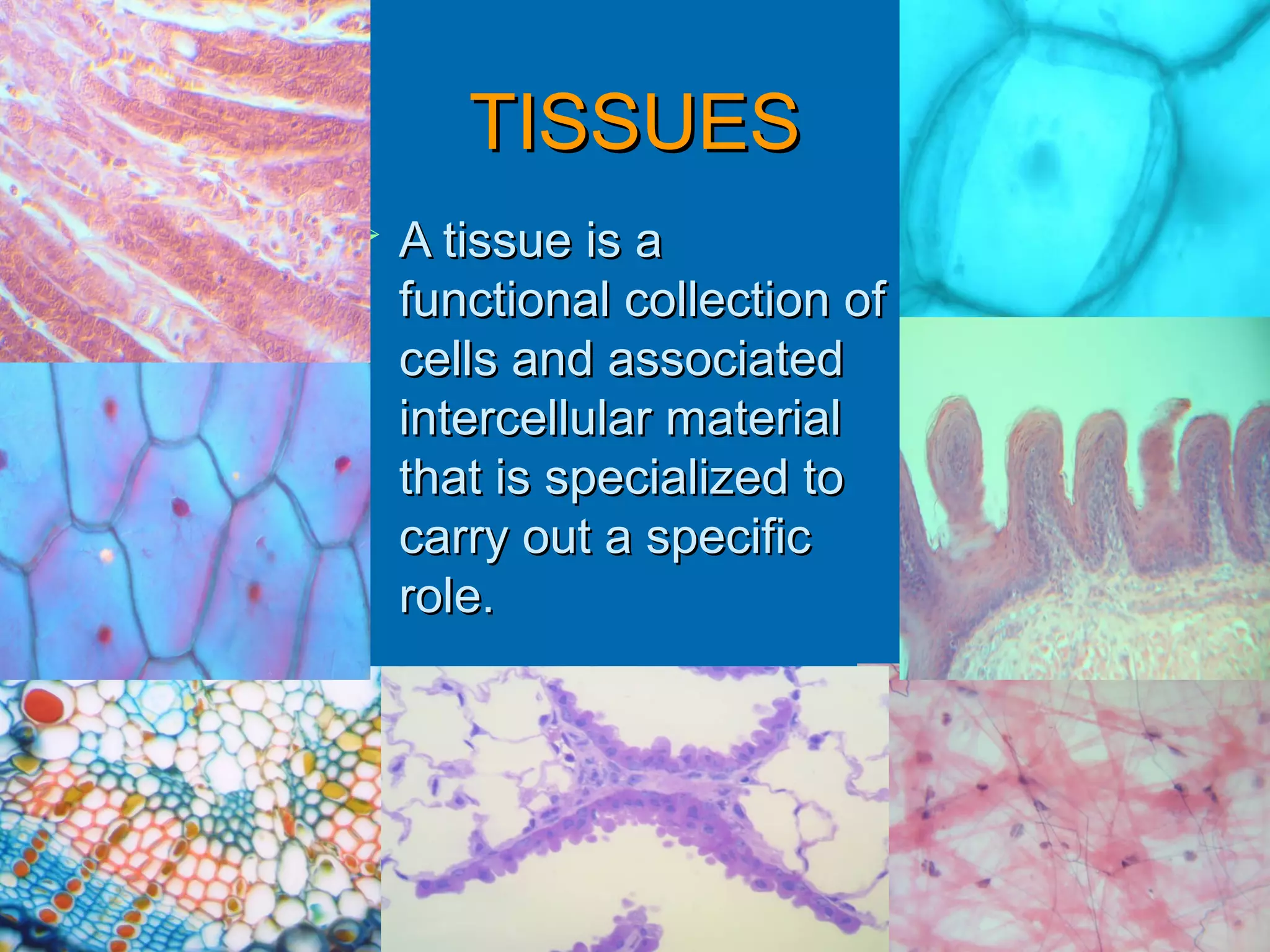
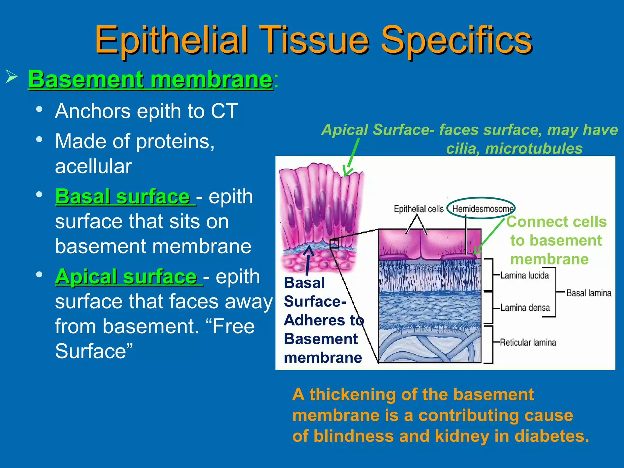
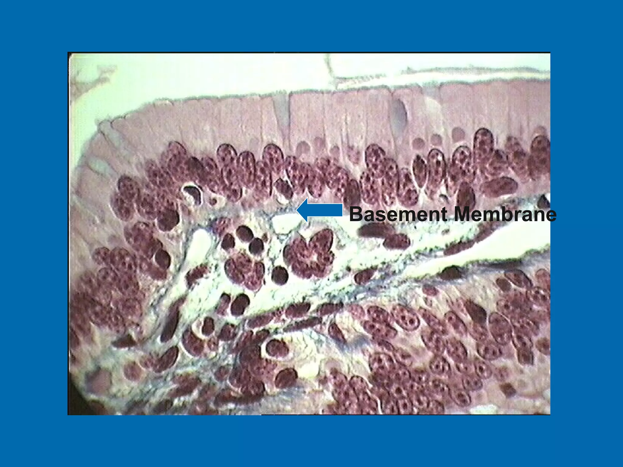
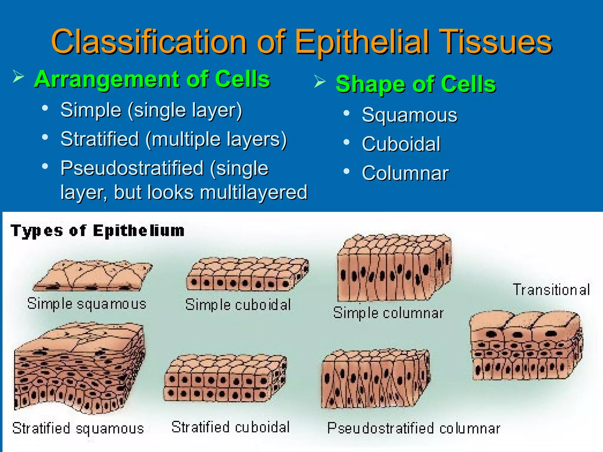
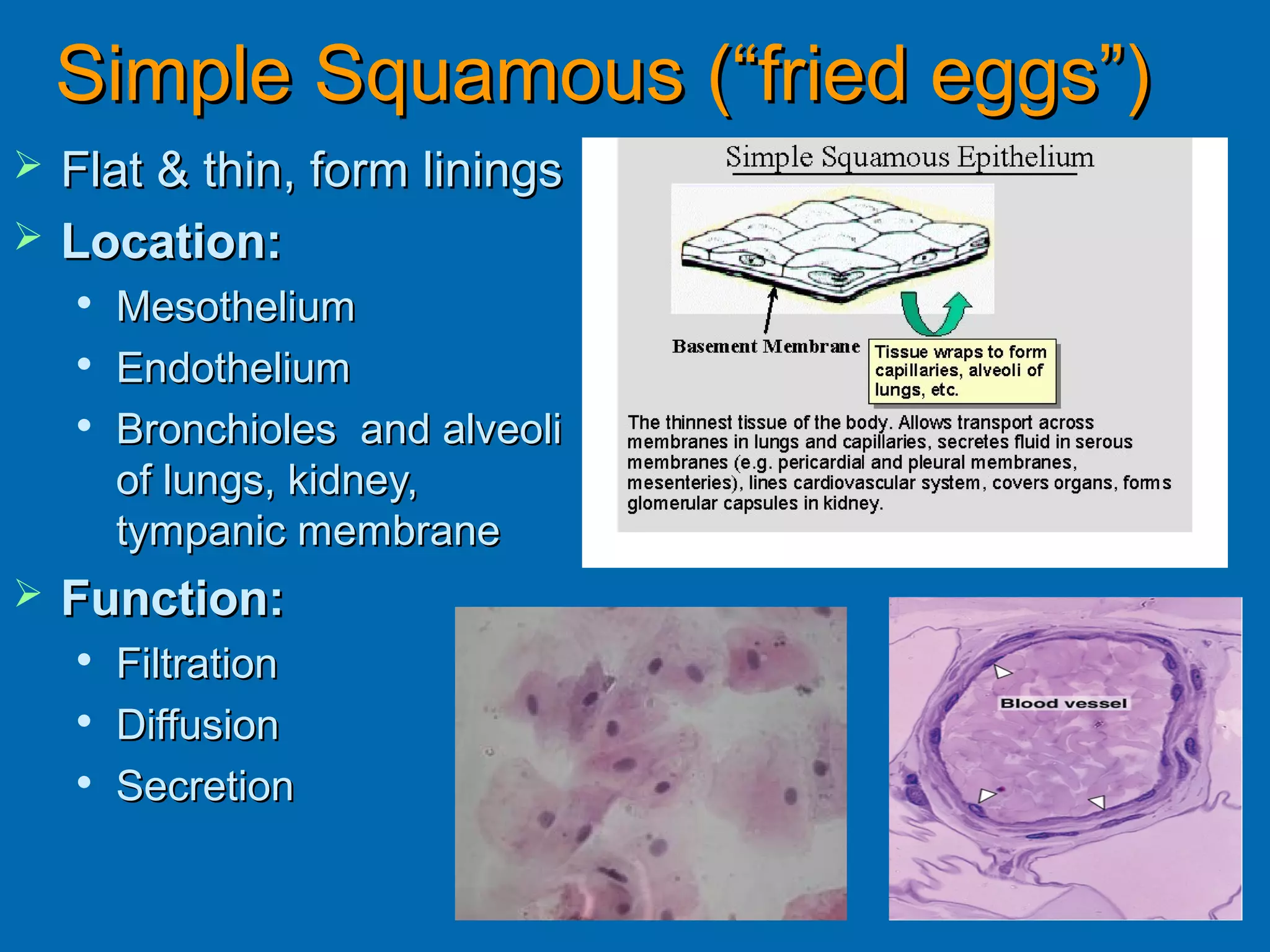
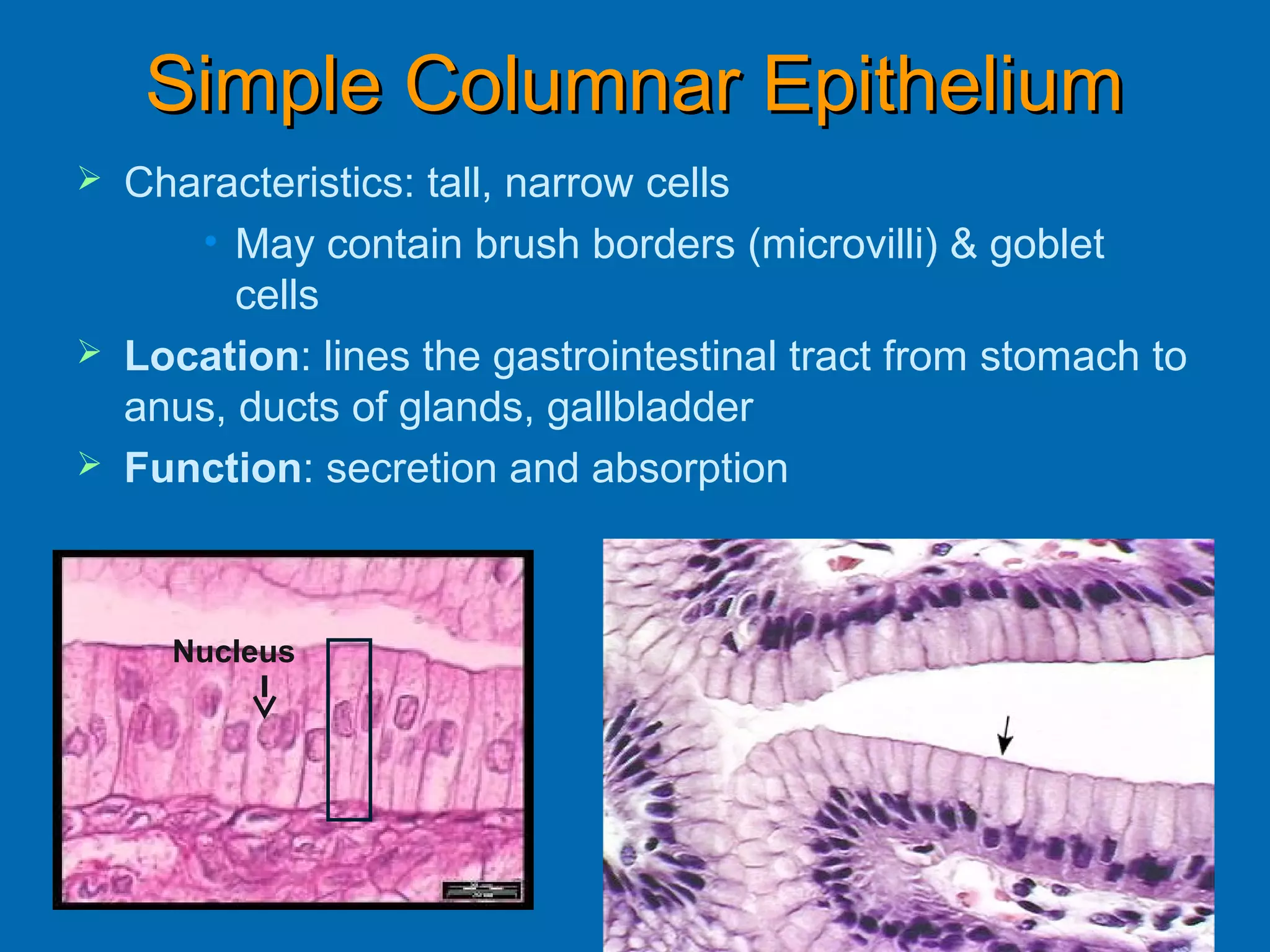
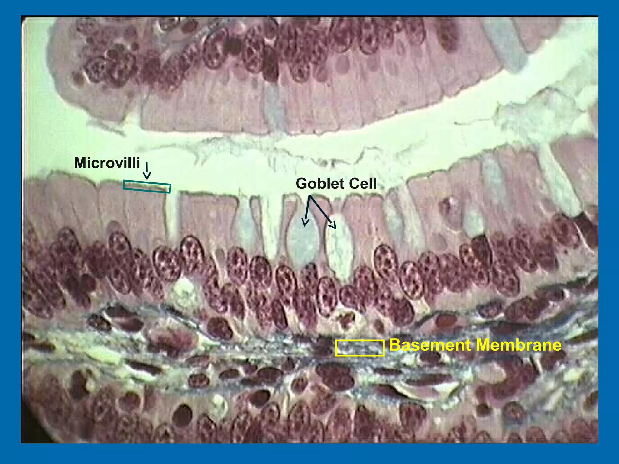
HISTOLOGY

 THE STUDY OF
   TISSUES
TISSUES
   A tissue is a
    functional collection of
    cells and associated
    intercellular material
    that is specialized to
    carry out a specific
    role.
Technique of
             Tissue
         Sectioning
1. Tissue is preserved in a fixative
   (chemical)
2. Cut into very thin slices by a special
   machine
   - slices are called histological
   sections
3. Sections are mounted on microscope
   slides
4. Sections on slides are stained
Classification of the FOUR basic
              tissues
          1- Epithelial
          2- Connective (CT)
          3- Muscular
          4- Nervous
Connective vs Epithelial
   Lots of extra-cellular      Little extra cellular
    material, few cells          material, lots of cells
   Lots of blood vessels       No blood vessels
   Covered by other            Usually form surface
    tissues                      layers and are NOT
                                 covered by another
                                 tissue (*except blood
                                 vessels)

                                  Epithelial tissue


                                  Connective tissue
Epithelial Tissues
   Flat sheet of cells with upper surface exposed to
    environment or internal space. (Barriers!
    Protection!)
   Covers body surface & lines body cavities
   Forms external & internal lining of organs
   Constitutes most gland tissues (Secretion!)
   Little extra cellular material
   No room for blood vessels (avascular) so
    depends on blood vessels in CT for food and
    waste movement
Epithelial Tissue Specifics
   Basement membrane:
             membrane
    
        Anchors epith to CT
                                      Apical Surface- faces surface, may have
    
        Made of proteins,                              cilia, microtubules
        acellular
       Basal surface - epith
        surface that sits on                                  Connect cells
        basement membrane                                     to basement
                                                              membrane
       Apical surface - epith    Basal
        surface that faces away   Surface-
                                  Adheres to
        from basement. “Free
                                  Basement
        Surface”                  membrane

                                   A thickening of the basement
                                   membrane is a contributing cause
                                   of blindness and kidney in diabetes.
Basement Membrane
Classification of Epithelial Tissues
   Arrangement of Cells                   Shape of Cells
    
        Simple (single layer)                  Squamous
    
        Stratified (multiple layers)        
                                                Cuboidal
       Pseudostratified (single            
                                                Columnar
        layer, but looks multilayered
Simple Squamous (“fried eggs”)
   Flat & thin, form linings
   Location:
    
        Mesothelium
    
        Endothelium
       Bronchioles and alveoli
        of lungs, kidney,
        tympanic membrane
   Function:
    
        Filtration
    
        Diffusion
       Secretion
Mesothelium is the simple squamous
  epithelium lining body cavities.
Endothelium is the simple squamous
  epithelium lining blood vessels

      Two blood vessels seen in cross section
Quick Review

                                 13. Identify the tissue.
                                 14. Identify the structure.
                                 15. What tissue makes up
                                     this (#14) structure?
                                 16. Identify the cellular
                                      structure.


     Click for Answers
13. Simple Cuboidal Epithelium
14. Blood Vessel
15. Simple Squamous epithelium
16. Nucleus
Stratified Squamous Epithelial
   Characteristics:
    
        Many layers becoming
        flatter from basal to
                                                  Stratified layers
        apical surfaces
                                                  of epithelial cells
    
        Basal cells are mitotic
       Apical cells are dead
                                                           Basal cells
   LOCATIONS:                                             Mitotic area
                                                           Cells are rounder
    
        Keritanized
         • skin
                                                         Stratified layers
    
        Nonkeritanized
         • Mouth, esophagus,
                                  Apical cells will be sloughed off and
           pharynx, vagina        replaced by cells in layer below.
   FUNCTION:
       protection
Simple Cuboidal Epithelium
 Characteristics: single layer, square or
  roundish
 Location: Ducts of many glands, lines
  kidney tubules, surface of ovary
   Function: secretion and absorption
Simple cuboidal epithelium lining a
    tubule (longitudinal cut).
Simple Columnar Epithelium
   Characteristics: tall, narrow cells
       • May contain brush borders (microvilli) & goblet
          cells
   Location: lines the gastrointestinal tract from stomach to
    anus, ducts of glands, gallbladder
   Function: secretion and absorption


      Nucleus
Simple columnar epithelium with very
     regular line-up of nuclei.
Pseudostratified Ciliated Columnar
                   Epithelium
   Location: Respiratory tract
   Function: secrete mucous to trap foreign particles
   Not all cells reach apical layer BUT all cells are connected
    to basement membrane!
   Cilia (sweep), Goblet cell (secrete mucous)


                                    Cilia
Microvilli
             Goblet Cell


                           Basement Membrane

Histology epithelial tissue

  • 1.
  • 2.
    TISSUES  A tissue is a functional collection of cells and associated intercellular material that is specialized to carry out a specific role.
  • 3.
    Technique of Tissue Sectioning 1. Tissue is preserved in a fixative (chemical) 2. Cut into very thin slices by a special machine - slices are called histological sections 3. Sections are mounted on microscope slides 4. Sections on slides are stained
  • 7.
    Classification of theFOUR basic tissues  1- Epithelial  2- Connective (CT)  3- Muscular  4- Nervous
  • 8.
    Connective vs Epithelial  Lots of extra-cellular  Little extra cellular material, few cells material, lots of cells  Lots of blood vessels  No blood vessels  Covered by other  Usually form surface tissues layers and are NOT covered by another tissue (*except blood vessels) Epithelial tissue Connective tissue
  • 9.
    Epithelial Tissues  Flat sheet of cells with upper surface exposed to environment or internal space. (Barriers! Protection!)  Covers body surface & lines body cavities  Forms external & internal lining of organs  Constitutes most gland tissues (Secretion!)  Little extra cellular material  No room for blood vessels (avascular) so depends on blood vessels in CT for food and waste movement
  • 10.
    Epithelial Tissue Specifics  Basement membrane: membrane  Anchors epith to CT Apical Surface- faces surface, may have  Made of proteins, cilia, microtubules acellular  Basal surface - epith surface that sits on Connect cells basement membrane to basement membrane  Apical surface - epith Basal surface that faces away Surface- Adheres to from basement. “Free Basement Surface” membrane A thickening of the basement membrane is a contributing cause of blindness and kidney in diabetes.
  • 11.
  • 12.
    Classification of EpithelialTissues  Arrangement of Cells  Shape of Cells  Simple (single layer)  Squamous  Stratified (multiple layers)  Cuboidal  Pseudostratified (single  Columnar layer, but looks multilayered
  • 13.
    Simple Squamous (“friedeggs”)  Flat & thin, form linings  Location:  Mesothelium  Endothelium  Bronchioles and alveoli of lungs, kidney, tympanic membrane  Function:  Filtration  Diffusion  Secretion
  • 14.
    Mesothelium is thesimple squamous epithelium lining body cavities.
  • 15.
    Endothelium is thesimple squamous epithelium lining blood vessels Two blood vessels seen in cross section
  • 16.
    Quick Review 13. Identify the tissue. 14. Identify the structure. 15. What tissue makes up this (#14) structure? 16. Identify the cellular structure. Click for Answers 13. Simple Cuboidal Epithelium 14. Blood Vessel 15. Simple Squamous epithelium 16. Nucleus
  • 17.
    Stratified Squamous Epithelial  Characteristics:  Many layers becoming flatter from basal to Stratified layers apical surfaces of epithelial cells  Basal cells are mitotic  Apical cells are dead Basal cells  LOCATIONS: Mitotic area Cells are rounder  Keritanized • skin Stratified layers  Nonkeritanized • Mouth, esophagus, Apical cells will be sloughed off and pharynx, vagina replaced by cells in layer below.  FUNCTION:  protection
  • 19.
    Simple Cuboidal Epithelium Characteristics: single layer, square or roundish  Location: Ducts of many glands, lines kidney tubules, surface of ovary  Function: secretion and absorption
  • 20.
    Simple cuboidal epitheliumlining a tubule (longitudinal cut).
  • 21.
    Simple Columnar Epithelium  Characteristics: tall, narrow cells • May contain brush borders (microvilli) & goblet cells  Location: lines the gastrointestinal tract from stomach to anus, ducts of glands, gallbladder  Function: secretion and absorption Nucleus
  • 22.
    Simple columnar epitheliumwith very regular line-up of nuclei.
  • 23.
    Pseudostratified Ciliated Columnar Epithelium  Location: Respiratory tract  Function: secrete mucous to trap foreign particles  Not all cells reach apical layer BUT all cells are connected to basement membrane!  Cilia (sweep), Goblet cell (secrete mucous) Cilia
  • 24.
    Microvilli Goblet Cell Basement Membrane