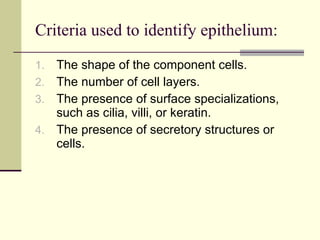
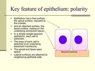
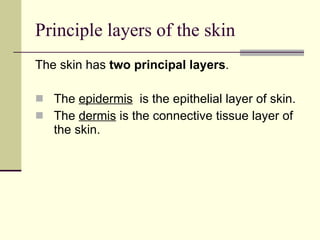
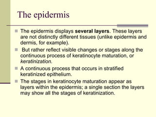
Downloaded 226 times
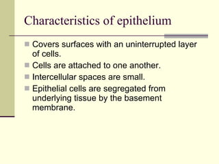
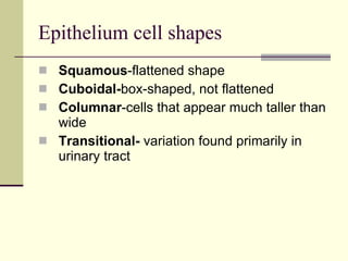
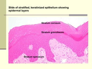
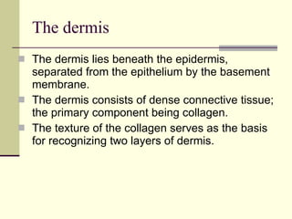
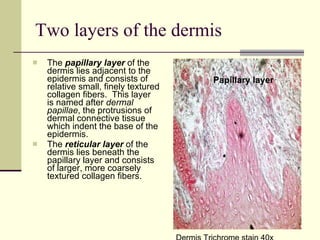
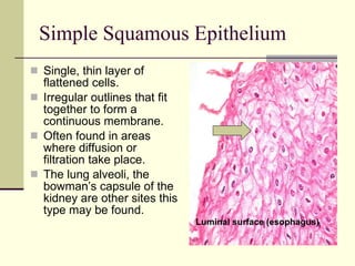

This document provides an overview of epithelium, including its characteristics, cell shapes, criteria used to identify it, key features like polarity, and examples of different types. The principal layers of skin are described as the epidermis and dermis. Various epithelial layers within the epidermis are defined, as are the two layers of the dermis. Examples of simple and stratified epithelia are given along with images to illustrate cell shapes and tissue organization. Specialized epithelial tissues like glands and hair follicles are also mentioned.


![Epithelium[1]](https://cdn.slidesharecdn.com/ss_thumbnails/epithelium1-200323141425-thumbnail.jpg?width=640&height=640&fit=bounds)


