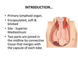
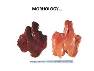
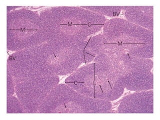
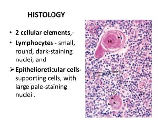
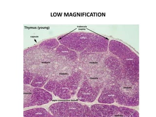
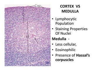
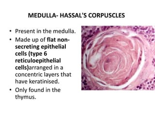
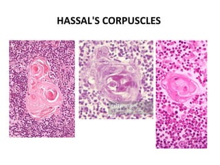
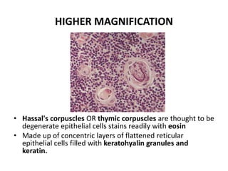
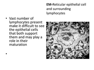
The thymus is a primary lymphoid organ located in the superior mediastinum. It plays a key role in T cell development and maturation. The thymus has an outer capsule and is divided into lobules containing cortex and medulla regions. The cortex contains densely packed developing T cells and epithelioreticular cells. The medulla is less cellular and contains Hassall's corpuscles. Developing lymphocytes interact with epithelioreticular cells, which provide structural support and signals to guide T cell maturation.
The lymph node is a small bean-shaped object which factors in the body's immune system. Lymph nodes clean out substances that travel through the lymphatic fluid, and they contain white blood cells that are the body's defense when fighting off ailments. There are many lymph nodes found throughout the body.
The lymphatic system has three functions:
Fluid recovery.
Immunity
Lipid absorption
The lymphatic vessels of the small intestine receive the special designation of lacteals or chyliferous vessels.
The components of the lymphatic system are :-
lymph, the recovered fluid;
Lymphatic vessels, which transport the lymph;
Lymphatic tissue, composed of aggregates of lymphocytes and macrophages that populate many organs of the body; and
Lymphatic organs, in which these cells are especially concentrated and which are set off from surrounding organs by connective tissue capsules.
Lymphatic organs of body
• Lymphoid organs are the organs, in which lymphocytes can differentiate and proliferate.
• They are part of lymphatic system.
• The lymphatic system is part of circulatory system and a vital part of the immune system, comprising a network of lymphatic vessels that carry a clear fluid called lymph (from Latin, lympha meaning "water" directionally towards the heart.
• Unlike the cardiovascular system, the lymphatic system is not a closed system.
• HISTORY –
• Hippocrates and Aristotle described lymph as white fluid.
• Gasparo aselli an Italian anatomist discovered lymphatic vessels in 1622.
• Van hook in 1652 demonstrated the presence of cisterna chyli and thoracic duct in humans.
• William hunter in the late 18th century was the first to describe the functions of lymphatic system.
• Olof Rudbeck of Swedish university described that lymphatic system constitute a circulatory system separate from blood circulation and this fact was accepted by Royal society of London.
This slide share deals with the elaborate knowledge about the primary lymphatic system which are bone marrow and thymus and we are going to study about maturation strategy of B cells and T cells at their different environments.
Lung Cancer: Artificial Intelligence, Synergetics, Complex System Analysis, S...Oleg Kshivets
RESULTS: Overall life span (LS) was 2252.1±1742.5 days and cumulative 5-year survival (5YS) reached 73.2%, 10 years – 64.8%, 20 years – 42.5%. 513 LCP lived more than 5 years (LS=3124.6±1525.6 days), 148 LCP – more than 10 years (LS=5054.4±1504.1 days).199 LCP died because of LC (LS=562.7±374.5 days). 5YS of LCP after bi/lobectomies was significantly superior in comparison with LCP after pneumonectomies (78.1% vs.63.7%, P=0.00001 by log-rank test). AT significantly improved 5YS (66.3% vs. 34.8%) (P=0.00000 by log-rank test) only for LCP with N1-2. Cox modeling displayed that 5YS of LCP significantly depended on: phase transition (PT) early-invasive LC in terms of synergetics, PT N0—N12, cell ratio factors (ratio between cancer cells- CC and blood cells subpopulations), G1-3, histology, glucose, AT, blood cell circuit, prothrombin index, heparin tolerance, recalcification time (P=0.000-0.038). Neural networks, genetic algorithm selection and bootstrap simulation revealed relationships between 5YS and PT early-invasive LC (rank=1), PT N0—N12 (rank=2), thrombocytes/CC (3), erythrocytes/CC (4), eosinophils/CC (5), healthy cells/CC (6), lymphocytes/CC (7), segmented neutrophils/CC (8), stick neutrophils/CC (9), monocytes/CC (10); leucocytes/CC (11). Correct prediction of 5YS was 100% by neural networks computing (area under ROC curve=1.0; error=0.0).
CONCLUSIONS: 5YS of LCP after radical procedures significantly depended on: 1) PT early-invasive cancer; 2) PT N0--N12; 3) cell ratio factors; 4) blood cell circuit; 5) biochemical factors; 6) hemostasis system; 7) AT; 8) LC characteristics; 9) LC cell dynamics; 10) surgery type: lobectomy/pneumonectomy; 11) anthropometric data. Optimal diagnosis and treatment strategies for LC are: 1) screening and early detection of LC; 2) availability of experienced thoracic surgeons because of complexity of radical procedures; 3) aggressive en block surgery and adequate lymph node dissection for completeness; 4) precise prediction; 5) adjuvant chemoimmunoradiotherapy for LCP with unfavorable prognosis.
Title: Sense of Taste
Presenter: Dr. Faiza, Assistant Professor of Physiology
Qualifications:
MBBS (Best Graduate, AIMC Lahore)
FCPS Physiology
ICMT, CHPE, DHPE (STMU)
MPH (GC University, Faisalabad)
MBA (Virtual University of Pakistan)
Learning Objectives:
Describe the structure and function of taste buds.
Describe the relationship between the taste threshold and taste index of common substances.
Explain the chemical basis and signal transduction of taste perception for each type of primary taste sensation.
Recognize different abnormalities of taste perception and their causes.
Key Topics:
Significance of Taste Sensation:
Differentiation between pleasant and harmful food
Influence on behavior
Selection of food based on metabolic needs
Receptors of Taste:
Taste buds on the tongue
Influence of sense of smell, texture of food, and pain stimulation (e.g., by pepper)
Primary and Secondary Taste Sensations:
Primary taste sensations: Sweet, Sour, Salty, Bitter, Umami
Chemical basis and signal transduction mechanisms for each taste
Taste Threshold and Index:
Taste threshold values for Sweet (sucrose), Salty (NaCl), Sour (HCl), and Bitter (Quinine)
Taste index relationship: Inversely proportional to taste threshold
Taste Blindness:
Inability to taste certain substances, particularly thiourea compounds
Example: Phenylthiocarbamide
Structure and Function of Taste Buds:
Composition: Epithelial cells, Sustentacular/Supporting cells, Taste cells, Basal cells
Features: Taste pores, Taste hairs/microvilli, and Taste nerve fibers
Location of Taste Buds:
Found in papillae of the tongue (Fungiform, Circumvallate, Foliate)
Also present on the palate, tonsillar pillars, epiglottis, and proximal esophagus
Mechanism of Taste Stimulation:
Interaction of taste substances with receptors on microvilli
Signal transduction pathways for Umami, Sweet, Bitter, Sour, and Salty tastes
Taste Sensitivity and Adaptation:
Decrease in sensitivity with age
Rapid adaptation of taste sensation
Role of Saliva in Taste:
Dissolution of tastants to reach receptors
Washing away the stimulus
Taste Preferences and Aversions:
Mechanisms behind taste preference and aversion
Influence of receptors and neural pathways
Impact of Sensory Nerve Damage:
Degeneration of taste buds if the sensory nerve fiber is cut
Abnormalities of Taste Detection:
Conditions: Ageusia, Hypogeusia, Dysgeusia (parageusia)
Causes: Nerve damage, neurological disorders, infections, poor oral hygiene, adverse drug effects, deficiencies, aging, tobacco use, altered neurotransmitter levels
Neurotransmitters and Taste Threshold:
Effects of serotonin (5-HT) and norepinephrine (NE) on taste sensitivity
Supertasters:
25% of the population with heightened sensitivity to taste, especially bitterness
Increased number of fungiform papillae
Ethanol (CH3CH2OH), or beverage alcohol, is a two-carbon alcohol
that is rapidly distributed in the body and brain. Ethanol alters many
neurochemical systems and has rewarding and addictive properties. It
is the oldest recreational drug and likely contributes to more morbidity,
mortality, and public health costs than all illicit drugs combined. The
5th edition of the Diagnostic and Statistical Manual of Mental Disorders
(DSM-5) integrates alcohol abuse and alcohol dependence into a single
disorder called alcohol use disorder (AUD), with mild, moderate,
and severe subclassifications (American Psychiatric Association, 2013).
In the DSM-5, all types of substance abuse and dependence have been
combined into a single substance use disorder (SUD) on a continuum
from mild to severe. A diagnosis of AUD requires that at least two of
the 11 DSM-5 behaviors be present within a 12-month period (mild
AUD: 2–3 criteria; moderate AUD: 4–5 criteria; severe AUD: 6–11 criteria).
The four main behavioral effects of AUD are impaired control over
drinking, negative social consequences, risky use, and altered physiological
effects (tolerance, withdrawal). This chapter presents an overview
of the prevalence and harmful consequences of AUD in the U.S.,
the systemic nature of the disease, neurocircuitry and stages of AUD,
comorbidities, fetal alcohol spectrum disorders, genetic risk factors, and
pharmacotherapies for AUD.
Explore natural remedies for syphilis treatment in Singapore. Discover alternative therapies, herbal remedies, and lifestyle changes that may complement conventional treatments. Learn about holistic approaches to managing syphilis symptoms and supporting overall health.
Report Back from SGO 2024: What’s the Latest in Cervical Cancer?bkling
Are you curious about what’s new in cervical cancer research or unsure what the findings mean? Join Dr. Emily Ko, a gynecologic oncologist at Penn Medicine, to learn about the latest updates from the Society of Gynecologic Oncology (SGO) 2024 Annual Meeting on Women’s Cancer. Dr. Ko will discuss what the research presented at the conference means for you and answer your questions about the new developments.
MANAGEMENT OF ATRIOVENTRICULAR CONDUCTION BLOCK.pdfJim Jacob Roy
Cardiac conduction defects can occur due to various causes.
Atrioventricular conduction blocks ( AV blocks ) are classified into 3 types.
This document describes the acute management of AV block.
Pulmonary Thromboembolism - etilogy, types, medical- Surgical and nursing man...VarunMahajani
Disruption of blood supply to lung alveoli due to blockage of one or more pulmonary blood vessels is called as Pulmonary thromboembolism. In this presentation we will discuss its causes, types and its management in depth.
Factory Supply Best Quality Pmk Oil CAS 28578–16–7 PMK Powder in Stockrebeccabio
Factory Supply Best Quality Pmk Oil CAS 28578–16–7 PMK Powder in Stock
Telegram: bmksupplier
signal: +85264872720
threema: TUD4A6YC
You can contact me on Telegram or Threema
Communicate promptly and reply
Free of customs clearance, Double Clearance 100% pass delivery to USA, Canada, Spain, Germany, Netherland, Poland, Italy, Sweden, UK, Czech Republic, Australia, Mexico, Russia, Ukraine, Kazakhstan.Door to door service
Hot Selling Organic intermediates
These lecture slides, by Dr Sidra Arshad, offer a quick overview of physiological basis of a normal electrocardiogram.
Learning objectives:
1. Define an electrocardiogram (ECG) and electrocardiography
2. Describe how dipoles generated by the heart produce the waveforms of the ECG
3. Describe the components of a normal electrocardiogram of a typical bipolar leads (limb II)
4. Differentiate between intervals and segments
5. Enlist some common indications for obtaining an ECG
Study Resources:
1. Chapter 11, Guyton and Hall Textbook of Medical Physiology, 14th edition
2. Chapter 9, Human Physiology - From Cells to Systems, Lauralee Sherwood, 9th edition
3. Chapter 29, Ganong’s Review of Medical Physiology, 26th edition
4. Electrocardiogram, StatPearls - https://www.ncbi.nlm.nih.gov/books/NBK549803/
5. ECG in Medical Practice by ABM Abdullah, 4th edition
6. ECG Basics, http://www.nataliescasebook.com/tag/e-c-g-basics
ARTIFICIAL INTELLIGENCE IN HEALTHCARE.pdfAnujkumaranit
Artificial intelligence (AI) refers to the simulation of human intelligence processes by machines, especially computer systems. It encompasses tasks such as learning, reasoning, problem-solving, perception, and language understanding. AI technologies are revolutionizing various fields, from healthcare to finance, by enabling machines to perform tasks that typically require human intelligence.
TEST BANK for Operations Management, 14th Edition by William J. Stevenson, Ve...kevinkariuki227
TEST BANK for Operations Management, 14th Edition by William J. Stevenson, Verified Chapters 1 - 19, Complete Newest Version.pdf
2. INTR0DUCTION…
• Primary lymphoid organ.
• Encapsulated, soft &
bilobed
• Site - Superior
Mediastinum
• Two parts are joined in
the midline by connective
tissue that merges with
the capsule of each lobe
3.
4. INTRODUCTION…
• Capsule may adhere to the fibrous pericardium,
which is thinner superiorly
• VIsible on CT and MRI
7. RELATIONS
• Largest in the early part of life, particularly
around puberty,
• Persists actively into old age
• As age advances fibrofatty degeneration (hiding
the existence of persistent thymic tissue.
• Greater part of the thymus lies in the superior
and anterior mediastinum;
• Inferior aspect of the thymus reaches the level of
the fourth costal cartilages
8. EMBRYOLOGY• In some children,
the thymus as
encircling the left
innominate vein* .
• Bilateral embryonic
origins from the
third pharyngeal
pouch.
• Superiorly,
extensions into the
neck are common.
10. RELATIONS…
• Reaches to the inferior poles of the thyroid
gland or even higher
• Connected to the thyroid gland bye thyrothymic
ligament.
• Shape is largely moulded by adjacent structures.
• Inferiorly, the right lobe commonly lies between
the right side of the ascending aorta and the
right lung, anterior to the superior V. cava.
11.
12. ANTERIOR RELATION…
From superior to inferior…
• Sternohyoid and sternothyroid,
• Cervical fascia,
• Manubrium sterni,
• Internal thoracic vessels,
• Upper three costal cartilages,
• Pleurae lie laterally
• Phrenic nerves are anterolateral and inferior;
(Last 2 structures may be injured during
thymectomy)
13. POSTERIOR RELATIONS…
• Lies is in contact with the vessels/Viscera of
the superior mediastinum…
Left brachiocephalic vein.
Inferior thyroid veins .
Superior part of the thoracic trachea.
Anterior cardiac surface (right atrium and
ventricle).
14. ECTOPIC THYMUS…
Often found…
Scattered around the gland
In unusual mediastinal locations.
• Accessory nodules may occur in the neck
• Representing separated portions, detached
during embryological descent.
• Sometimes reaching more superiorly than the
thyroid cartilage.
• Ectopic intrathyroidal thymi have been
reported in children.
15.
16. VASCULAR SUPPLY…
• Thymic branches originating from…
Internal thoracic,
Inferior and
Superior thyroid arteries.
• As no definite hilum exists
• Arterial branches either travel along the
interlobar septa before entering the thymus at
the junction of the cortex and medulla
• Reach the thymic tissue directly through the
capsule.
17. VENOUS DRAINAGE…
Thymic veins drain to …
Left brachiocephalic v.,
Internal thoracic v.and
Inferior thyroid veins, and occasionally
directly into the superior vena cava.
• One or more veins often emerge medially
from each lobe of the thymus to form a
common trunk opening into the left
brachiocephalic v*.
18. LYMPHATIC DRAINAGE…
• No afferent lymphatics.
• Efferent lymphatics arise from the medulla and
corticomedullary junction,
• Drainage through the extravascular spaces,
accompany the supplying arteries and veins, d
• Drained in…
Brachiocephalic,
Tracheobronchial
Parasternal nodes.
19. INNERVATION…
Sympathetic -
• Cervicothoracic (stellate) ganglia or ansa
subclavia
• Branches from the phrenic and descending
cervical nerves (inferior roots of the ansa
cervicalis) are distributed mainly to the
capsule.
Parasympathetic - Vagi.
• Two lobes are innervated separately through
their dorsal, lateral and medial aspects.
20. INNERVATION…
• During development and before its descent
into the thorax , by the vagi in the neck.
• After its descent, receives a sympathetic
innervation via fibres travel along the vessels;
• Postganglionic sympathetic terminations -
branch radially and form a plexus with the
vagal fibres at the cortico-medullary junction.
21. FUNCTION…
Organ is important for…
• Development of immunocompetent T-cells,
• Proliferation of clones of of mature T-cells,
• Developing immunological self-tolerance,
• Secretion of hormones for T-cell development.
(thymosin, thymulin and thymopoietin )
• Hormones are produced by reticular epithelial
cells in the cortex.
22. HISTOLOGY…
• Two lobes divided up into many incomplete
lobules.
• CORTEX - outer, more darkly staining region is
the cortex, and this is highly cellular.
• MEDULLA - inner lighter staining region,less
cellular.
• An outer connective tissue capsule and septa
divides organ into incomplete lobules.
23. CAPSULE AND TRABECULAE
Contain …
• Blood vessels…
• Efferent (but not afferent) lymphatic vessels
• Nerves.
• Collagen fibers and fibroblasts,
• Variable numbers of plasma cells,
granulocytes, lymphocytes, mast cells, adipose
cells,and macrophages.
24.
25. CORTEX…
• Outer portion of the parenchyma,
• Markedly basophilic in (H&E) preparations
because of the closely packed developing T
lymphocytes with their intensely staining
nuclei.
Thymic parenchyma contains developing T cells
in an Extensive meshwork formed by
epithelioreticular cells.
26. HISTOLOGY
• 2 cellular elements,-
• Lymphocytes - small,
round, dark-staining
nuclei, and
Epithelioreticular cells-
supporting cells, with
large pale-staining
nuclei .
27. EPITHELIAO-RETICULAR CELLS
OR
RETICULAR STROMA
• Arises from Endodermal epithelium and
produces a cellular reticulum.
• No reticular fibers associated with these cells;
• Cells, designated epithelio-reticular cells,
serve as the stroma.
• Lymphocytes come to lie in the interstices of
the cellular reticulum
28. EPITHELIORETICULAR CELLS
• Have features of both epithelial and reticular
cells.
• Provide a framework for the developing T cells;
• Correspond to the reticular cells and their
associated reticular fibers in other lymphatic
tissues and organs.
• Reticular connective tissue cells and their fibers,
however, are not present in the thymic
parenchyma.
• Cells exhibit characteristic of epithelium such as
intercellular junctions and intermediate
filaments.
29. LYMPHOCYTES
Multipotential lymphoid stem cells:
• Migrate into the endodermal rudiment in the
embryo
• Derived from the yolk sac and red bone
marrow.
• Proliferate and become immunologically
competent.
• Differentiating into the thymus-dependent
lymphocytes (i.e.T lymphocytes)
30. FATE OF LYMPHOCYTES
• Migrate to lymph nodes and spleen. to
populate the thymus-dependent portions .
• Reside in the loose connective tissue.
• Many die or are destroyed in the thymus.
• Programmed against “self” antigens - Random
process by which they acquire the ability to
recognize and react to antigens
• Numerous macrophages are present to
phagocytize these destroyed lymphocytes.
32. CORTEX..
• Stains more darkly (basophilic) as it contains
more lymphocytes than the medulla.
• Epithelial cell network is more finely branched
than in the medulla - and this gives this
network the name 'reticular'.
• Epithelial cells are connected to each other by
Desmosomes,
Intermediate filament protein keratin is
present in their cytoplasm.
34. MEDULLA- HASSAL'S CORPUSCLES
• Present in the medulla.
• Made up of flat non-
secreting epithelial
cells (type 6
reticuloepithelial
cells)arranged in a
concentric layers that
have keratinised.
• Only found in the
thymus.
36. HIGHER MAGNIFICATION
• Hassal's corpuscles OR thymic corpuscles are thought to be
degenerate epithelial cells stains readily with eosin
• Made up of concentric layers of flattened reticular
epithelial cells filled with keratohyalin granules and
keratin.
37. BLOOD THYMUS BARRIER.
• Blood from the inferior thyroid, and internal
thoracic arteries enters the thymus.
• Epithelial cells form a sheath around the
capillaries, to form a barrier to the entry of
antigenic material into the spaces between
the epithelial cells in the reticular network..
• Efferent lymphatics transport lymph and
lymphocytes away from this organ.
38.
39.
40. EM-Reticular epithelial cell
and surrounding
lymphocytes
• Vast number of
lymphocytes present
make it difficult to see
the epithelial cells
that both support
them and may play a
role in their
maturation
•
41. CROSSTALK
• Epithelioreticular cells of the thymic cortex play
an important role in the development of
immunocompetent T cells,
• Recent evidence shows that T cells at the
different stages of differentiation control the
microarchitecture of the thymic epithelioreticular
cells,
Developing lymphocytes and epithelioreticular cells
thus influence each other during T-cell
development.