Download to read offline

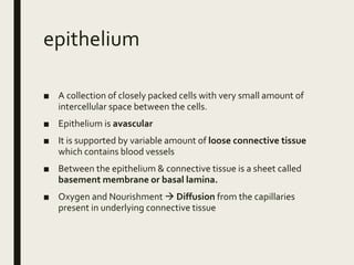
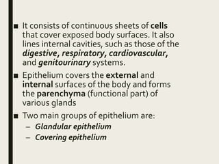
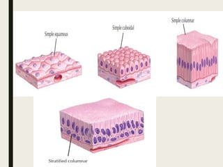

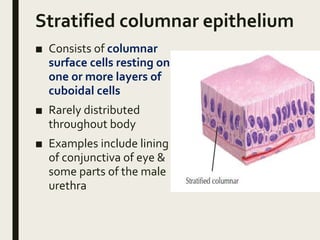
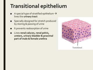

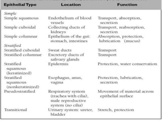


Epithelium is a tissue that consists of closely packed cells that form a continuous layer. It covers external surfaces of the body and internal cavities. There are two main types of epithelium - glandular epithelium which produces secretions or excretions, and covering epithelium which forms protective layers. Covering epithelium is classified based on cell layers (simple vs stratified), cell shape (squamous, cuboidal, columnar), and cellular modifications like cilia or microvilli. Epithelium serves functions like protection, secretion, absorption, excretion, and transport.