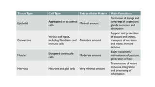
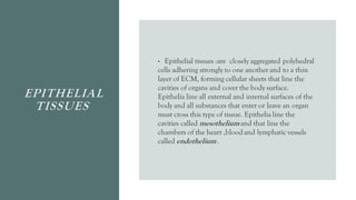
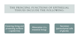
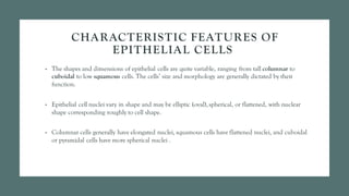
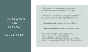
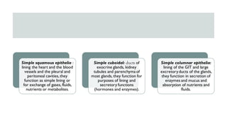
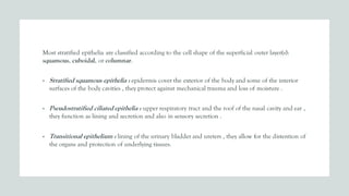
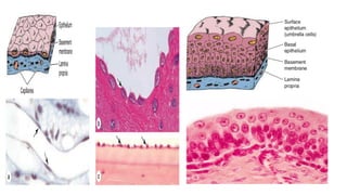
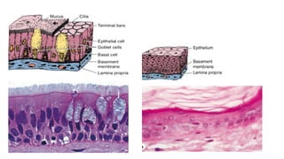
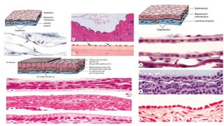
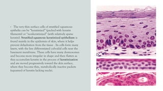
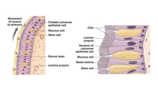
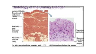
SlideShare a Scribd company logo
EPITHELIAL TISSUES
BY
DR.BUSHRA
M.JARALLAH
WE HAVE FOUR MAJOR TYPES OF TISSUES
The majority of organs consistof parenchyma which are the functioning cells of
the organand the stromawhich is the connective tissue
Nervous
Muscular
Connective
tissues
Epithelial
TissueType CellType Extracellular Matrix Main Functions
Epithelial
Aggregated or scattered
cells
Minimal amount
Formation of linings and
coverings of organs and
glands, secretion and
absorption
Connective
Various cell types,
including fibroblasts and
immune cells
Abundant amount
Support and protection
of tissues and organs,
transport of nutrients
and waste, immune
defense
Muscle
Elongated contractile
cells
Moderate amount
Body movement,
maintenance of posture,
generation of heat
Nervous Neurons and glial cells Very minimal amount
Transmission of nerve
impulses, integration
and processing of
information
EPITHELIAL
TISSUES
• Epithelial tissues :are closely aggregated polyhedral
cells adhering strongly to one another and to a thin
layer of ECM, forming cellular sheets that line the
cavities of organs and cover the body surface.
Epithelia line all external and internal surfaces of the
body and all substances that enter or leave an organ
must cross this type of tissue. Epithelia line the
cavities called mesotheliumand that line the
chambers of the heart ,bloodand lymphatic vessels
called endothelium.
THE PRINCIPAL FUNCTIONS OF EPITHELIAL
TISSUES INCLUDE THE FOLLOWING:
Covering, lining, and
protecting surfaces
( epidermis)
Absorption (the
intestinal lining)
Secretion
(parenchymal cells
of glands)
CHARACTERISTIC FEATURES OF
EPITHELIAL CELLS
• The shapes and dimensions of epithelial cells are quite variable, ranging from tall columnar to
cuboidal to low squamous cells. The cells’ size and morphology are generally dictated by their
function.
• Epithelial cell nuclei vary in shape and may be elliptic (oval), spherical, or flattened, with nuclear
shape corresponding roughly to cell shape.
• Columnar cells generally have elongated nuclei, squamous cells have flattened nuclei, and cuboidal
or pyramidal cells have more spherical nuclei .
• It is difficult to see all the layers by the light
microscope (because of the lipid rich membrane) so
these nuclei can be used to know the shape of the cell
and the number of the cell layers . The connective
tissues near the epithelia called the lamina
propria from which the epithelia get it’s blood and
nerve supply .
• Sometimes we see evaginations from the c.t to the
epithelia and extending to the external surface called
papillae , these are most seen in surfaces subjected to
more friction as the tongue.
• Epithelial cells also have polarity , they have basal and apical
surfaces .
• Regions of cuboidal or columnar cells that adjoin neighboring
cells comprise the cells’ lateral surfaces; cell membranes here
often have numerous folds which increase the area and
functional capacity of that surface.
BASEMENT
MEMBRANE
• The basal surface of all epithelia rests on a thin
extracellular, felt-like sheet of macromolecules referred
to as the basement membrane, a semipermeable filter
for substances reaching epithelial cells from below.
• Glycoproteins and other components in this structure
can often be stained and visualized with the light
microscope .
• With the transmission electron microscope (TEM) two
parts of the basement membrane may be resolved.
• Nearest the epithelial cells is an electron-dense layer,
20-100 nm thick, consisting of a network of fine fibrils
that comprise the basal lamina and beneath this layer
is a . more diffuse and fibrous reticular lamina.
BASEMENT MEMBRANE
TYPES OF
EPITHELIA
Epithelia can be divided into two main groups:
1. covering (or lining) epithelia
2. secretory(glandular) epithelia.
This is functional division for there are lining
epithelia in which all the cells also secrete (eg, the
lining of the stomach) or in which glandular cells are
distributed among the lining cells (eg, mucous cells in
the small intestine or trachea).
COVERING
OR
LINING
EPITHELIA
Cells covering the surfaces or lining the inner
surfaces arrange into one or more cell layers .
Epithelia are classified according to the number of
the layers and the morphology of the cells into :
• Simple epithelia contain only one cell layer
• Stratified epithelia contain two or more layers
Based on cell shape, simple epithelia are
further classified as squamous(thin
cells), cuboidal (cell width and thickness roughly
similar) or columnar (cells taller than they are wide).
Simple squamous epithelia :
lining the heart and the blood
vessels and the pleural and
peritoneal cavities, they
function as simple lining or
for exchange of gases, fluids,
nutrients or metabolites.
Simple cuboidal: ducts of
exocrine glands, kidney
tubules and parenchyma of
most glands, they function for
purposes of lining and
secretory functions
(hormones and enzymes).
Simple columnar epithelia:
lining of the GIT and large
excretoryducts of the glands,
they function in secretion of
enzymes and mucus and
absorption of nutrients and
fluids.
Most stratified epithelia are classified according to the cell shape of the superficial outer layer(s):
squamous, cuboidal, or columnar.
• Stratified squamous epithelia : epidermis cover the exterior of the body and some of the interior
surfaces of the body cavities , they protect against mechanical trauma and loss of moisture .
• Pseudostratified ciliated epithelia : upper respiratory tract and the roof of the nasal cavity and ear ,
they function as lining and secretion and also in sensory secretion .
• Transitional epithelium : lining of the urinary bladder and ureters , they allow for the distention of
the organs and protection of underlying tissues.
• The very thin surface cells of stratified squamous
epithelia can be “keratinized” (packed with keratin
filaments) or “nonkeratinized” (with relatively sparse
keratin). Stratified squamous keratinized epithelium is
found mainly in the epidermis of skin, where it helps
prevent dehydration from the tissue . Its cells form many
layers, with the less differentiated cuboidal cells near the
basement membrane. These cells have many desmosomes
and become more irregular in shape and then flatten as
they accumulate keratin in the process of keratinization
and are moved progressively toward the skin surface,
where they become thin, metabolically inactive packets
(squames) of keratin lacking nuclei.
This surface layer of cells helps protect against water loss
across this epithelium.
Stratified squamous nonkeratinized epithelium
lines moist internal cavities (eg, mouth,esophagus,and
vagina) where water loss is not a problem.Here the
flattened cells of the surface layer retain their nuclei and
most metabolic functions.
Stratified cuboidal and stratified columnar
epithelia are both relatively rare.Stratified cuboidal
epithelium occurs in the excretory ducts of salivary and
sweat glands . Stratified columnar epithelium is seen in
the conjunctiva lining the eyelids,where it is both
protective and mucus secreting.
• Unique transitional epithelium or urothelium lines
much of the urinary tract, extending from the kidneys to
the proximal part of the urethra, and is characterized by a
superficial layer of large, dome-like cells sometimes called
umbrella cells , these cells are specialized to protect
underlying tissues from the hypertonic and potentially
cytotoxic effects of urine. Importantly, unique
morphological features of the cells allow distension of
transitional epithelium as the urinary bladder fills.
• A final morphological type of epithelium is called
pseudostratified columnar epithelium , here tall irregular
cells all are attached to the basement membrane , but ,
their nuclei are at different levels and not all the cells
reaching the free surface , giving the stratified appearance .
A good example is the lining of upper respiratory tract ,
the cells also here are heavy ciliated .
Major Feature Cell Form
Examples of
Distribution Main Function
Simple (one layer of cells) Squamous
Lining of vessels
(endothelium);
covering the ovary,
thyroid
Facilitates the movement of the viscera (mesothelium), active transport by pinocytosis (mesothelium and endothelium), secretion of
biologically active molecules (mesothelium)
Cuboidal
Covering the ovary,
thyroid
Covering, secretion
Columnar
Lining of intestine,
gallbladder
Protection, lubrication, absorption, secretion
Stratified (two or more layers of
cells)
Squamous keratinized
(dry)
Epidermis Protection; prevents water loss
Squamous
nonkeratinized (moist)
Mouth, esophagus,
larynx, vagina, anal
canal
Protection, secretion; prevents water loss
Cuboidal
Sweat glands,
developing ovarian
follicles
Protection, secretion
Transitional
Bladder, ureters, renal
calyces
Protection, distensibility
Columnar Coniunctiva Protection
Pseudostratified (layers of cells
with nuclei at different levels; not
all cells reach surface but all
adhere to basal lamina)
Columnar
Lining of trachea,
bronchi, nasal cavity
Protection, secretion; cilia-mediated transport of particles trapped in mucus out of the air passages
INTERCELLULAR ADHESION & OTHER JUNCTIONS
• Epithelial cells adhere strongly to neighboring cells and basal laminae, particularly in epithelia
subject to friction or other mechanical forces.
• Tight or occluding junctions form a seal between adjacent cells. also called zonulae occludens, are
the most apical of the junctions. The term “zonula” indicates that the junction forms a band
completely encircling each cell.
• Adherent or anchoring junctions are sites of strong cell adhesion. or zonula adherens , which also
encircles the epithelial cell, usually immediately below the tight junction.
• Gap junctions are channels for communication between adjacent cells. mediate inter- cellular
communication rather than adhesion or occlusion between cells. Abundant in many epithelia, gap
junctions are also functionally important in nearly all mammalian tissues.
• Desmosomes : or macula adherens . As the name implies this junction resembles a single “spot-
weld” and does not form a belt around the cell. Desmosomes are disc-shaped structures at the
surface of one cell that are matched with identical structures at an adjacent cell surface
• Hemidesmosomes , which can be seen by TEM . These adhesive structures resemble a half-
desmosome ultra structurally, they found on the basal surface of the cell , attaching the cells to the
basal lamina .
SPECIALIZATIONS OF THE APICAL CELL
SURFACE
• Microvilli : cytoplasmic projections , actin filaments binding to the plasma membrane by a protein,
increase the surface area , The average microvillus is about 1-μm long and 0.1-μm wide.
• Stereocilia : found in the lining of the male reproductive tract , also have microfilaments connected
to actin , but are longer than the microvilli and less mobile , and show branching distally .
• Cilia : are long, highly motile apical structures, larger than microvilli, containing internal arrays of
microtubules not microfilaments . In addition to cilia on epithelial cells, most (if not all) other cell
types have at least one short projection called a primary cilium, which is not motile but is enriched
with receptors and signal transduction complexes for detection of light, odors, motion, and flow of
liquid past the cells. They are abundant on columnar and cuboidal cells, Typical cilia are 5-10 μm
long and 0.2 μm in diameter, which is much longer and two times wider than a typical microvilli .
• Regeneration of epithelial cells :
• Epithelial tissues are relatively labile structures whose
cells are renewed continuously by mitotic activity and
stem cell populations. The rate of renewal varies
widely; it can be fast in tis- sues such as the intestinal
epithelium, which is replaced every week, or slow, as in
the large glands. In stratified epithelial tis- sues, stem
cells and mitosis occur only within the basal layer in
contact with the basal lamina. In some functionally
complex epithelia, stem cells are located only in
restricted niches some distance from the transit
amplifying cells and differentiating cells
• For example, the epithelium lining the small intestine
is derived completely from stem cells found in the simple
glands between the intestinal villi. In the epidermis, many
stem cells are located at a characteristic position along the
wall of hair follicles.
• Epithelia are normally capable of rapid repair and
replacement of apoptotic or damaged cells. In some large
glands, most notably the liver, mitotic activity is normally
rare but is actively renewed following major damage to the
organ. When a portion of liver tissue is removed surgically
or lost by the acute effects of toxic substances, cells of
undamaged regions quickly begin active proliferation and
a mass of liver tissue with normal function is regenerated.
• Metaplasia
Under certain abnormal conditions, one type of epithelial
tissue may undergo transformation into another type in
another reversible process called metaplasia. In heavy
cigarette smokers, the ciliated pseudostratified epithelium
lining the bronchi can be transformed into stratified
squamous epithelium.
• Neoplasia
Some epithelial cells are prone to abnormal growth or
dysplasia, which can progress to precancerous growth
called neoplasia. Early neoplastic growth is often
reversible and does not always result in cancer. Neoplasia
of epithelial cells called carcinoma .
BODY SURFACES
The externalsurface of the body is
covered with a continuous layer of skin of
variable thickness and color .The
epidermis,the outermost layer of skin,
providesa waterproof barrier and creates
our skin tone.
The dermis,beneath the epidermis,contains
tough connective tissue, hair follicles,and
sweat glands.
The deeper subcutaneoustissue
(hypodermis)is made of fat and connective
tissue.This is the same structure in different
parts of the skin . However the thickness of
the skin differ resulting into thin and thick
skin .
MUCUS
MEMBRANES
1- mucosa : consist of epithelia ,
lamina properia ,and muscularis mucosa
2- submucosa : which is the connective
tissues
3- muscularis externa : consist of
smooth muscles arranged in cicular and
longitudinal layers
4- serosa or adventitia
Typical mucus membrane consist of four layers:
THANK YOU

More Related Content

What's hot

Cell junction
Cell junctionCell junction
Cell junction
AanshiSrivastava2
 
Transport across cell membrane
Transport across cell membraneTransport across cell membrane
Transport across cell membrane
Anu Priya
 
Skin and fascial medicos notes.com
Skin and fascial medicos notes.comSkin and fascial medicos notes.com
Skin and fascial medicos notes.com
MedicosNotes
 
Histology of cns.dk.2014
Histology of cns.dk.2014Histology of cns.dk.2014
Histology of cns.dk.2014
Deepak Khedekar
 
Tight junctions workshop - By Vaishnavi Charanya Sundar - Physiology
Tight junctions workshop - By Vaishnavi Charanya Sundar - PhysiologyTight junctions workshop - By Vaishnavi Charanya Sundar - Physiology
Tight junctions workshop - By Vaishnavi Charanya Sundar - Physiology
VaishnavicharanyaSun
 
Ch14 nervous tissue
Ch14 nervous tissueCh14 nervous tissue
Ch14 nervous tissue
KemUnited
 
Cell junctions ppt
Cell junctions pptCell junctions ppt
Cell junctions ppt
shaimaaf12
 
Cell membrane (Plasma Membrane) & Cellular Junctions
Cell membrane (Plasma Membrane) & Cellular JunctionsCell membrane (Plasma Membrane) & Cellular Junctions
Cell membrane (Plasma Membrane) & Cellular Junctions
Pradeep Singh Narwat
 
Inter cellular junctions
Inter cellular junctionsInter cellular junctions
Inter cellular junctions
Ambika Jawalkar
 
Smooth muscle
Smooth muscleSmooth muscle
Smooth muscle
Ramya Aravind
 
3. cardiac muscle tissue
3.  cardiac muscle tissue3.  cardiac muscle tissue
3. cardiac muscle tissueMichael Wrock
 
Golgi complex
Golgi complexGolgi complex
Golgi complex
Dr. T. A. Gitte
 
Intercellular junctions
Intercellular junctionsIntercellular junctions
Intercellular junctions
Jilsha Cecil
 
Cell Junctions
Cell JunctionsCell Junctions
Cell Junctions
Dr ABU SURAIH SAKHRI
 
Nerve. ppt
Nerve. pptNerve. ppt
Nerve. ppt
Sukesh Vangeti
 
Golgi complex structure and functions
Golgi complex structure and functionsGolgi complex structure and functions
Golgi complex structure and functions
bikashsingh2510
 
Endoplasmic reticulum by amita
Endoplasmic reticulum by amitaEndoplasmic reticulum by amita
Endoplasmic reticulum by amitaAmita Mevada
 
Muscular tissue Histology
Muscular tissue HistologyMuscular tissue Histology
Muscular tissue Histology
Dr. Devi Shankar
 
epithelium
epitheliumepithelium
Tissue
TissueTissue
Tissue
Poojakate4
 

What's hot (20)

Cell junction
Cell junctionCell junction
Cell junction
 
Transport across cell membrane
Transport across cell membraneTransport across cell membrane
Transport across cell membrane
 
Skin and fascial medicos notes.com
Skin and fascial medicos notes.comSkin and fascial medicos notes.com
Skin and fascial medicos notes.com
 
Histology of cns.dk.2014
Histology of cns.dk.2014Histology of cns.dk.2014
Histology of cns.dk.2014
 
Tight junctions workshop - By Vaishnavi Charanya Sundar - Physiology
Tight junctions workshop - By Vaishnavi Charanya Sundar - PhysiologyTight junctions workshop - By Vaishnavi Charanya Sundar - Physiology
Tight junctions workshop - By Vaishnavi Charanya Sundar - Physiology
 
Ch14 nervous tissue
Ch14 nervous tissueCh14 nervous tissue
Ch14 nervous tissue
 
Cell junctions ppt
Cell junctions pptCell junctions ppt
Cell junctions ppt
 
Cell membrane (Plasma Membrane) & Cellular Junctions
Cell membrane (Plasma Membrane) & Cellular JunctionsCell membrane (Plasma Membrane) & Cellular Junctions
Cell membrane (Plasma Membrane) & Cellular Junctions
 
Inter cellular junctions
Inter cellular junctionsInter cellular junctions
Inter cellular junctions
 
Smooth muscle
Smooth muscleSmooth muscle
Smooth muscle
 
3. cardiac muscle tissue
3.  cardiac muscle tissue3.  cardiac muscle tissue
3. cardiac muscle tissue
 
Golgi complex
Golgi complexGolgi complex
Golgi complex
 
Intercellular junctions
Intercellular junctionsIntercellular junctions
Intercellular junctions
 
Cell Junctions
Cell JunctionsCell Junctions
Cell Junctions
 
Nerve. ppt
Nerve. pptNerve. ppt
Nerve. ppt
 
Golgi complex structure and functions
Golgi complex structure and functionsGolgi complex structure and functions
Golgi complex structure and functions
 
Endoplasmic reticulum by amita
Endoplasmic reticulum by amitaEndoplasmic reticulum by amita
Endoplasmic reticulum by amita
 
Muscular tissue Histology
Muscular tissue HistologyMuscular tissue Histology
Muscular tissue Histology
 
epithelium
epitheliumepithelium
epithelium
 
Tissue
TissueTissue
Tissue
 

Similar to Epithelial Tissue

Histology basics of sciences and microanatomy.pdf
Histology basics of sciences and microanatomy.pdfHistology basics of sciences and microanatomy.pdf
Histology basics of sciences and microanatomy.pdf
gargshivansh14
 
5566.pptx
5566.pptx5566.pptx
Basic Histology no.2 epithelium tissue.pptx
Basic Histology no.2  epithelium tissue.pptxBasic Histology no.2  epithelium tissue.pptx
Basic Histology no.2 epithelium tissue.pptx
YousifAhmedDA
 
Anatomy :O
Anatomy :OAnatomy :O
Anatomy :OSteven S
 
The Tissue level of organization .pptx
The Tissue level of organization   .pptxThe Tissue level of organization   .pptx
The Tissue level of organization .pptx
shraddhaparab530
 
study of epithelium.pptx
study of epithelium.pptxstudy of epithelium.pptx
study of epithelium.pptx
Dinraj Kulkarni
 
General principles of tissue organization
General principles of tissue organizationGeneral principles of tissue organization
General principles of tissue organization
Eneutron
 
Epithelia tissue.pptx
Epithelia tissue.pptxEpithelia tissue.pptx
Epithelia tissue.pptx
NavyaPS2
 
Basic Tissues 1.pptx
Basic Tissues 1.pptxBasic Tissues 1.pptx
Basic Tissues 1.pptx
FrancisEtseyDushie
 
HUMAN ANATOMY AND PHYSIOLOGY.PPT.pdf.pdf
HUMAN ANATOMY AND PHYSIOLOGY.PPT.pdf.pdfHUMAN ANATOMY AND PHYSIOLOGY.PPT.pdf.pdf
HUMAN ANATOMY AND PHYSIOLOGY.PPT.pdf.pdf
souvickmaity618
 
3.tissues of body
3.tissues of body3.tissues of body
3.tissues of body
M Humayun Jamil
 
epithelia Tissues.pptx
epithelia Tissues.pptxepithelia Tissues.pptx
epithelia Tissues.pptx
FrancisEtseyDushie
 
section 1, chapter 5
section 1, chapter 5section 1, chapter 5
section 1, chapter 5
Michael Walls
 
Animal Tissues And Homeostasis
Animal Tissues And HomeostasisAnimal Tissues And Homeostasis
Animal Tissues And Homeostasis
sum_itbhu
 
4. Tissues: Living Communities
4. Tissues: Living Communities4. Tissues: Living Communities
4. Tissues: Living CommunitiesSUNY Ulster
 
Tissues
Tissues Tissues
Tissues
Sara Khan
 
Histology 1 fro medicine and science student
Histology 1 fro medicine and science student Histology 1 fro medicine and science student
Histology 1 fro medicine and science student
Hussain Alsaady
 
Epithelium
EpitheliumEpithelium
EpitheliumIIDC
 

Similar to Epithelial Tissue (20)

Histology basics of sciences and microanatomy.pdf
Histology basics of sciences and microanatomy.pdfHistology basics of sciences and microanatomy.pdf
Histology basics of sciences and microanatomy.pdf
 
5566.pptx
5566.pptx5566.pptx
5566.pptx
 
Basic Histology no.2 epithelium tissue.pptx
Basic Histology no.2  epithelium tissue.pptxBasic Histology no.2  epithelium tissue.pptx
Basic Histology no.2 epithelium tissue.pptx
 
Anatomy :O
Anatomy :OAnatomy :O
Anatomy :O
 
The Tissue level of organization .pptx
The Tissue level of organization   .pptxThe Tissue level of organization   .pptx
The Tissue level of organization .pptx
 
study of epithelium.pptx
study of epithelium.pptxstudy of epithelium.pptx
study of epithelium.pptx
 
General principles of tissue organization
General principles of tissue organizationGeneral principles of tissue organization
General principles of tissue organization
 
Epithelia tissue.pptx
Epithelia tissue.pptxEpithelia tissue.pptx
Epithelia tissue.pptx
 
Basic Tissues 1.pptx
Basic Tissues 1.pptxBasic Tissues 1.pptx
Basic Tissues 1.pptx
 
HUMAN ANATOMY AND PHYSIOLOGY.PPT.pdf.pdf
HUMAN ANATOMY AND PHYSIOLOGY.PPT.pdf.pdfHUMAN ANATOMY AND PHYSIOLOGY.PPT.pdf.pdf
HUMAN ANATOMY AND PHYSIOLOGY.PPT.pdf.pdf
 
3.tissues of body
3.tissues of body3.tissues of body
3.tissues of body
 
epithelia Tissues.pptx
epithelia Tissues.pptxepithelia Tissues.pptx
epithelia Tissues.pptx
 
section 1, chapter 5
section 1, chapter 5section 1, chapter 5
section 1, chapter 5
 
Histology
HistologyHistology
Histology
 
Tissues
TissuesTissues
Tissues
 
Animal Tissues And Homeostasis
Animal Tissues And HomeostasisAnimal Tissues And Homeostasis
Animal Tissues And Homeostasis
 
4. Tissues: Living Communities
4. Tissues: Living Communities4. Tissues: Living Communities
4. Tissues: Living Communities
 
Tissues
Tissues Tissues
Tissues
 
Histology 1 fro medicine and science student
Histology 1 fro medicine and science student Histology 1 fro medicine and science student
Histology 1 fro medicine and science student
 
Epithelium
EpitheliumEpithelium
Epithelium
 

Recently uploaded

SCHIZOPHRENIA Disorder/ Brain Disorder.pdf
SCHIZOPHRENIA Disorder/ Brain Disorder.pdfSCHIZOPHRENIA Disorder/ Brain Disorder.pdf
SCHIZOPHRENIA Disorder/ Brain Disorder.pdf
SELF-EXPLANATORY
 
PRESENTATION ABOUT PRINCIPLE OF COSMATIC EVALUATION
PRESENTATION ABOUT PRINCIPLE OF COSMATIC EVALUATIONPRESENTATION ABOUT PRINCIPLE OF COSMATIC EVALUATION
PRESENTATION ABOUT PRINCIPLE OF COSMATIC EVALUATION
ChetanK57
 
filosofia boliviana introducción jsjdjd.pptx
filosofia boliviana introducción jsjdjd.pptxfilosofia boliviana introducción jsjdjd.pptx
filosofia boliviana introducción jsjdjd.pptx
IvanMallco1
 
NuGOweek 2024 Ghent - programme - final version
NuGOweek 2024 Ghent - programme - final versionNuGOweek 2024 Ghent - programme - final version
NuGOweek 2024 Ghent - programme - final version
pablovgd
 
Unveiling the Energy Potential of Marshmallow Deposits.pdf
Unveiling the Energy Potential of Marshmallow Deposits.pdfUnveiling the Energy Potential of Marshmallow Deposits.pdf
Unveiling the Energy Potential of Marshmallow Deposits.pdf
Erdal Coalmaker
 
general properties of oerganologametal.ppt
general properties of oerganologametal.pptgeneral properties of oerganologametal.ppt
general properties of oerganologametal.ppt
IqrimaNabilatulhusni
 
Astronomy Update- Curiosity’s exploration of Mars _ Local Briefs _ leadertele...
Astronomy Update- Curiosity’s exploration of Mars _ Local Briefs _ leadertele...Astronomy Update- Curiosity’s exploration of Mars _ Local Briefs _ leadertele...
Astronomy Update- Curiosity’s exploration of Mars _ Local Briefs _ leadertele...
NathanBaughman3
 
Mammalian Pineal Body Structure and Also Functions
Mammalian Pineal Body Structure and Also FunctionsMammalian Pineal Body Structure and Also Functions
Mammalian Pineal Body Structure and Also Functions
YOGESH DOGRA
 
Lateral Ventricles.pdf very easy good diagrams comprehensive
Lateral Ventricles.pdf very easy good diagrams comprehensiveLateral Ventricles.pdf very easy good diagrams comprehensive
Lateral Ventricles.pdf very easy good diagrams comprehensive
silvermistyshot
 
What is greenhouse gasses and how many gasses are there to affect the Earth.
What is greenhouse gasses and how many gasses are there to affect the Earth.What is greenhouse gasses and how many gasses are there to affect the Earth.
What is greenhouse gasses and how many gasses are there to affect the Earth.
moosaasad1975
 
platelets- lifespan -Clot retraction-disorders.pptx
platelets- lifespan -Clot retraction-disorders.pptxplatelets- lifespan -Clot retraction-disorders.pptx
platelets- lifespan -Clot retraction-disorders.pptx
muralinath2
 
Deep Behavioral Phenotyping in Systems Neuroscience for Functional Atlasing a...
Deep Behavioral Phenotyping in Systems Neuroscience for Functional Atlasing a...Deep Behavioral Phenotyping in Systems Neuroscience for Functional Atlasing a...
Deep Behavioral Phenotyping in Systems Neuroscience for Functional Atlasing a...
Ana Luísa Pinho
 
Orion Air Quality Monitoring Systems - CWS
Orion Air Quality Monitoring Systems - CWSOrion Air Quality Monitoring Systems - CWS
Orion Air Quality Monitoring Systems - CWS
Columbia Weather Systems
 
in vitro propagation of plants lecture note.pptx
in vitro propagation of plants lecture note.pptxin vitro propagation of plants lecture note.pptx
in vitro propagation of plants lecture note.pptx
yusufzako14
 
The ASGCT Annual Meeting was packed with exciting progress in the field advan...
The ASGCT Annual Meeting was packed with exciting progress in the field advan...The ASGCT Annual Meeting was packed with exciting progress in the field advan...
The ASGCT Annual Meeting was packed with exciting progress in the field advan...
Health Advances
 
Hemoglobin metabolism_pathophysiology.pptx
Hemoglobin metabolism_pathophysiology.pptxHemoglobin metabolism_pathophysiology.pptx
Hemoglobin metabolism_pathophysiology.pptx
muralinath2
 
Leaf Initiation, Growth and Differentiation.pdf
Leaf Initiation, Growth and Differentiation.pdfLeaf Initiation, Growth and Differentiation.pdf
Leaf Initiation, Growth and Differentiation.pdf
RenuJangid3
 
role of pramana in research.pptx in science
role of pramana in research.pptx in sciencerole of pramana in research.pptx in science
role of pramana in research.pptx in science
sonaliswain16
 
THE IMPORTANCE OF MARTIAN ATMOSPHERE SAMPLE RETURN.
THE IMPORTANCE OF MARTIAN ATMOSPHERE SAMPLE RETURN.THE IMPORTANCE OF MARTIAN ATMOSPHERE SAMPLE RETURN.
THE IMPORTANCE OF MARTIAN ATMOSPHERE SAMPLE RETURN.
Sérgio Sacani
 
Multi-source connectivity as the driver of solar wind variability in the heli...
Multi-source connectivity as the driver of solar wind variability in the heli...Multi-source connectivity as the driver of solar wind variability in the heli...
Multi-source connectivity as the driver of solar wind variability in the heli...
Sérgio Sacani
 

Recently uploaded (20)

SCHIZOPHRENIA Disorder/ Brain Disorder.pdf
SCHIZOPHRENIA Disorder/ Brain Disorder.pdfSCHIZOPHRENIA Disorder/ Brain Disorder.pdf
SCHIZOPHRENIA Disorder/ Brain Disorder.pdf
 
PRESENTATION ABOUT PRINCIPLE OF COSMATIC EVALUATION
PRESENTATION ABOUT PRINCIPLE OF COSMATIC EVALUATIONPRESENTATION ABOUT PRINCIPLE OF COSMATIC EVALUATION
PRESENTATION ABOUT PRINCIPLE OF COSMATIC EVALUATION
 
filosofia boliviana introducción jsjdjd.pptx
filosofia boliviana introducción jsjdjd.pptxfilosofia boliviana introducción jsjdjd.pptx
filosofia boliviana introducción jsjdjd.pptx
 
NuGOweek 2024 Ghent - programme - final version
NuGOweek 2024 Ghent - programme - final versionNuGOweek 2024 Ghent - programme - final version
NuGOweek 2024 Ghent - programme - final version
 
Unveiling the Energy Potential of Marshmallow Deposits.pdf
Unveiling the Energy Potential of Marshmallow Deposits.pdfUnveiling the Energy Potential of Marshmallow Deposits.pdf
Unveiling the Energy Potential of Marshmallow Deposits.pdf
 
general properties of oerganologametal.ppt
general properties of oerganologametal.pptgeneral properties of oerganologametal.ppt
general properties of oerganologametal.ppt
 
Astronomy Update- Curiosity’s exploration of Mars _ Local Briefs _ leadertele...
Astronomy Update- Curiosity’s exploration of Mars _ Local Briefs _ leadertele...Astronomy Update- Curiosity’s exploration of Mars _ Local Briefs _ leadertele...
Astronomy Update- Curiosity’s exploration of Mars _ Local Briefs _ leadertele...
 
Mammalian Pineal Body Structure and Also Functions
Mammalian Pineal Body Structure and Also FunctionsMammalian Pineal Body Structure and Also Functions
Mammalian Pineal Body Structure and Also Functions
 
Lateral Ventricles.pdf very easy good diagrams comprehensive
Lateral Ventricles.pdf very easy good diagrams comprehensiveLateral Ventricles.pdf very easy good diagrams comprehensive
Lateral Ventricles.pdf very easy good diagrams comprehensive
 
What is greenhouse gasses and how many gasses are there to affect the Earth.
What is greenhouse gasses and how many gasses are there to affect the Earth.What is greenhouse gasses and how many gasses are there to affect the Earth.
What is greenhouse gasses and how many gasses are there to affect the Earth.
 
platelets- lifespan -Clot retraction-disorders.pptx
platelets- lifespan -Clot retraction-disorders.pptxplatelets- lifespan -Clot retraction-disorders.pptx
platelets- lifespan -Clot retraction-disorders.pptx
 
Deep Behavioral Phenotyping in Systems Neuroscience for Functional Atlasing a...
Deep Behavioral Phenotyping in Systems Neuroscience for Functional Atlasing a...Deep Behavioral Phenotyping in Systems Neuroscience for Functional Atlasing a...
Deep Behavioral Phenotyping in Systems Neuroscience for Functional Atlasing a...
 
Orion Air Quality Monitoring Systems - CWS
Orion Air Quality Monitoring Systems - CWSOrion Air Quality Monitoring Systems - CWS
Orion Air Quality Monitoring Systems - CWS
 
in vitro propagation of plants lecture note.pptx
in vitro propagation of plants lecture note.pptxin vitro propagation of plants lecture note.pptx
in vitro propagation of plants lecture note.pptx
 
The ASGCT Annual Meeting was packed with exciting progress in the field advan...
The ASGCT Annual Meeting was packed with exciting progress in the field advan...The ASGCT Annual Meeting was packed with exciting progress in the field advan...
The ASGCT Annual Meeting was packed with exciting progress in the field advan...
 
Hemoglobin metabolism_pathophysiology.pptx
Hemoglobin metabolism_pathophysiology.pptxHemoglobin metabolism_pathophysiology.pptx
Hemoglobin metabolism_pathophysiology.pptx
 
Leaf Initiation, Growth and Differentiation.pdf
Leaf Initiation, Growth and Differentiation.pdfLeaf Initiation, Growth and Differentiation.pdf
Leaf Initiation, Growth and Differentiation.pdf
 
role of pramana in research.pptx in science
role of pramana in research.pptx in sciencerole of pramana in research.pptx in science
role of pramana in research.pptx in science
 
THE IMPORTANCE OF MARTIAN ATMOSPHERE SAMPLE RETURN.
THE IMPORTANCE OF MARTIAN ATMOSPHERE SAMPLE RETURN.THE IMPORTANCE OF MARTIAN ATMOSPHERE SAMPLE RETURN.
THE IMPORTANCE OF MARTIAN ATMOSPHERE SAMPLE RETURN.
 
Multi-source connectivity as the driver of solar wind variability in the heli...
Multi-source connectivity as the driver of solar wind variability in the heli...Multi-source connectivity as the driver of solar wind variability in the heli...
Multi-source connectivity as the driver of solar wind variability in the heli...
 

Epithelial Tissue

  • 2. WE HAVE FOUR MAJOR TYPES OF TISSUES The majority of organs consistof parenchyma which are the functioning cells of the organand the stromawhich is the connective tissue Nervous Muscular Connective tissues Epithelial
  • 3. TissueType CellType Extracellular Matrix Main Functions Epithelial Aggregated or scattered cells Minimal amount Formation of linings and coverings of organs and glands, secretion and absorption Connective Various cell types, including fibroblasts and immune cells Abundant amount Support and protection of tissues and organs, transport of nutrients and waste, immune defense Muscle Elongated contractile cells Moderate amount Body movement, maintenance of posture, generation of heat Nervous Neurons and glial cells Very minimal amount Transmission of nerve impulses, integration and processing of information
  • 4. EPITHELIAL TISSUES • Epithelial tissues :are closely aggregated polyhedral cells adhering strongly to one another and to a thin layer of ECM, forming cellular sheets that line the cavities of organs and cover the body surface. Epithelia line all external and internal surfaces of the body and all substances that enter or leave an organ must cross this type of tissue. Epithelia line the cavities called mesotheliumand that line the chambers of the heart ,bloodand lymphatic vessels called endothelium.
  • 5. THE PRINCIPAL FUNCTIONS OF EPITHELIAL TISSUES INCLUDE THE FOLLOWING: Covering, lining, and protecting surfaces ( epidermis) Absorption (the intestinal lining) Secretion (parenchymal cells of glands)
  • 6. CHARACTERISTIC FEATURES OF EPITHELIAL CELLS • The shapes and dimensions of epithelial cells are quite variable, ranging from tall columnar to cuboidal to low squamous cells. The cells’ size and morphology are generally dictated by their function. • Epithelial cell nuclei vary in shape and may be elliptic (oval), spherical, or flattened, with nuclear shape corresponding roughly to cell shape. • Columnar cells generally have elongated nuclei, squamous cells have flattened nuclei, and cuboidal or pyramidal cells have more spherical nuclei .
  • 7. • It is difficult to see all the layers by the light microscope (because of the lipid rich membrane) so these nuclei can be used to know the shape of the cell and the number of the cell layers . The connective tissues near the epithelia called the lamina propria from which the epithelia get it’s blood and nerve supply . • Sometimes we see evaginations from the c.t to the epithelia and extending to the external surface called papillae , these are most seen in surfaces subjected to more friction as the tongue.
  • 8. • Epithelial cells also have polarity , they have basal and apical surfaces . • Regions of cuboidal or columnar cells that adjoin neighboring cells comprise the cells’ lateral surfaces; cell membranes here often have numerous folds which increase the area and functional capacity of that surface.
  • 9. BASEMENT MEMBRANE • The basal surface of all epithelia rests on a thin extracellular, felt-like sheet of macromolecules referred to as the basement membrane, a semipermeable filter for substances reaching epithelial cells from below. • Glycoproteins and other components in this structure can often be stained and visualized with the light microscope . • With the transmission electron microscope (TEM) two parts of the basement membrane may be resolved. • Nearest the epithelial cells is an electron-dense layer, 20-100 nm thick, consisting of a network of fine fibrils that comprise the basal lamina and beneath this layer is a . more diffuse and fibrous reticular lamina.
  • 11.
  • 12. TYPES OF EPITHELIA Epithelia can be divided into two main groups: 1. covering (or lining) epithelia 2. secretory(glandular) epithelia. This is functional division for there are lining epithelia in which all the cells also secrete (eg, the lining of the stomach) or in which glandular cells are distributed among the lining cells (eg, mucous cells in the small intestine or trachea).
  • 13. COVERING OR LINING EPITHELIA Cells covering the surfaces or lining the inner surfaces arrange into one or more cell layers . Epithelia are classified according to the number of the layers and the morphology of the cells into : • Simple epithelia contain only one cell layer • Stratified epithelia contain two or more layers Based on cell shape, simple epithelia are further classified as squamous(thin cells), cuboidal (cell width and thickness roughly similar) or columnar (cells taller than they are wide).
  • 14. Simple squamous epithelia : lining the heart and the blood vessels and the pleural and peritoneal cavities, they function as simple lining or for exchange of gases, fluids, nutrients or metabolites. Simple cuboidal: ducts of exocrine glands, kidney tubules and parenchyma of most glands, they function for purposes of lining and secretory functions (hormones and enzymes). Simple columnar epithelia: lining of the GIT and large excretoryducts of the glands, they function in secretion of enzymes and mucus and absorption of nutrients and fluids.
  • 15. Most stratified epithelia are classified according to the cell shape of the superficial outer layer(s): squamous, cuboidal, or columnar. • Stratified squamous epithelia : epidermis cover the exterior of the body and some of the interior surfaces of the body cavities , they protect against mechanical trauma and loss of moisture . • Pseudostratified ciliated epithelia : upper respiratory tract and the roof of the nasal cavity and ear , they function as lining and secretion and also in sensory secretion . • Transitional epithelium : lining of the urinary bladder and ureters , they allow for the distention of the organs and protection of underlying tissues.
  • 16.
  • 17.
  • 18.
  • 19. • The very thin surface cells of stratified squamous epithelia can be “keratinized” (packed with keratin filaments) or “nonkeratinized” (with relatively sparse keratin). Stratified squamous keratinized epithelium is found mainly in the epidermis of skin, where it helps prevent dehydration from the tissue . Its cells form many layers, with the less differentiated cuboidal cells near the basement membrane. These cells have many desmosomes and become more irregular in shape and then flatten as they accumulate keratin in the process of keratinization and are moved progressively toward the skin surface, where they become thin, metabolically inactive packets (squames) of keratin lacking nuclei.
  • 20. This surface layer of cells helps protect against water loss across this epithelium. Stratified squamous nonkeratinized epithelium lines moist internal cavities (eg, mouth,esophagus,and vagina) where water loss is not a problem.Here the flattened cells of the surface layer retain their nuclei and most metabolic functions. Stratified cuboidal and stratified columnar epithelia are both relatively rare.Stratified cuboidal epithelium occurs in the excretory ducts of salivary and sweat glands . Stratified columnar epithelium is seen in the conjunctiva lining the eyelids,where it is both protective and mucus secreting.
  • 21. • Unique transitional epithelium or urothelium lines much of the urinary tract, extending from the kidneys to the proximal part of the urethra, and is characterized by a superficial layer of large, dome-like cells sometimes called umbrella cells , these cells are specialized to protect underlying tissues from the hypertonic and potentially cytotoxic effects of urine. Importantly, unique morphological features of the cells allow distension of transitional epithelium as the urinary bladder fills.
  • 22. • A final morphological type of epithelium is called pseudostratified columnar epithelium , here tall irregular cells all are attached to the basement membrane , but , their nuclei are at different levels and not all the cells reaching the free surface , giving the stratified appearance . A good example is the lining of upper respiratory tract , the cells also here are heavy ciliated .
  • 23. Major Feature Cell Form Examples of Distribution Main Function Simple (one layer of cells) Squamous Lining of vessels (endothelium); covering the ovary, thyroid Facilitates the movement of the viscera (mesothelium), active transport by pinocytosis (mesothelium and endothelium), secretion of biologically active molecules (mesothelium) Cuboidal Covering the ovary, thyroid Covering, secretion Columnar Lining of intestine, gallbladder Protection, lubrication, absorption, secretion Stratified (two or more layers of cells) Squamous keratinized (dry) Epidermis Protection; prevents water loss Squamous nonkeratinized (moist) Mouth, esophagus, larynx, vagina, anal canal Protection, secretion; prevents water loss Cuboidal Sweat glands, developing ovarian follicles Protection, secretion Transitional Bladder, ureters, renal calyces Protection, distensibility Columnar Coniunctiva Protection Pseudostratified (layers of cells with nuclei at different levels; not all cells reach surface but all adhere to basal lamina) Columnar Lining of trachea, bronchi, nasal cavity Protection, secretion; cilia-mediated transport of particles trapped in mucus out of the air passages
  • 24.
  • 25. INTERCELLULAR ADHESION & OTHER JUNCTIONS • Epithelial cells adhere strongly to neighboring cells and basal laminae, particularly in epithelia subject to friction or other mechanical forces. • Tight or occluding junctions form a seal between adjacent cells. also called zonulae occludens, are the most apical of the junctions. The term “zonula” indicates that the junction forms a band completely encircling each cell. • Adherent or anchoring junctions are sites of strong cell adhesion. or zonula adherens , which also encircles the epithelial cell, usually immediately below the tight junction.
  • 26. • Gap junctions are channels for communication between adjacent cells. mediate inter- cellular communication rather than adhesion or occlusion between cells. Abundant in many epithelia, gap junctions are also functionally important in nearly all mammalian tissues. • Desmosomes : or macula adherens . As the name implies this junction resembles a single “spot- weld” and does not form a belt around the cell. Desmosomes are disc-shaped structures at the surface of one cell that are matched with identical structures at an adjacent cell surface • Hemidesmosomes , which can be seen by TEM . These adhesive structures resemble a half- desmosome ultra structurally, they found on the basal surface of the cell , attaching the cells to the basal lamina .
  • 27.
  • 28. SPECIALIZATIONS OF THE APICAL CELL SURFACE • Microvilli : cytoplasmic projections , actin filaments binding to the plasma membrane by a protein, increase the surface area , The average microvillus is about 1-μm long and 0.1-μm wide. • Stereocilia : found in the lining of the male reproductive tract , also have microfilaments connected to actin , but are longer than the microvilli and less mobile , and show branching distally . • Cilia : are long, highly motile apical structures, larger than microvilli, containing internal arrays of microtubules not microfilaments . In addition to cilia on epithelial cells, most (if not all) other cell types have at least one short projection called a primary cilium, which is not motile but is enriched with receptors and signal transduction complexes for detection of light, odors, motion, and flow of liquid past the cells. They are abundant on columnar and cuboidal cells, Typical cilia are 5-10 μm long and 0.2 μm in diameter, which is much longer and two times wider than a typical microvilli .
  • 29.
  • 30. • Regeneration of epithelial cells : • Epithelial tissues are relatively labile structures whose cells are renewed continuously by mitotic activity and stem cell populations. The rate of renewal varies widely; it can be fast in tis- sues such as the intestinal epithelium, which is replaced every week, or slow, as in the large glands. In stratified epithelial tis- sues, stem cells and mitosis occur only within the basal layer in contact with the basal lamina. In some functionally complex epithelia, stem cells are located only in restricted niches some distance from the transit amplifying cells and differentiating cells
  • 31. • For example, the epithelium lining the small intestine is derived completely from stem cells found in the simple glands between the intestinal villi. In the epidermis, many stem cells are located at a characteristic position along the wall of hair follicles. • Epithelia are normally capable of rapid repair and replacement of apoptotic or damaged cells. In some large glands, most notably the liver, mitotic activity is normally rare but is actively renewed following major damage to the organ. When a portion of liver tissue is removed surgically or lost by the acute effects of toxic substances, cells of undamaged regions quickly begin active proliferation and a mass of liver tissue with normal function is regenerated.
  • 32. • Metaplasia Under certain abnormal conditions, one type of epithelial tissue may undergo transformation into another type in another reversible process called metaplasia. In heavy cigarette smokers, the ciliated pseudostratified epithelium lining the bronchi can be transformed into stratified squamous epithelium. • Neoplasia Some epithelial cells are prone to abnormal growth or dysplasia, which can progress to precancerous growth called neoplasia. Early neoplastic growth is often reversible and does not always result in cancer. Neoplasia of epithelial cells called carcinoma .
  • 33. BODY SURFACES The externalsurface of the body is covered with a continuous layer of skin of variable thickness and color .The epidermis,the outermost layer of skin, providesa waterproof barrier and creates our skin tone. The dermis,beneath the epidermis,contains tough connective tissue, hair follicles,and sweat glands. The deeper subcutaneoustissue (hypodermis)is made of fat and connective tissue.This is the same structure in different parts of the skin . However the thickness of the skin differ resulting into thin and thick skin .
  • 34.
  • 35. MUCUS MEMBRANES 1- mucosa : consist of epithelia , lamina properia ,and muscularis mucosa 2- submucosa : which is the connective tissues 3- muscularis externa : consist of smooth muscles arranged in cicular and longitudinal layers 4- serosa or adventitia Typical mucus membrane consist of four layers:
  • 36.
  • 37.
  • 38.