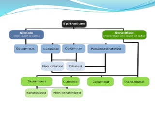
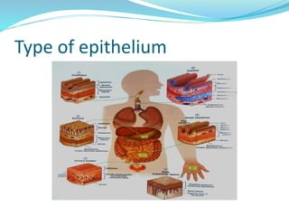
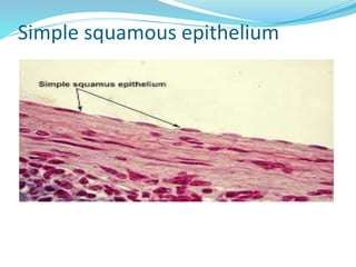
What is a Gland?
An organised collection of secretory epithelial cells. Most glands are formed during development by proliferation of epithelial cells so that they project into the underlying connective tissue. Some glands retain their continuity with the surface via a duct and are known as EXOCRINE GLANDS. Other glands lose this direct continuity with the surface when their ducts degenerate during development. These glands are known as ENDOCRINE glands.
The Indian Dental Academy is the Leader in continuing dental education , training dentists in all aspects of dentistry and
offering a wide range of dental certified courses in different formats.for more details please visit
www.indiandentalacademy.com
Epithelium cellstissues histology
1. Chapter 4 Tissues and Histology • Tissues - collections of similar cells and the substances surrounding them • Tissue classification based on structure of cells, composition of noncellular extracellular matrix, and cell function • Major types of adult tissues – Epithelial – Connective – Muscle – Nervous • Histology: Microscopic Study of Tissues – Biopsy: removal of tissues for diagnostic purposes – Autopsy: examination of organs of a dead body to determine cause of death
https://userupload.net/3ppacneii1wj
Toxicologic Pathology (Second Edition), 2010
INTRODUCTION
The oral mucosa is, in many ways, similar to the skin in its architecture, function, and reaction patterns. This section only emphasizes those characteristics of the oral mucosa that influence or result in a distinct group of pathologic entities.
Because of its location at the entrance of the digestive and respiratory tracts and its proximity to the teeth, the oral mucosa is subjected to numerous natural and man-made xenobiotics. The peculiar architecture and absorption characteristics of the oral mucosa, especially in areas of extreme thinness, coupled with the rich microorganism flora of the mouth, makes the oral mucosa a peculiar site deserving separate discussion.
Epithelial tissue is a type of tissue that covers the surfaces of organs, lines body cavities and forms glands. This PDF provides a comprehensive overview of epithelial tissue, including its types, structure, function, and location in the body. The document covers the different types of epithelial cells, their shapes, and layers, as well as their functions in different organs and tissues. The PDF also delves into the unique features of epithelial tissue, such as its polarity, cell junctions, and the basement membrane. Additionally, Whether you are a student, medical professional, or someone interested in learning about the human body, this PDF provides a detailed understanding of epithelial tissue and its importance in the body.
B.Pharm-Ist sem-HAP-Chapter 3-tissue level of organization.pptxSheetal Patil
Introduction
The term tissue is used to describe a group of cells found together in the body.
Types of tissues
1. Epithelial tissue:]
2. Connective tissue: ]
3. Muscular tissue: ]
4. Nervous tissue: ]
-Development of Tissues
Tissues of the body develop from three primary germ layers: Ectoderm, Endoderm and Mesoderm.
1. Epithelial cell
Epithelial tissue consists of cells arranged in continuous sheets, in either single or multiple layers.
Closely packed and held tightly together.
-General Features of Epithelial Cells:
1. Apical (free) surface
2. Lateral surfaces
3. Basal surface
Basement membrane
Basal lamina
Reticular lamina
-Classification of epithelial tissues
Epithelial tissues are classified according to :
Number of the cell layers formed
1. Simple epithelium (one layer)
2. Stratified epithelium(several layer)
-The shape of the cells
1. Squamous (flat cell)
2. Cuboidal (cube like)
3. Columnar (rectangular)
4. Transitional (variable)
- Glandular Epithelium and Glands
There are main two types of glands
1. Endocrine Glands
2. Exocrine Glands
-Structural Classification of Exocrine Glands
1. Simple gland
2. Compound gland
-Functional Classification of 1. Exocrine Glands
2. Merocrine glands
3. Aprocrine glands
4. Holocrine glands
2. Connective Tissue
-Functions of connective tissues
-Classification of Connective Tissues
Embryonic connective tissue
Mesenchyme
Mucous connective tissue
Mature connective tissue
Loose connective tissue
Areolar connective tissue
Adipose tissue
Reticular connective tissue
Dense connective tissue
Dense regular connective tissue
Dense irregular connective tissue
Elastic connective tissue
Cartilage
Hyaline cartilage
Fibrocartilage
Elastic cartilage
Bone tissue
-Blood and Lymph
--Characteristics of Connective Tissue
1. Extra cellular matrix
2. Fibers
3. Cells of various types
Extracellular matrix of Connective Tissue
-Connective Tissue Cells
1. Fibroblasts
2. Adipocytes (fat cells)
3. Mast cells
4. White blood cells
5. Macrophages
6. Plasma cells
-Connective Tissue Extracellular Matrix;
Ground substance and fibres make up the ECM.
a. Ground substance
-Complex combination of proteins and polysaccharides (hyaluronic acid, chondroitin sulphate dermatan sulphate and keratan sulphate).
b. Fibres
-Collagen fibers
-Elastic fibers
-Reticular fibers
3. Muscular Tissue
-Skeletal muscle tissue (Attached to bone by tendons)
-Cardiac muscle tissue (Heart wall)
-Smooth muscle tissue (Iris of eyes, walls of hollow internal structures such as blood vessels, airways of lungs, stomach, intestine, gall bladder, urinary bladder and uterus)
--Consists of elongated cells called muscle fibers or myocytes for contraction.
--Cells use ATP to generate force.
4. Nervous Tissue
-Neurons or nerve cells
-Neuroglia
--Exhibit sensitivity to various types of stimuli, converts them into nerve impulses (action potentials) and conducts nerve impulses to other neurons.
Report Back from SGO 2024: What’s the Latest in Cervical Cancer?bkling
Are you curious about what’s new in cervical cancer research or unsure what the findings mean? Join Dr. Emily Ko, a gynecologic oncologist at Penn Medicine, to learn about the latest updates from the Society of Gynecologic Oncology (SGO) 2024 Annual Meeting on Women’s Cancer. Dr. Ko will discuss what the research presented at the conference means for you and answer your questions about the new developments.
Title: Sense of Taste
Presenter: Dr. Faiza, Assistant Professor of Physiology
Qualifications:
MBBS (Best Graduate, AIMC Lahore)
FCPS Physiology
ICMT, CHPE, DHPE (STMU)
MPH (GC University, Faisalabad)
MBA (Virtual University of Pakistan)
Learning Objectives:
Describe the structure and function of taste buds.
Describe the relationship between the taste threshold and taste index of common substances.
Explain the chemical basis and signal transduction of taste perception for each type of primary taste sensation.
Recognize different abnormalities of taste perception and their causes.
Key Topics:
Significance of Taste Sensation:
Differentiation between pleasant and harmful food
Influence on behavior
Selection of food based on metabolic needs
Receptors of Taste:
Taste buds on the tongue
Influence of sense of smell, texture of food, and pain stimulation (e.g., by pepper)
Primary and Secondary Taste Sensations:
Primary taste sensations: Sweet, Sour, Salty, Bitter, Umami
Chemical basis and signal transduction mechanisms for each taste
Taste Threshold and Index:
Taste threshold values for Sweet (sucrose), Salty (NaCl), Sour (HCl), and Bitter (Quinine)
Taste index relationship: Inversely proportional to taste threshold
Taste Blindness:
Inability to taste certain substances, particularly thiourea compounds
Example: Phenylthiocarbamide
Structure and Function of Taste Buds:
Composition: Epithelial cells, Sustentacular/Supporting cells, Taste cells, Basal cells
Features: Taste pores, Taste hairs/microvilli, and Taste nerve fibers
Location of Taste Buds:
Found in papillae of the tongue (Fungiform, Circumvallate, Foliate)
Also present on the palate, tonsillar pillars, epiglottis, and proximal esophagus
Mechanism of Taste Stimulation:
Interaction of taste substances with receptors on microvilli
Signal transduction pathways for Umami, Sweet, Bitter, Sour, and Salty tastes
Taste Sensitivity and Adaptation:
Decrease in sensitivity with age
Rapid adaptation of taste sensation
Role of Saliva in Taste:
Dissolution of tastants to reach receptors
Washing away the stimulus
Taste Preferences and Aversions:
Mechanisms behind taste preference and aversion
Influence of receptors and neural pathways
Impact of Sensory Nerve Damage:
Degeneration of taste buds if the sensory nerve fiber is cut
Abnormalities of Taste Detection:
Conditions: Ageusia, Hypogeusia, Dysgeusia (parageusia)
Causes: Nerve damage, neurological disorders, infections, poor oral hygiene, adverse drug effects, deficiencies, aging, tobacco use, altered neurotransmitter levels
Neurotransmitters and Taste Threshold:
Effects of serotonin (5-HT) and norepinephrine (NE) on taste sensitivity
Supertasters:
25% of the population with heightened sensitivity to taste, especially bitterness
Increased number of fungiform papillae
NVBDCP.pptx Nation vector borne disease control programSapna Thakur
NVBDCP was launched in 2003-2004 . Vector-Borne Disease: Disease that results from an infection transmitted to humans and other animals by blood-feeding arthropods, such as mosquitoes, ticks, and fleas. Examples of vector-borne diseases include Dengue fever, West Nile Virus, Lyme disease, and malaria.
TEST BANK for Operations Management, 14th Edition by William J. Stevenson, Ve...kevinkariuki227
TEST BANK for Operations Management, 14th Edition by William J. Stevenson, Verified Chapters 1 - 19, Complete Newest Version.pdf
Acute scrotum is a general term referring to an emergency condition affecting the contents or the wall of the scrotum.
There are a number of conditions that present acutely, predominantly with pain and/or swelling
A careful and detailed history and examination, and in some cases, investigations allow differentiation between these diagnoses. A prompt diagnosis is essential as the patient may require urgent surgical intervention
Testicular torsion refers to twisting of the spermatic cord, causing ischaemia of the testicle.
Testicular torsion results from inadequate fixation of the testis to the tunica vaginalis producing ischemia from reduced arterial inflow and venous outflow obstruction.
The prevalence of testicular torsion in adult patients hospitalized with acute scrotal pain is approximately 25 to 50 percent
The prostate is an exocrine gland of the male mammalian reproductive system
It is a walnut-sized gland that forms part of the male reproductive system and is located in front of the rectum and just below the urinary bladder
Function is to store and secrete a clear, slightly alkaline fluid that constitutes 10-30% of the volume of the seminal fluid that along with the spermatozoa, constitutes semen
A healthy human prostate measures (4cm-vertical, by 3cm-horizontal, 2cm ant-post ).
It surrounds the urethra just below the urinary bladder. It has anterior, median, posterior and two lateral lobes
It’s work is regulated by androgens which are responsible for male sex characteristics
Generalised disease of the prostate due to hormonal derangement which leads to non malignant enlargement of the gland (increase in the number of epithelial cells and stromal tissue)to cause compression of the urethra leading to symptoms (LUTS
Knee anatomy and clinical tests 2024.pdfvimalpl1234
This includes all relevant anatomy and clinical tests compiled from standard textbooks, Campbell,netter etc..It is comprehensive and best suited for orthopaedicians and orthopaedic residents.
3. INTRODUCTION
TISSUES ARE GROUP OF CELLS OR AGGREGATES TO
PERFORM ONE OR MORE FUNCTIONS.
EPITHELIUM (epi + thele + ium) is one of the four basic
types of animal tissue, along with
A-CONNECTIVE TISSUE
B-MUSCLE TISSUE
C-NEURAL TISSUE
4. The term EPITHELIUM is applied to the layer of cells
covering the body surfaces.
Epithelium is an avascular tissue comprised of cells that
line both the internal cavities and external surfaces.
5. DEFINITION
Epithelium is an avascular tissue composed of cells that
cover the exterior body surfaces and line internal closed
cavities (including the vascular system) and body tubes
that communicate with the exterior (the alimentary,
respiratory, and genitourinary tracts). Epithelium also
forms the secretory portion (parenchyma) of glands and
their ducts. In addition, specialized epithelial cells function
as receptors for the special senses (smell, taste, hearing,
and vision).
6. DEVELOPMENT
EPITHELIUM IS DERIVED FROM THREE GERM
LAYERS
ECTODERM
MESODERM
ENDODERM
ALTHOUGH MOST OF THE EPITHELIA ARE
DERIVED FROM ECTODERM AND ENDODEREM
7.
8. ECTODERM: Oral and nasal mucosa, cornea, epidermis
of the skin & glands of the skin & the mammary glands.
ENDODERM: The liver, the pancreas & the lining of the
respiratory and GIT.
9. MESODERM: Uriniferous tubules of the kidney, the lining
of the male and female reproductive systems,the endothelial
lining of the circulatory system and the mesothelium of the
body cavity.
10. FUNCTIONS OF EPITHELIUM
Selective Barriers
Protection
Secretory function
Sensory function
Characteristic of Epithelia
11. Predominantly Cellular
The Basal surface of an epithelium is in contact with
Basal Lamina.
Avascular
Can regenerate when injured
14. Epithelium can be grouped mainly into
Lining Epithelium
Glandular Epithelium
The lining epithelium is further classified into
A-Simple epithelium (unilaminar)
B-Stratified epithelium (multilaminar)
17. SIMPLE SQUAMOUS EPITHELIUM
Composed of flattened, irregularly shaped cells
forming a continuous surface which may be refered
to as pavemented epithelium.
Term ‘squamous’ derives from the comparison of the
cells to the scales of a fish.
Supported by an underlying delicate membrane.
Involved in passive transport of either gases or fluids.
18. PRESENT IN:
Lining- pulmonary alveoli, loop of Henle, parietal layer of
Bawman capsule, inner and middle ear, blood and lymphatic
vessels, pleural and peritoneal cavities.
FUNCTION:
Limiting membrane
Fluid transport
Gaseous exchange
Lubrication
Reducing friction
Lining membrane
22. Single layer of cube like cells with large , spherical
central nuclei.
Location- Kidney tubules , Ducts and secretory
portions of small glands , ovary surface , Intercalated
ducts of salivary glands.
FUNCTIONS- Secretion and absorption.
23. SIMPLE COLUMNAR EPITHELIUM
• Cells are taller and appear columnar in sections at right
angles to the basement membrane.
• Nuclei are elongated and may be located towards the
base, the centre or occasionally the apex of the
cytoplasm, this is known as polarity.
PRESENT IN:
• Small intestine & colon
• Stomach lining and gastric glands
• Gall bladder
25. SIMPLE COLUMNAR EPITHELIUM
Single layer of tall cells with round nuclei.
CILIATED – most of respiratory tract small bronchi ,
uterine tubes , and some regions 0f the uterus.
29. PSEUDO STRATIFIED EPITHELIUM
Simple columnar epithelium , nuclei at different levels in
a vertical section.
Migrating lymphocytes and mast cells give this
appearance.
Seen in ciliated lining of the respiratory tract and
sensory epithelium of the olfactory areas.
Excretory ducts of salivary glands.
Multi-laminar epithelium.
30. STRATIFIED SQUAMOUS
Stratified squamous epithelium
Basal cells are cuboidal or columnar
Surface cells squamous
Occur in sites exposed to constant damage
Two types keratinized and non- keratinized
Location : Non-keratinized forms the moist linings of
the esophagus , mouth, and vagina;
Keratinized variety forms the epidermis
32. STRATIFIED CUBOIDAL
STRATIFIED CUBOIDAL EPITHELIUM
Thin, stratified epithelium which usually consists of
only two or three layers of cuboidal or low columnar
cells.
Not involved in significant absorptive or secretory
activity
PRESENT IN:
Ducts of sweat glands
Large ducts of exocrine glands
Anorectal junction
37. Transitional Epithelium
Resembles both stratified squamous and stratified cuboidal ;
basal cells cuboidal or columnar surface cells dome shaped or
squamous like ,depending on degree of organ stretch.
Stretches readily and permits distension of urinary organ by contained
urine
Lines the ureters, urinary bladder and part of the urethra
Transitional epithelium of ureter
38. Transitional Epithelium
In the stretched state, it appears 2-3 cells thick (although
the actual number of layers remains constant) and the
intermediate and surface layers are extremely flattened.
PRESENT IN:
Urethra
Ureters
Bladders
Renal calyces
FUNCTION:
Protection
Distensible
39. Oral mucosa
Mucous membrane : moist lining of the gastrointestinal
tract, nasal passages ,Other body cavities that
communicate with the exterior.
In the oral cavity the lining is called as oral mucous
membrane or oral mucosa.
40. Functions of oral mucosa
Protection: Barrier for mechanical trauma and
microbiological insults
Sensation: Temperature (heat and cold) , touch ,pain,
taste buds, thirst; reflexes such as swallowing , etching,
gagging and salivating .
Secretion : Salivary secretion
41. Development and structure of oral epithelium
The epithelium of the oral cavity derived from the
embryonic ectoderm.
Most of the mucosal surface is lined by a non-
keratinized stratified squamous epithelium.
Except for Gingiva , hard palate and dorsal surface of
the tongue where the epithelium is keratinized.
42. FUNCTIONAL CHARACTERISTICS
As the cells migrate from the basal layer to the
surface layer, differentiation produces a surface layer
that is either
Non-keratinized
Ortho Keratinized
Para keratinized
43. Non-Keratinized Epithelium
The surface cells retain their nuclei and cytoplasm does not
contain keratin filaments.
Stratum spinosum and granulosum is absent.
Lining mucosa of the oral cavity.
Clinical considerations:
Prosthetic devices thin the epithelium
Linea alba
Angina pectoris- Nitroglycerine tablets are placed
sublingually and absorbed quickly through the thin non-
keratinized epithelium and into large veins of the ventral
surface of the tongue.
44. Ortho Keratinized epithelium
Stratum basale- tall columnar or cuboidal.
Stratum corneum elliptical / spherical.
In contact only by Desmosomes.
Stratum granulosum- flat cells with keratohyaline
granules.
Stratum spinosum- squamous cells with no nuclei.
Masticatory mucosa-Ortho keratinized.
45. ULTRA STRUCTURE OF THE EPITHELIAL CELL
Cells of the basal layer are least differentiated.
They contain organelles and typical filamentous strands
called tonofilaments.
Tonofilaments aggregate- tonofibrills.
Chemically they are cytokeratins.
Epithelial cells are named as keratinocyte
Keratins
Epithelial cells produce proteins- carbohydrate complex
which provide cohesion.
Desmosomes.
Hemi desmosomes.
46. APICAL CELL SPECIALISATION
Microvilli
Finger like cytoplasmic processes.
Number and shape correlates with absorptive capacity.
Contain actin filament.
Cilia:
Stereocilia
47. GLANDULAR EPITHELIUM
Classification of Glands
ENDOCRINE GLANDS:-
Hormones are secreted directly into blood.
No ducts.
Under the microscope , they look like any stratified
epithelial tissue with one big difference: THEY DO NOT
HAVE A FREE SURFACE AND SURROUNDED DIRECTLY BY
OTHER TISSUES.
48. EXOCRINE GLANDS
Release their products onto the free surface of the skin or of
the open cavities of the body such as the digestive ,
respiratory or reproductive tracts.
Their products are NOT released into the blood.
THESE ARE OF TWO TYPES:
UNICELLULAR:
They are scattered amongst other non-secretory epithelial
cells.
They have NO ducts, but they secrete their products directly
on the free surface.
The most common unicellular exocrine glands are the
Goblet cells(mucous secreting)
51. SECRETORY CELLS
SEROUS CELLS
Pyramidal cells with basal nuclei Microvilli on the
luminal surface.
Spherical nuclei basally.
Lateral surface have folds interdigitating with same
from adjacent cells.
Joined together by cell junctions.
52. MUCOUS CELLS
Tubular configuration with mucous cells surroundings
a central lumen.
Demi Lune- serous cells associated with them at the
end of the tubule.
Apical cytoplasm has accumulated mucous
53. Ducts – Intercalated ducts
Lined by simple cuboidal epithelium.
Centrally placed nuclei.
Small cytoplasm , few ER and a small Golgi complex.
Apically few secretory granules.
Few short microvilli
Unidentified in routine histology.
54. Striated duct cells
Cells are columnar , centrally placed nucleus and
acidophilic cytoplasm.
Striation appear in the basal cytoplasm, mitochondria
in cytoplasmic partition separated by highly folded cell
membrane.
55. Excretory ducts
Pseudo stratified epithelium with columnar cells.
Larger than striated ducts.
Scattered Goblet cells seen.
56. Clinical importance
Cystic fibrosis (CF) is a genetic disorder of certain
exocrine glands including the salivary glands, the sweat
glands, the pancreas and the mucous glands of the
respiratory tract.
A person with CF has clogged bronchial tubes, frequent
episodes of pneumonia and ultimately lungs that can
not carry out gas exchange.
CF –mutations in the gene that encodes the CF
transmembrane conductance regular
57. SPECIAL EPITHELIA
Epithelium of sense organs:olfactory, gustatory and
vestibulocochlear epithelium.
Myoepithelial cells/basket cells.
Seminiferous epithelium
61. Forms tight seals between cells such as the epithelial
cells that comprise the inner lining of the stomach,
intestine, and urinary bladder.
They prevent the passage of substances between cells
62. ADHERENCES JUNCTIONS
Strongly fasten cells to each other or to the extracellular
matrix.
They help epithelial surfaces resist separation.
Cardiac muscle
63. Desmosomes (cell to cell)
Desmoglein and Desmocollin interaction with adjacent
cells forms a dense middle line.
65. Gap junctions
Formed by minute, fluid filled tunnels that permit
passage of electrical signals or chemicals (i.e. ions and
small molecules) from one cell to a neighbouring cell,
located in some parts of the nervous system, in heart
muscle and in the gastrointestinal tract.
67. SUMMARY
Epithelial is an avascular tissue derived from all the
three germ layers.
They line the external surface of the body , occur as
membranes and as glands.
All epithelia lie upon a basal lamina.
68. Cells are connected to each other by cell junctions.
May possess cilia or microvilli.
They are protective , secretory , absorptive , sensory
depending on the location
69. BIBILOGRAPHY
Freshney, R.I. (2002). “Introduction “. In Freshney,
R .Ian; Freshney, Mary. Culture of epithelial
cells.ISBN 978-0-471-6.
Orban’s Oral Histology and Embryology-10th
Edition
Essential of Oral Histology and Embryology-
James K Avery, 2nd Edition
Oral Histology Development, Structures &
Fundamental – Ten Cate’s, 7th Edition
70. Histology Text and Atlas- Michael H Ross,Lynn J
Romrell, Gorden I Kaye, 3rd Edition
Gray’s Anatomy- The Anatomical Basis of Clinical
Practice ,40th Edition Susan Standing
Bloom & Fawcett’s Concise Histology- Don W
Essentials of Anatomy and Physiology ,5th Edition,
Valerie Scalon ,PhD.