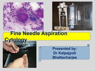
This document provides an overview of fine needle aspiration cytology (FNAC). It discusses the history, advantages, limitations, equipment, techniques, processing, complications, and applications of FNAC for diagnosing lesions in the salivary gland, oral cavity, and other areas. FNAC is a simple, economical technique that can provide a rapid diagnosis without the need for an open biopsy. It describes the evaluation of FNAC samples under the microscope for diagnosing various conditions.
Fine Needle Aspiration Cytology (FNAC) is a simple, quick and inexpensive method that is used to sample superficial masses like those found in the neck and is usually performed in the outpatient clinic.
Bone marrow aspiration & trephine biopsySanjeev Kumar
Bone marrow aspiration & trephine biopsy, Complication of BM Aspiration, Clinical significance, Indication of Bone Marrow Aspiration and Biopsy, Types Of Needles, Site for Bone Marrow Biopsy And Aspiration, types Of Smear for Bone Marrow, Advantages of Bone Marrow Trephine Biopsy
FNAC of breast - definition, history, purpose, preparations, basic equipment, procedure, smear preparation, fixatives, staining solutions, rapid stains - toluidine blue, difference between air dried and wet fixed slides, complications and contraindications, advantages, general criteris for malignancy, nuclear size and pleomorphism, nuclear membrane, irregularity and extranuclear chromatin, nuclear fragility and mitotic figures, types of breast carcinoma.
FNAC of breast - definition, history, purpose, preparations, basic equipment, procedure, smear preparation, fixatives, staining solutions, rapid stains - toluidine blue, difference between air dried and wet fixed slides, complications and contraindications, advantages, general criteris for malignancy, nuclear size and pleomorphism, nuclear membrane, irregularity and extranuclear chromatin, nuclear fragility and mitotic figures, types of breast carcinoma.
HOT NEW PRODUCT! BIG SALES FAST SHIPPING NOW FROM CHINA!! EU KU DB BK substit...GL Anaacs
Contact us if you are interested:
Email / Skype : kefaya1771@gmail.com
Threema: PXHY5PDH
New BATCH Ku !!! MUCH IN DEMAND FAST SALE EVERY BATCH HAPPY GOOD EFFECT BIG BATCH !
Contact me on Threema or skype to start big business!!
Hot-sale products:
NEW HOT EUTYLONE WHITE CRYSTAL!!
5cl-adba precursor (semi finished )
5cl-adba raw materials
ADBB precursor (semi finished )
ADBB raw materials
APVP powder
5fadb/4f-adb
Jwh018 / Jwh210
Eutylone crystal
Protonitazene (hydrochloride) CAS: 119276-01-6
Flubrotizolam CAS: 57801-95-3
Metonitazene CAS: 14680-51-4
Payment terms: Western Union,MoneyGram,Bitcoin or USDT.
Deliver Time: Usually 7-15days
Shipping method: FedEx, TNT, DHL,UPS etc.Our deliveries are 100% safe, fast, reliable and discreet.
Samples will be sent for your evaluation!If you are interested in, please contact me, let's talk details.
We specializes in exporting high quality Research chemical, medical intermediate, Pharmaceutical chemicals and so on. Products are exported to USA, Canada, France, Korea, Japan,Russia, Southeast Asia and other countries.
Lung Cancer: Artificial Intelligence, Synergetics, Complex System Analysis, S...Oleg Kshivets
RESULTS: Overall life span (LS) was 2252.1±1742.5 days and cumulative 5-year survival (5YS) reached 73.2%, 10 years – 64.8%, 20 years – 42.5%. 513 LCP lived more than 5 years (LS=3124.6±1525.6 days), 148 LCP – more than 10 years (LS=5054.4±1504.1 days).199 LCP died because of LC (LS=562.7±374.5 days). 5YS of LCP after bi/lobectomies was significantly superior in comparison with LCP after pneumonectomies (78.1% vs.63.7%, P=0.00001 by log-rank test). AT significantly improved 5YS (66.3% vs. 34.8%) (P=0.00000 by log-rank test) only for LCP with N1-2. Cox modeling displayed that 5YS of LCP significantly depended on: phase transition (PT) early-invasive LC in terms of synergetics, PT N0—N12, cell ratio factors (ratio between cancer cells- CC and blood cells subpopulations), G1-3, histology, glucose, AT, blood cell circuit, prothrombin index, heparin tolerance, recalcification time (P=0.000-0.038). Neural networks, genetic algorithm selection and bootstrap simulation revealed relationships between 5YS and PT early-invasive LC (rank=1), PT N0—N12 (rank=2), thrombocytes/CC (3), erythrocytes/CC (4), eosinophils/CC (5), healthy cells/CC (6), lymphocytes/CC (7), segmented neutrophils/CC (8), stick neutrophils/CC (9), monocytes/CC (10); leucocytes/CC (11). Correct prediction of 5YS was 100% by neural networks computing (area under ROC curve=1.0; error=0.0).
CONCLUSIONS: 5YS of LCP after radical procedures significantly depended on: 1) PT early-invasive cancer; 2) PT N0--N12; 3) cell ratio factors; 4) blood cell circuit; 5) biochemical factors; 6) hemostasis system; 7) AT; 8) LC characteristics; 9) LC cell dynamics; 10) surgery type: lobectomy/pneumonectomy; 11) anthropometric data. Optimal diagnosis and treatment strategies for LC are: 1) screening and early detection of LC; 2) availability of experienced thoracic surgeons because of complexity of radical procedures; 3) aggressive en block surgery and adequate lymph node dissection for completeness; 4) precise prediction; 5) adjuvant chemoimmunoradiotherapy for LCP with unfavorable prognosis.
micro teaching on communication m.sc nursing.pdfAnurag Sharma
Microteaching is a unique model of practice teaching. It is a viable instrument for the. desired change in the teaching behavior or the behavior potential which, in specified types of real. classroom situations, tends to facilitate the achievement of specified types of objectives.
These simplified slides by Dr. Sidra Arshad present an overview of the non-respiratory functions of the respiratory tract.
Learning objectives:
1. Enlist the non-respiratory functions of the respiratory tract
2. Briefly explain how these functions are carried out
3. Discuss the significance of dead space
4. Differentiate between minute ventilation and alveolar ventilation
5. Describe the cough and sneeze reflexes
Study Resources:
1. Chapter 39, Guyton and Hall Textbook of Medical Physiology, 14th edition
2. Chapter 34, Ganong’s Review of Medical Physiology, 26th edition
3. Chapter 17, Human Physiology by Lauralee Sherwood, 9th edition
4. Non-respiratory functions of the lungs https://academic.oup.com/bjaed/article/13/3/98/278874
Couples presenting to the infertility clinic- Do they really have infertility...Sujoy Dasgupta
Dr Sujoy Dasgupta presented the study on "Couples presenting to the infertility clinic- Do they really have infertility? – The unexplored stories of non-consummation" in the 13th Congress of the Asia Pacific Initiative on Reproduction (ASPIRE 2024) at Manila on 24 May, 2024.
Acute scrotum is a general term referring to an emergency condition affecting the contents or the wall of the scrotum.
There are a number of conditions that present acutely, predominantly with pain and/or swelling
A careful and detailed history and examination, and in some cases, investigations allow differentiation between these diagnoses. A prompt diagnosis is essential as the patient may require urgent surgical intervention
Testicular torsion refers to twisting of the spermatic cord, causing ischaemia of the testicle.
Testicular torsion results from inadequate fixation of the testis to the tunica vaginalis producing ischemia from reduced arterial inflow and venous outflow obstruction.
The prevalence of testicular torsion in adult patients hospitalized with acute scrotal pain is approximately 25 to 50 percent
Ethanol (CH3CH2OH), or beverage alcohol, is a two-carbon alcohol
that is rapidly distributed in the body and brain. Ethanol alters many
neurochemical systems and has rewarding and addictive properties. It
is the oldest recreational drug and likely contributes to more morbidity,
mortality, and public health costs than all illicit drugs combined. The
5th edition of the Diagnostic and Statistical Manual of Mental Disorders
(DSM-5) integrates alcohol abuse and alcohol dependence into a single
disorder called alcohol use disorder (AUD), with mild, moderate,
and severe subclassifications (American Psychiatric Association, 2013).
In the DSM-5, all types of substance abuse and dependence have been
combined into a single substance use disorder (SUD) on a continuum
from mild to severe. A diagnosis of AUD requires that at least two of
the 11 DSM-5 behaviors be present within a 12-month period (mild
AUD: 2–3 criteria; moderate AUD: 4–5 criteria; severe AUD: 6–11 criteria).
The four main behavioral effects of AUD are impaired control over
drinking, negative social consequences, risky use, and altered physiological
effects (tolerance, withdrawal). This chapter presents an overview
of the prevalence and harmful consequences of AUD in the U.S.,
the systemic nature of the disease, neurocircuitry and stages of AUD,
comorbidities, fetal alcohol spectrum disorders, genetic risk factors, and
pharmacotherapies for AUD.
Ozempic: Preoperative Management of Patients on GLP-1 Receptor Agonists Saeid Safari
Preoperative Management of Patients on GLP-1 Receptor Agonists like Ozempic and Semiglutide
ASA GUIDELINE
NYSORA Guideline
2 Case Reports of Gastric Ultrasound
Title: Sense of Taste
Presenter: Dr. Faiza, Assistant Professor of Physiology
Qualifications:
MBBS (Best Graduate, AIMC Lahore)
FCPS Physiology
ICMT, CHPE, DHPE (STMU)
MPH (GC University, Faisalabad)
MBA (Virtual University of Pakistan)
Learning Objectives:
Describe the structure and function of taste buds.
Describe the relationship between the taste threshold and taste index of common substances.
Explain the chemical basis and signal transduction of taste perception for each type of primary taste sensation.
Recognize different abnormalities of taste perception and their causes.
Key Topics:
Significance of Taste Sensation:
Differentiation between pleasant and harmful food
Influence on behavior
Selection of food based on metabolic needs
Receptors of Taste:
Taste buds on the tongue
Influence of sense of smell, texture of food, and pain stimulation (e.g., by pepper)
Primary and Secondary Taste Sensations:
Primary taste sensations: Sweet, Sour, Salty, Bitter, Umami
Chemical basis and signal transduction mechanisms for each taste
Taste Threshold and Index:
Taste threshold values for Sweet (sucrose), Salty (NaCl), Sour (HCl), and Bitter (Quinine)
Taste index relationship: Inversely proportional to taste threshold
Taste Blindness:
Inability to taste certain substances, particularly thiourea compounds
Example: Phenylthiocarbamide
Structure and Function of Taste Buds:
Composition: Epithelial cells, Sustentacular/Supporting cells, Taste cells, Basal cells
Features: Taste pores, Taste hairs/microvilli, and Taste nerve fibers
Location of Taste Buds:
Found in papillae of the tongue (Fungiform, Circumvallate, Foliate)
Also present on the palate, tonsillar pillars, epiglottis, and proximal esophagus
Mechanism of Taste Stimulation:
Interaction of taste substances with receptors on microvilli
Signal transduction pathways for Umami, Sweet, Bitter, Sour, and Salty tastes
Taste Sensitivity and Adaptation:
Decrease in sensitivity with age
Rapid adaptation of taste sensation
Role of Saliva in Taste:
Dissolution of tastants to reach receptors
Washing away the stimulus
Taste Preferences and Aversions:
Mechanisms behind taste preference and aversion
Influence of receptors and neural pathways
Impact of Sensory Nerve Damage:
Degeneration of taste buds if the sensory nerve fiber is cut
Abnormalities of Taste Detection:
Conditions: Ageusia, Hypogeusia, Dysgeusia (parageusia)
Causes: Nerve damage, neurological disorders, infections, poor oral hygiene, adverse drug effects, deficiencies, aging, tobacco use, altered neurotransmitter levels
Neurotransmitters and Taste Threshold:
Effects of serotonin (5-HT) and norepinephrine (NE) on taste sensitivity
Supertasters:
25% of the population with heightened sensitivity to taste, especially bitterness
Increased number of fungiform papillae
Recomendações da OMS sobre cuidados maternos e neonatais para uma experiência pós-natal positiva.
Em consonância com os ODS – Objetivos do Desenvolvimento Sustentável e a Estratégia Global para a Saúde das Mulheres, Crianças e Adolescentes, e aplicando uma abordagem baseada nos direitos humanos, os esforços de cuidados pós-natais devem expandir-se para além da cobertura e da simples sobrevivência, de modo a incluir cuidados de qualidade.
Estas diretrizes visam melhorar a qualidade dos cuidados pós-natais essenciais e de rotina prestados às mulheres e aos recém-nascidos, com o objetivo final de melhorar a saúde e o bem-estar materno e neonatal.
Uma “experiência pós-natal positiva” é um resultado importante para todas as mulheres que dão à luz e para os seus recém-nascidos, estabelecendo as bases para a melhoria da saúde e do bem-estar a curto e longo prazo. Uma experiência pós-natal positiva é definida como aquela em que as mulheres, pessoas que gestam, os recém-nascidos, os casais, os pais, os cuidadores e as famílias recebem informação consistente, garantia e apoio de profissionais de saúde motivados; e onde um sistema de saúde flexível e com recursos reconheça as necessidades das mulheres e dos bebês e respeite o seu contexto cultural.
Estas diretrizes consolidadas apresentam algumas recomendações novas e já bem fundamentadas sobre cuidados pós-natais de rotina para mulheres e neonatos que recebem cuidados no pós-parto em unidades de saúde ou na comunidade, independentemente dos recursos disponíveis.
É fornecido um conjunto abrangente de recomendações para cuidados durante o período puerperal, com ênfase nos cuidados essenciais que todas as mulheres e recém-nascidos devem receber, e com a devida atenção à qualidade dos cuidados; isto é, a entrega e a experiência do cuidado recebido. Estas diretrizes atualizam e ampliam as recomendações da OMS de 2014 sobre cuidados pós-natais da mãe e do recém-nascido e complementam as atuais diretrizes da OMS sobre a gestão de complicações pós-natais.
O estabelecimento da amamentação e o manejo das principais intercorrências é contemplada.
Recomendamos muito.
Vamos discutir essas recomendações no nosso curso de pós-graduação em Aleitamento no Instituto Ciclos.
Esta publicação só está disponível em inglês até o momento.
Prof. Marcus Renato de Carvalho
www.agostodourado.com
Pulmonary Thromboembolism - etilogy, types, medical- Surgical and nursing man...VarunMahajani
Disruption of blood supply to lung alveoli due to blockage of one or more pulmonary blood vessels is called as Pulmonary thromboembolism. In this presentation we will discuss its causes, types and its management in depth.
3. INTRODUCTION
Fine Needle Aspiration Cytology (FNAC) is a
technique whereby cells are obtained from a
lesion using a thin bore needle and smears
are made for cytopathological diagnosis.
This technique is based on the fact that tumor
cells are less cohesive and are easily
aspirated.
Used in the diagnosis of breast lumps, thyroid
nodules, liver disease, subcutaneous soft
tissue mass, salivary gland diseases and oral
diseases.
4. Oral cavity is a site where mucosa is very vascular and an
open biopsy leads to a lot of bleeding which is difficult to
control.
In recent times FNAC solves these problems, adequate
material can easily be obtained by using a 10 ml. syringe
from an intraoral or extraoral site without any discomfort to
the patient and with no bleeding.
In some cases a subsequent surgery is not needed and
patient can be put on appropriate treatment.
FNAC report is prepared within 24 hours of sampling gives
early, quick information to the surgeon about the type of
lesion he is dealing with.
5. HISTORICAL PERSPECTIVE
The technique was introduced in the 1930s by Martin
and Ellis in the United States, but it never became
widespread.
Since the 1950s it has been used extensively in
Scandinavia and in Holland.
Fine Needle for aspiration were first introduced in
Europe in the 1950’s by Lopez-Cardozo in the
Netherlands and Soderstrom in Sweden
Publication by Zajicek from Karolinska Hospital in
Stockholm that brought aspiration cytology to
international alterations.
6. ADVANTAGE
Simple office technique
Rapid diagnosis
Economical
Sampling from multiple sites in the same sitting
High diagnostic accuracy
Many techniques such as bacterial culture,
immunocytochemistry, flow cytometry, cytogenetics,
polymerase chain reaction, etc. are possible from
FNAC material.
7. LIMITATIONS
Loss of tissue architecture
Capsular invasion and lymphovascular invasions
cannot be detected
Difficult to differentiate in situ versus invasive
carcinoma
Considerable training is needed for accurate
interpretation.
8. FNAC AS A TOOL IN CLINICAL
INVESTIGATION
Initially used as a mean to confirm a clinical suspicion
of local recurrence or metastasis of known cancer
without subjecting the patient to further surgical
intervention.
Inflammation, infection, degenerative conditions, in
diagnosis and monitoring of graft rejection in
transplantation surgery
Alternative or complement to frozen section
Intraoperative cytology
9. THE PRACTICE OF FNAC
Success of FNAC depends on four fundamental
requirement:
1. Samples must be representative of the lesion
investigated.
2. Samples must be adequate in terms of cells & other
tissue components
3. Samples must be correctly smeared and processed
4. Biopsy must be accompanied by relevant and
correct clinical/radiological information.
11. 4) Sterile container: Physiological saline or Hank’s
balanced solution
5) Slides: clean, dry & free of grease.
6) A 0.4 mm haemocytometer coverslip gives better
control over smearing pressure & a more perfect
spread
7) Fixatives: 70-90% ethanol, Carnoy’s fixative, 10%
buffered formalin, gluteraldehyde used
8) Stains
9) Microscopes
12. FINE NEEDLE ASPIRATION TECHNIQUE
2 techniques:
1. FNAC with aspiration
2. FNAC without aspiration
13. Site of FNAC should be
cleaned by spirit swab
Needle is introduced in the swelling and is gently
moving to and fro. Simultaneously negative suction
is also created by withdrawing the piston
Air is taken in the syringe and needle is reattachedThe aspirated material is expelled and the
smear is made by gently pressing the upper
slide on the lower one
FNAC with aspiration
14.
15. -Introduced by Zajdela in 1987
-based on the observation that the capillary pressure in a
fine needle is sufficient to keep the detached cell inside the
lumen of the needle
FNAC without aspiration
16. FAILURE TO OBTAIN A REPRESENTATIVE
SAMPLE
Needle has missed the target tangentially
Needle in central cystic/necrotic/hemorrhagic area
devoid of diagnostic cells.
Needle in dominant benign mass missing a small
adjacent malignant lesions.
Fibrotic/desmoplastic target tissue giving a scant cell
yield.
17.
18. PROCESSING THE SAMPLE
Sample expelled on to a clean & dry
microscope slide using air in a syringe.
SMEARING
DIRECT
INDIRECT
20. Indirect smearing: Thin fluid samples are
best processed by centrifugation on the
cytocentrifuge.
Milipore nucleopore filtration is an alternative
Thinprep technique
21. FIXATION & STAINING
2 fundamentally different methods of fixation &
staining are used in FNAC:
1. Air drying followed by staining with a
haematological stain such as MAY GRUNWALD-
GIEMSA STAIN , Jenner-Giesma, Diff-Quik
2. Alcohol fixation and staining according to PAP or
with H&E.
23. Romanowsky stain
Contents:
Methylene blue/azure B and eosin, dissoved in
acetone-free methanol, include jenner, Giesma, May
Grunwald and Leishman stain
Results:
Nuclei- purple/blue
Cytoplasm- pink/blue
Eosinophils- pink/red
24. Diff-Quik is a commercial Romanowsky stain variant,
commonly used in histological staining to rapidly stain and
differentiate a variety of smears, commonly blood and non-
gynecological smears, including those of fine needle
aspirates.
25. MAY GRUNWALD- GIEMSA STAIN
commonly used staining of blood smears
Contents:
methylene blue (a basic dye)
Azures (also basic dyes)
Eosin (an acid dye)
Results:
Nuclei of white blood cells and the granules of
basophil granulocytes- blue
Red blood cells and eosinophil granules – red
cytoplasm of white blood cells - light blue
26. SPECIAL STAINS
1. PAS or Alcian blue - mucins, glycogen
2. Prussian blue - iron
3. Masson-Fontana - melanin
4. Congo red - amyloid
5. Ziehl-neelson - acid fast bacilli
6. Bile pigment- Fauchet’s reagent counterstained with
sirus red.
7. Gram, PAS or Gomori’s silver stain for microorganism
27. COMPLICATIONS
Usually free of complications
Bleedings, hematoma, emphysema (in lung).
Rarely anaphylactic reaction- accidental
rupture of hydrated cyst
28. FNAC OF SALIVARY GLAND
FNAC of the salivary gland lesions has gained wide
clinical recognition.
The incisional biopsy of the salivary gland may cause
fistula formation and other complications that can be
avoided in FNAC.
For accurate diagnosis of salivary gland lesions, an
adequate sample stained by both May Grunwald-
Giemsa stain and Papanicolaou’s stain (or H and E)
along with detailed clinical history are needed.
29. Cytology of Normal Salivary Glands
Benign ductal cells: These cells are usually in
small clusters or monolayered sheets. The cells
are round to oval, with scanty cytoplasm having
monomorphic nuclei.
Acinar cells: These are commonly present as
small ball like clusters. The individual cells are
round with abundant foamy cytoplasm and small
round nuclei. The acinar cells may also be present
discretely and cells with bare nuclei may be
mistaken as lymphocytes.
30. Myoepithelial cells: These are oval to
spindle shaped cells present near the
basement membrane of the ductules. Oval
plasmacytoid myoepithelial cells may also be
seen.
Fibrous tissue: Fragments of fibrous and
adipose tissue may also be seen in the
background of the normal salivary aspirate.
31. Aspirate of normal salivary gland tissue showing
grapelike clusters of epithelial cells composed of spherical
acini and branching ducts (Romanowsky’s stain).
32. Benign acinar cells arranged in ‘‘rosette’’ formation. The cells have
abundant foamy vacuolated cytoplasm with indistinct cell borders, and
eccentric nuclei (Papanicolaou stain).
34. Fatty Infiltration
Presents as a diffuse enlargement
Addition to normal salivary gland elements, there is
significant increase in the amount of adipose tissue
situated between ductal and acinar cells
Associated with diabetes, cirrhosis, alcoholism,
medications, nutritional deficiencies and hormonal
disturbances
35. Chronic sialadenitis
Commonly results from stones or postsurgical scarring
More frequently seen in the submandibular gland
Cytology:
low cellularity and show predominance of ductal cells,
background has variable number of lymphocytes,
occasional plasma cells and neutrophils, and spindle
fibroblasts
Squamous and mucinous metaplasia may be focally
encountered
Acute sialadenitis: seldom sampled, many neutrophils
are admixed with benign salivary gland elements, reactive
and reparative changes may be seen
36. Short tubular segments of ductal epithelium composed of small hyperchromatic
cells characteristic of chronic sialadenitis
(Romanowsky’s stain).
37. Benign Lymphoepithelial Cysts of the Parotid
Glands
Cytology:
Smears - characterized by a mixed lymphoid infiltrate
with a predominance of small mature lymphocytes
Characteristically has a watery proteinaceous
background in which the lymphoid elements are
distributed
Cuboidal or mucin-containing cells may be found
distributed singly in the smear
38. Aspirates of benign lymphoepithelial cysts are characterized by a
mixed population of lymphoid cells dispersed in a watery
background (Romanowsky’s stain).
39. Pleomorphic Adenoma
Pleomorphic adenoma (PA) commonly involves the parotid
gland (more than 75%).
painless, slow growing, firm to hard swelling of the salivary
gland.
Cytology
• Pinkish fibrillar chondromyxoid matrix material with frayed
indistinct margins
• Clusters of round, ovoid or plasmacytoid epithelial cells with
moderate amount of dense cytoplasm
• Clusters and discrete spindle shaped myoepithelial cells
embedded in mesenchymal stroma
40. Immunochemistry:
Epithelial cells are positive for cytokeratin (CK)
Myoepithelial cells are positive for CK and vimentin
(co-expressed), S-100, Glial fibrillary acid protein
(GFAP) and Calponin
41. Long strands of spindle cells
embedded in the connective tissue
stroma
(H & E)
Cellular pleomorphic adenoma
showing discrete and cluster of
epithelial cells
(H & E)
42. Clusters of epithelial and myoepithelial
cells associated with fragments of
myxoid and chondroid stroma are
characteristic of pleomorphic
adenomas
(Papanicolaou’s stain)
The myxoid-chondroid stroma
of pleomorphic adenomas often
has a fibrillary character along
its edges (Romanowsky’s stain)
The myoepithelial cell
component of pleomorphic
adenomas
frequently has a plasmacytoid
appearance
(Romanowsky’s stain)
43. Warthin’s Tumor
2nd most common tumor, occurs almost exclusively in
parotid gland.
Cytology:
The aspirate has a thin watery mucoid appearance,
and consists of a mixed population of lymphocytes,
occasional plasma cells, and variable number of
oncocytes
mucoid material or greenish-brown dirty fluid
Many cohesive sheets of oncocytes
Squamous and mucinous metaplasia.
44. There is admixed population of large
flat sheets of oncocytes and lymphoid
cells.
The epithelial cells have abundant
dense eosinophilic cytoplasm, well
defined cytoplasmic border, enlarged
centrally placed nuclei, and prominent
nucleoli (Papanicolaou stain)
Aspirates of Warthin tumors are
characterized by a mixed population
of lymphocytes and clusters of
epithelial cells with abundant
granular cytoplasm. The cells lie
within a dirty proteinaceous
background
(Romanowsky’s stain).
45. Basal Cell Adenoma (BCA)
Basal cell adenoma (BCA) is an uncommon salivary
gland tumor
The majority of BCA arises in the major salivary gland.
The parotid gland is the predominant site of
occurrence and more than 75 percent of BCA arise in
parotid gland.
46. Cytology:
Cohesive groups of basaoid cells
Peripheral palisading arrangement
Round nucleus, bland nuclear chromatin and scanty
cytoplasm
Squamous morules
Scanty homogeneous acellular stromal material.
47. Tightly cohesive clusters of basaloid
cells and many background stripped
nuclei
(DQ stain)
Membranous variant of basal cell
adenoma. There is dense hyaline
extracellular material surrounding the
basaloid cell clusters. (Papanicolaou stain)
48. Oncocytoma
This is a benign salivary gland neoplasm that
predominantly involves parotid gland (about 75%).
The tumor predominantly presents as a painless
mobile mass.
Occurs exclusively among elderly people
Pain is generally absent.
49. Cytology:
Three-dimensional clusters of oncocytes
Polygonal cells with abundant densely granular
eosinophilic cytoplasm
Central to eccentric monomorphic round nucleus.
50. Predominantly discrete oncocytic
cells in FNAC smear
(MGG)
Oncocytomas contain epithelial
cells with abundant granular
cytoplasm identical to those seen
in Warthin tumors, but
oncocytomas lack the lymphoid
component
(Romanowsky’s stain).
51. Myoepithelial Tumors
This tumor comprises only 11.5 percent of all salivary
gland neoplasms.
The patient usually presents as a slow growing
painless mass in the parotid or minor salivary gland
regions.
52. Cytology:
Spindle cell type
• Abundant clusters and dissociated spindle cells
• Elongated nuclei, fine nuclear chromatin and
inconspicuous nucleoli
Hyaline myoepithelial cells
• Dissociated round to oval cells
• Plasmacytoid cells with abundant cytoplasm and
eccentric nucleus.
53. The hyaline variant of myoepithelioma showing dissociated
plasmacytoid cells with abundant cytoplasm and eccentric
nucleus
(MGG)
54. Adenoid cystic carcinoma
Adenoid cystic carcinoma (ACC) is a slow growing
tumor.
Tendency to recurrence
ACC infiltrates local nerves, causes paralysis of the
motor nerves and produces pain in the ear.
Most common in parotid
55. Cytology:
• Multiple variable sized globular, spherical or tubular
homogeneous, acellular magenta colored matrix
material
• These globules are surrounded by cells
• Clusters and dissociated small cells with scanty
cytoplasm
• Round monomorphic hyperchromatic nuclei with
coarse chromatin.
IHC:
High Ki67 index.
c-Kit overexpression noted.
56. Small round cells with scanty
cytoplasm along with pinkish hyaline
globules
(MGG)
Cells arranged around the pinkish
globules
(MGG)
57. Adenoid cystic carcinoma.
The neoplastic cells have oval to round hyperchromatic nuclei with finely to
coarsely granular chromatin and scant cytoplasm.
They are associated with hyaline globules, which stain pale gray-green in this
preparation.
(Papanicolaou stain)
58. Acinic Cell Carcinoma
Malignant epithelial neoplasm of the duct apparatus,
but occasional lesions seem to show acinar
differentiation
Cytology:
low-grade malignancy characterized by sheets of
large cells with abundant cytoplasm
The neoplastic cells have foamy/vacuolated
cytoplasm with ill-defined borders, eccentrically placed
nuclei, small inconspicuous nucleoli, and lack
significant nuclear atypia or pleomorphism
Occasionally lymphocytes and psammoma bodies
Clean background.
59. A, Acinic cell carcinomas are characterized cytologically by irregular clusters and sheets
of epithelial cells with foamy or granular cytoplasm and bland nuclei (Romanowsky’s
stain).
B, Some cells of acinic cell carcinomas contain characteristic red granules within their
cytoplasm (Romanowsky’s stain).
C, Cells from acinic cell carcinomas have a finely vacuolated or foamy cytoplasm
(Romanowsky’s stain).
60. Mucoepidermoid Carcinoma
Mucoepidermoid carcinoma (MEC) originates from the
ductal cells of the salivary gland and is commonly
located in the parotid gland.
Low-grade MEC is commonly cystic and is
cytologically characterized by an admixture of
glandular and metaplastic squamous cells.
The background demonstrates mucinous material and
debris
The glandular cells may have a ductal appearance
(intermediate cells) or may resemble macrophages
(mucin-producing cells)
61. High-grade MEC has a less prominent cystic
component and greater degree of atypia, compared
with low-grade MEC.
High-grade MEC is cytologically characterized by
large pleomorphic cells with predominantly epidermoid
or undifferentiated cell features
Glandular cells are rarely seen in high-grade MEC,
but their presence in association with squamous cells
establishes the diagnosis.
62. Abundant dissociated squamoid
cells with nuclear enlargement and
pleomorphism. Occasional mucus
secreting cells are also seen
(H & E)
High-grade mucoepidermoid
carcinoma. The cells are large,
pleomorphic and show severe
cytologic atypia. The cytoplasm has a
dense quality with squamoid features
(Papanicolaou stain)
63. Salivary Duct Carcinoma
Salivary duct carcinoma (SDC) is a rare primary salivary
gland malignancy, high and low grades.
High-grade SDC is the most common subtype (>90% of
cases) and considered one of the most aggressive salivary
gland malignancies.
Moderate to severe nuclear atypia and pleomorphism,
presence of associated necrosis in the background
constitutes an important clue to the diagnosis of high-grade
SDC.
In low-grade SDC, the neoplastic cells have a uniform
appearance and show minimal atypia
64. High-grade salivary duct carcinoma. There are flat sheets of epithelial cells with
abundant delicate cytoplasm and moderate nuclear atypia.
The nuclei are round to oval in shape and show finely granular chromatin and
prominent nucleoli. Abundant necrosis is associated with the malignant cells
(Papanicolaou stain)
65. Epithelial-myoepithelial carcinoma
Epithelialmyoepithelial carcinoma (EMC) is regarded
as a low to intermediate-grade malignancy with
propensity for local recurrence
FNA is usually of high cellularity and shows two
distinct cell populations, ductal and myoepithelial
66. Epithelial-myoepithelial carcinoma. Dual populations of ductal and myoepithelial
cells.
The ductal cells are more cohesive, three-dimensional, and show less amount of
cytoplasm. The myoepithelial cells are arranged as loosely cohesive groups of
large cells with pale vacuolated (clear) cytoplasm and have round-elongated
nuclei and small nucleoli (Papanicolaou stain)
67. Polymorphous Low Grade Adenocarcinoma
Polymorphous low grade adenocarcinoma (PLGA)
arises exclusively in the minor salivary glands and 60
percent of the tumor arises from the palate.
The patient usually presents as painless mass in the
palate.
Infiltrative growth and perineural invasion seen.
68. Cytology:
Clusters and papillary arrangement of cells
Small round to oval cells with scanty to moderate
cytoplasm
Round nucleus, fine stippled nuclear chromatin and
inconspicuous nucleoli
Magenta colored matrix material.
69. Cells are in tight cluster with scanty to moderate cytoplasm
(H & E)
71. Hemangioma
Cytology:
Aspirates of hemangiomas are characterized by a
highly bloody background in which are dispersed
single, plump, spindle shaped cells and clusters of
spindle-shaped to ovoid cells
most aspirates from hemangiomas are of scant
cellularity
The aspirated tissue fragments often show a three-
dimensional “arcade” architecture with preserved
lumen
The nuclei often run in a streaming pattern or may
form complex coils or tubular structures
72. Aspirates from hemangiomas have a bloody background in
which are scattered a small number of clusters of spindle cells.
These clusters may have a tubular appearance. The nuclei are
bland and elongated (Romanowsky’s stain)
73. Lipoma
Cytology:
Aspirates of lipomas grossly yield an oily fluid that,
when smeared, has a glistening appearance.
Microscopically lipomas are characterized by lacelike
sheets of adipose tissue.
Free fat may be seen in the background and is
especially prominent in air-dried material.
The tissue fragments have a three-dimensional
character and are composed of large adipocytes with
abundant clear cytoplasm and small round to ovoid
nuclei
74. Lipomas are characterized cytologically by tissue fragments with a
lacelike appearance. The individual cells have abundant clear
cytoplasm and small dark nuclei
(hematoxylin and eosin stain).
75. Granular Cell Tumor
Cytology:
cellular with a background of granular debris
Cells lie both singly and in syncytial groups.
The cytoplasmic borders are indistinct, and the
cytoplasmic membranes frequently rupture, resulting
in scattered naked nuclei
Within the syncytial aggregates, the nuclei may form a
pseudofollicular pattern or present as flat sheets of
cells.
76. Aspirates from granular cell tumors are characterized by syncytial groups
of cells with abundant granular cytoplasm surrounding bland round to
oval nuclei. The background of the smear has a dirty granular
appearance
(Papanicolaou’s stain).
77. intraoral Cysts and Tumors
Odontogenic cysts, both developmental and
inflammatory types, share an essentially identical
cytomorphology.
Aspirates contain abundant anucleated squamous
cells similar in appearance to superficial and
intermediate cells of stratified squamous epithelium
Occasional odontogenic keratocysts will show
evidence of mineralization within the squamous cells
Odontogenic Cysts, Including Dentigerous Cysts,
Eruption Cysts, Odontogenic Keratocysts, and
Periapical Cysts
78. The cytoplasm of the squamous cells is abundant and
brightly eosinophilic on Papanicolaou staining
The cells often have a glassy refractile appearance
The nuclei are round or slightly ovoid and centrally
located and possess a granular chromatin
Occasional aggregates of keratinized eosinophilic bodies
are seen consistent with squamous pearls
Developmental cysts usually contain little inflammation,
whereas inflammatory cysts contain prominent infiltrates
of neutrophils and histiocytes.
79. Ameloblastomas
Cytology:
moderately cellular and composed of two types of
epithelial cells.
The basaloid or ameloblast-like cells and the squamous
epithelial cells are present in varying proportions
Basaloid cells - homogeneous and arranged in tight
clusters with well-defined edges, cells show nuclear
palisading
The palisading cells have basophilic cytoplasm with poorly
defined cytoplasmic outlines, nuclei are round to oval
80. Squamous epithelial cells - loosely cohesive and
demonstrates a dense glassy cytoplasm.
The aggregates of squamous cells may contain spherical
keratinized bodies, round or oval nuclei that are centrally
placed, presence of spherical keratinized bodies in the
squamous cells is a common feature in ameloblastomas
Foamy macrophages present
82. Schwannoma (Neurilemmoma)
Cytology:
Generally hypocellular, with the majority of cells forming
small- to moderate-size tissue aggregates
These tissue aggregates have a filamentous
appearance and indistinct cytoplasmic membranes
low-power examination- the tissue clusters often have a
jigsaw puzzle appearance
High-power examination- reveals these tissue clusters to
be composed of spindle-shaped cells with filamentous
cytoplasm
Indistinct nuclear palisading is seen, but the cytologic
demonstration of Verocay bodies is rare
83. Aspirates from schwannoma contain tissue aggregates with
indistinct cytoplasmic membranes (Romanowsky’s stain).
Schwannomas contain spindle cells with filamentous cytoplasm
(inset). Nuclei are blunt or pointed with nuclear bends
(Romanowsky’s stain).
84. Calcifying Odontogenic Cysts
Cytology:
Smears - moderate cellularity.
Cells have a basaloid appearance with scant cytoplasm.
A high proportion of the tissue fragments contain a
population of ghost cells characterized by densely
eosinophilic, homogeneous, glassy cytoplasmic structures
Scattered MGCs, calcific debris, and ribbonlike fragments
of dense acellular material are scattered in the background
visible nuclei
85. Cementifying Fibroma
Cytology:
Smears - cellular and contain single cells or clusters of
ovoid to spindle-shaped fibroblast-like cells, associated
with a large number of spherical calcified psammoma
bodies
The cytoplasm of the spindle cells is scant with poorly
defined cell borders
Presence of numerous spherical calcified psammoma
body–like structures admixed with a predominantly
fibroblastic population of cells should suggest a diagnosis
of benign fibro-osseous lesion
86. Odontogenic Myxoma
Cytology:
Smear- yields abundant glistening viscous material that is
sparsely cellular
Romanowsky stained, air-dried preparations show abundant
red-purple extracellular matrix with a granular or fibrillary
appearance
Scattered within this matrix are a small number of plump
ovoid cells of uniform appearance, have moderate to
abundant pale blue cytoplasm that may be finely vacuolated
in some cases, nuclei are round to oval with a finely granular
chromatin
No epithelial component is identified
87. Squamous Cell Carcinoma
Most common oral cancer
Cytology:
Divided into keratinizing and nonkeratinizing types
Keratinizing squamous cell carcinoma- predominant single
cell presentation, single cells and clusters of neoplastic cells
are present.
Keratinized malignant cell: cytoplasm refractile and
eosinophilic (PAP, H&E), dense blue (MGG)
Perinuclear halo seen, irregular angular, densely
hyperchromatic nuclei.
Squamous pearls are often seen
Necrosis
88. Non keratinising squamous cell carcinoma-
Irregular solid cohesive fragments
Elongated or spindle shaped nuclei
Variable chromatin density in adjacent cells
89. Well differentiated kertinizing
squamous cell carcinoma: Smears
are characterized by high cellularity
with abundant anucleated and
nucleated keratinized squamous
cells.
(DQ stain)
Poorly differentiated squamous
cell carcinoma.
Smears show cohesive and
dyscohesive group of cells with
vesicular nuclei, coarsely granular
chromatin, and large prominent
nucleoli. No evidence of
squamous keratinization is seen in
the specimen
(Papanicolaou stain)
90. Melanoma
Cytology:
Smear: highly cellular and characterized by marked
pleomorphism, most cells lie in a dyscohesive pattern
Characterized by a mixture of epithelioid cells and smaller
numbers of malignant giant cells containing multiple
pleomorphic nuclei.
Background rich in red blood cells, but a prominent tumor
diathesis with granular debris is frequent
Romanowsky stain- deep, dark blue to nearly black pigment
Papanicolaou staining- dark brown finely granular pigment.
91. Melanomas are characterized by dyscohesive spindle or epithelioid cells with
prominent nucleoli (Papanicolaou’s stain).
Melanin pigment may be seen in melanoma (hematoxylin and eosin stain).
92. Kaposi Sarcoma
Cytology:
Usually hemorrhagic and contain scant tissue
fragments.
The individual tissue fragments are of small to moderate
size.
The larger tissue aggregates may display ill-defined
vascular spaces and a radial or parallel arrangement of
the elongated spindle-shaped cells
Individual cells are represented by naked nuclei with
loss of cytoplasm.
Fragments of pink- mauve stroma may be found in the
background.
94. Conclusion
FNAC is a highly accurate procedure for differentiating
benign and malignant lesions.
However, specific cytological diagnosis may be
difficult to make in the absence of characteristic
architectural patterns.
Diagnosis of aspirates from cystic lesions may be less
specific than the solid lesions due to paucity of
specific lesional cells in the former and also due to
superimposed infection
95. Reference
Gnepp, diagnostic surgical pathology of the head and
neck, 2nd edition.
Surgical Pathology of the Head and Neck, Third
Edition, LEON BARNES
Fine needle aspiration cytology, 4TH edition, Svante R
Orell.
Fine needle aspiration cytology, Review article, JV
LEVER, PA TROTT, AJ WEBBA, J Clin Pathol
1985;38: 1-11.