Download as PDF, PPTX
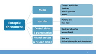

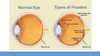
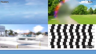


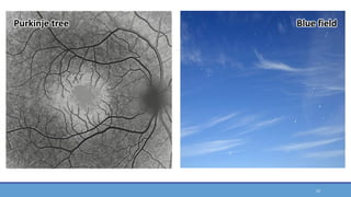


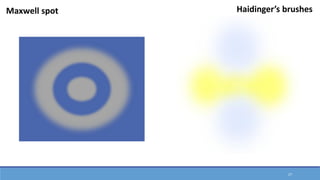


Entoptic phenomena are visual effects and sensations that occur from causes within the human eye itself. Some key entoptic phenomena include floaters and flashes caused by vitreous changes, the Purkinje tree and blue field phenomenon caused by viewing retinal blood vessels, Haidinger's brushes and Maxwell's spot related to macular pigmentation, and blue arcs and phosphenes resulting from retinal or neural stimulation. These phenomena are caused by normal eye anatomy or may result from various pathological conditions. The observer cannot share a direct view of entoptic phenomena with others as they are caused by structures within one's own eye.


