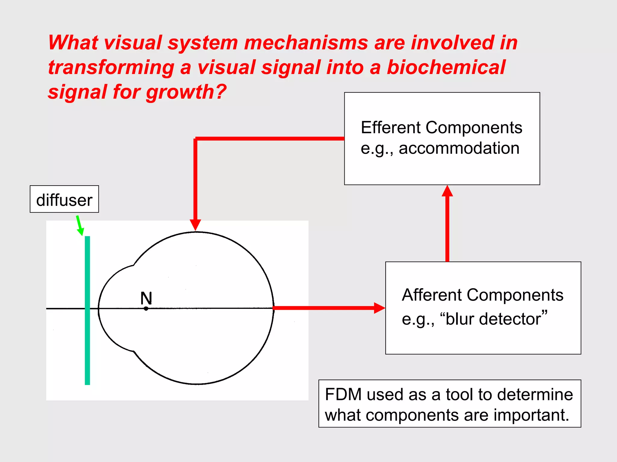
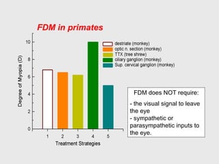
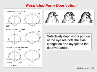
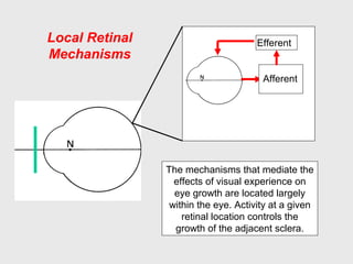
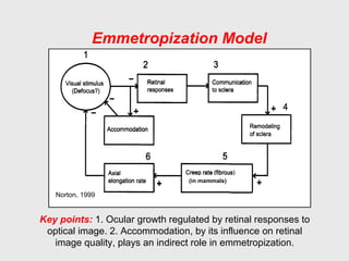
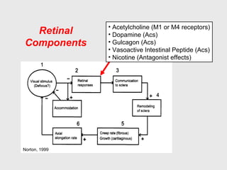
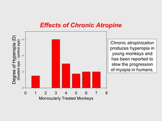
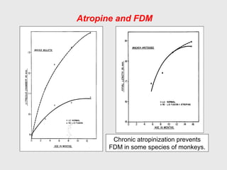
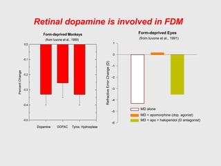
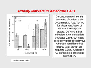
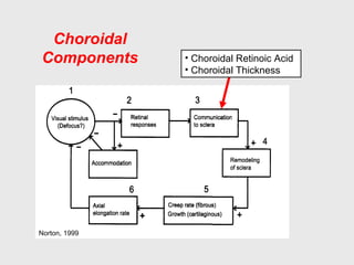
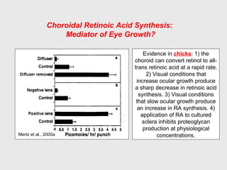
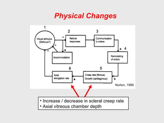
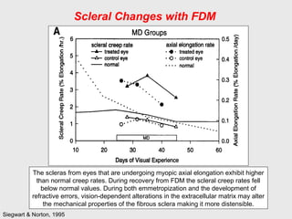
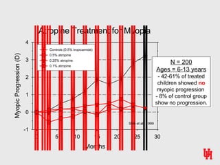
What visual system mechanisms are involved in
transforming a visual signal into a biochemical
signal for growth?
Afferent Components
e.g., “blur detector”
Efferent Components
e.g., accommodation
diffuser
FDM used as a tool to determine
what components are important.
FDM in primates
FDM does NOT require:
- the visual signal to leave
the eye
- sympathetic or
parasympathetic inputs to
the eye.
Restricted Form Deprivation
Selectively depriving a portion
of the eye restricts the axial
elongation and myopia to the
deprived areas.
Wallman et al. 1978
Local Retinal
Mechanisms
Afferent
Efferent
The mechanisms that mediate the
effects of visual experience on
eye growth are located largely
within the eye. Activity at a given
retinal location controls the
growth of the adjacent sclera.
Emmetropization Model
Norton, 1999
Key points: 1. Ocular growth regulated by retinal responses to
optical image. 2. Accommodation, by its influence on retinal
image quality, plays an indirect role in emmetropization.
(in mammals)
Retinal
Components
Norton, 1999
‱ Acetylcholine (M1 or M4 receptors)
‱ Dopamine (Acs)
‱ Gulcagon (Acs)
‱ Vasoactive Intestinal Peptide (Acs)
‱ Nicotine (Antagonist effects)
Chronic atropinization
produces hyperopia in
young monkeys and
has been reported to
slow the progression
of myopia in humans.
Effects of Chronic Atropine
Atropine and FDM
Chronic atropinization prevents
FDM in some species of monkeys.
Neurochemical Transmission in
the Parasympathetic System
Atropine produces cycloplegia by blocking the action of
acetylcholine on muscarinic receptors in ciliary muscle.
Cholinergic Receptor Subtypes
M1 CNS, nerves
M2 Heart, smooth muscle, ciliary muscle
M3 Smooth muscle, exocrine glands, ciliary muscle
M4 CNS, nerves
M5 CNS, ciliary muscle
Atropine blocks all
muscarinic receptor
subtypes.
Effects of Muscarinic Agents
Treatment Regimen
InterocularDifference(mm)
0.0
0.1
0.2
0.3
0.4
on Form-deprivation Myopia
(Stone et al.)
MD control
MD + atropine
MD + pirenzepine (M1)
MD + 4 DAMP (smooth muscle)
Blocking actions:
atropine - all muscarinic sites
4-DAMP - smooth muscle
pirenzepine - neural ganglia
Atropine and pirenzepine
are effective in preventing
FDM in tree shrews. Other
selective muscarinic
antagonists (M2,
gallamine; M3, P-f-HHSid)
were not effective in
blocking FDM. Hence, the
M1 receptor appears to
have potential therapeutic
value. M1 blockers do not
eliminate accommodation.
McBrien et al., 2000
Tree Shrew: Pirenzepine & FDM
Form-deprived Eyes
RefractiveErrorChange(D)
-6
-5
-4
-3
-2
-1
0
1
(from Iuvone et al., 1991)
MD alone
MD + apomorphine (dop. agonist)
MD + apo + haloperidol (D antagonist)
Retinal dopamine is involved in FDM
Activity Markers in Amacrine Cells
Glucagon amacrine cells
are more abundant than
dopaminergic Acs. Tested
for visual regulation of
several transcription
factors. Conditions that
stimulate axial elongation
decrease ZENK synthesis
(basically glucagon activity)
whereas conditions that
reduce axial growth up-
regulate ZENK. Glucagon
AC exhibit sign of defocus
information.
Seltner & Stell, 1995
Choroidal
Components
Norton, 1999
‱ Choroidal Retinoic Acid
‱ Choroidal Thickness
Choroidal Retinoic Acid Synthesis:
Mediator of Eye Growth?
Evidence in chicks: 1) the
choroid can convert retinol to all-
trans retinoic acid at a rapid rate.
2) Visual conditions that
increase ocular growth produce
a sharp decrease in retinoic acid
synthesis. 3) Visual conditions
that slow ocular growth produce
an increase in RA synthesis. 4)
application of RA to cultured
sclera inhibits proteoglycan
production at physiological
concentrations.Mertz et al., 2000a
Choroidal Mechanisms
Changes in choroid thickness move the retina toward the
appropriate focal point.
from Wallman et al., 1995
normal chick chick recovering
from induced myopia
Scleral
Components
Norton, 1999
‱ bFGF & TGF beta (growth factors)
‱ For a myopic stimulus:
‱ Decrease proteoglycan synthesis
‱ Decrease sulfated GAGs
‱ Increase gelatinolytic enzymes
Daily Dose of bFGF (g)
1e-10 1e-9 1e-8 1e-7
-0.1
0.0
0.1
0.2
0.3
0.4
0.5
0.6
0.7
Daily Dose of TGF-beta
1e-14 1e-13 1e-12 1e-11 1e-10 1e-9 1e-8 1e-7
AxiallengthDifference(mm)
-0.1
0.0
0.1
0.2
0.3
0.4
0.5
0.6
0.7
MD and bFGF MD and TGF-beta and bFGF
Biochemical "stop" and "go" Signals
Rhorer and Stell, 1994
(basic Fibrobast Growth Factor) (Transforming Growth Factor Beta)
Possible growth factors involved in FDM
bFGF = basic fibroblast growth factor. TGF-beta = transforming growth factor beta.
The broad dose response curve suggests that more than one type of FGF receptor
is involved.
Matrix metalloproteinase (MMP-2) appears to be the major gelatinolytic
enzyme in the tree shrew sclera. Form deprivation increases catabolism in
the sclera. Hyperopic defocus reduces the degree of scleral catabolism.
Scleral Changes with FDM
Guggenheim & McBrien, 1996
Decorin is the major proteoglycan in the marmoset sclera. The rate
of proteoglycan synthesis is reduced in the posterior pole of FDM.
Rada et al., 2000
Scleral Changes with FDM
Physical Changes
Norton, 1999
‱ Increase / decrease in scleral creep rate
‱ Axial vitreous chamber depth
The scleras from eyes that are undergoing myopic axial elongation exhibit higher
than normal creep rates. During recovery from FDM the scleral creep rates fell
below normal values. During both emmetropization and the development of
refractive errors, vision-dependent alterations in the extracellular matrix may alter
the mechanical properties of the fibrous sclera making it more distensible.
Siegwart & Norton, 1995
Scleral Changes with FDM
Perspective on MyopiaPerspective on Myopia
“The“The aetiologyaetiology of myopia has excited an immenseof myopia has excited an immense
amount of speculation and controversy...and theamount of speculation and controversy...and the
theories which have been put forward to explain itstheories which have been put forward to explain its
development are as ingenious, fanciful anddevelopment are as ingenious, fanciful and
contradictory as have accumulated around any subjectcontradictory as have accumulated around any subject
in medicine. Unfortunately their enthusiasticin medicine. Unfortunately their enthusiastic
implementation in practice has too often involved far-implementation in practice has too often involved far-
reaching social and economic consequences, thereaching social and economic consequences, the
rational basis for which has usually been insubstantial.”rational basis for which has usually been insubstantial.”
- Sir Stewart Duke-Elder, 1970- Sir Stewart Duke-Elder, 1970
Why Worry About Myopia?
‱ Myopia is common.
– 36% of all prescriptions in USA.
‱ Myopia is expensive.
– Total direct costs ($ billions) – estimated for 2000 in USA
‱ $5 to $6 Spectacles & contact lenses
‱ $1.6 to $1.9 Professional Services
‱ $2.2 Refractive Surgery
‱ Inconvenience and complications of correcting
strategies.
Ocular Sequelae of Myopia
(Curtin, 1985)
Posterior
Subcapsular Cataract
2 to 5 X
Idiopathic Retinal
Detachment
4 to 10 X
Open-Angle Glaucoma
2.2 X
Chorioretinal
Degeneration
Health ConcernsHealth Concerns
Myopia is the 7th leading cause of
legal blindness in the U.S.A. (Zadnik,
2001).
The second highest cause of
blindness in India (Edwards, 1998).
Myopic retinal degeneration is the
second highest cause of low vision
in asians (Yap et al., 1990).
The idea that something about near
work causes myopia has dominated
thinking for centuries.
Theoretical basis for traditional therapy
- Increased IOP
- Excessive convergence &/or accommodation
- Gravity & posture
Duke-Elder, 1970
Levinson, 1919
Traditional Treatment MethodsTraditional Treatment Methods
Vision Therapy; biofeedback trainingVision Therapy; biofeedback training
Bifocals; distance over & under correctionBifocals; distance over & under correction
Base-in prismsBase-in prisms
Pharmaceutical agentsPharmaceutical agents
–– cycloplegiacycloplegia
–– intraocular pressureintraocular pressure
Lag of Accommodation
Myopic children
accommodate
significantly less than
emmetropic children
for real targets at
near distances.
Gwiazda et al, 1993
Investigative Ophthalmology & Vision Research, September 2002
Randomized, double-masked clinical trial to
determine whether progressive addition lenses
(SOLA MC lenses with a near addition of +1.50
D) reduce the progression of myopia in children
over a 2 year period.
Do bifocals reduce the rate of
myopic progression?
Time (months)
0 6 12 18 24
CycloplegicRefraction(D)
-4.5
-4.0
-3.5
-3.0
-2.5
PAL
Single Vision
Time (months)
0 6 12 18 24
AxialLength(mm)
24.0
24.5
25.0
25.5
Edwards et al., 2002
Longitudinal Changes in Refractive Error
and Axial Length
Mean ± SEM
At the end of the treatment period, the PAL
group was on average 0.25 D less myopic.
The Comet Study
Investigative Ophthalmology & Vision Science 44:1492, 2003
Randomized, double-masked clinical trial to determine
whether progressive addition lenses (Varilux Comfort
Lenses with a near addition of +2.00D) reduce the
progression of myopia in children over a 3 year period.
The Comet Study
Gwiazda et al., 2003
Myopic Progression
PALs
SV
Gwiazda et al., 2004
Phoria
Eso Ortho Exo
3-YearTreatmentEffect(D)
-0.2
0.0
0.2
0.4
0.6
0.8
Larger Acc Lag
Smaller Acc Lag
PALs reduce progression
rate by about 50% (about
0.75 D in 3 years) in
esophores with large lags of
accommodation.
The Comet Study
Do Near Adds Eliminate
Accommodative Errors?
Subjects typically fail to relax
accommodation by an amount
equal to the add. Near adds
may actually increase the
degree of retinal defocus.
Optimal Add?
Rosenfield & Carrel, 2001
Randomized, controlled clinical trial to determine
the effects of undercorrection on the rate of
progression of myopia.
Does undercorrection slow
myopic progression?
Methods
Subject Selection Criteria
‱ Age: 9-14 years.
‱ At least –0.5 D of myopia (sph equiv) in
both eyes & myopic in all meridians.
‱ < 2.0 D of astigmatism in each eye.
‱ Corrected VA = 20/20 or better in each
eye.
‱ No significant binocular vision problems.
‱ Normal ocular health.
‱ No previous contact lens wear.
Methods
Chung, Mohidin and O’Leary
‱ Spectacle Corrections:
– Full Correction: Maximum plus to obtain
best VA in each eye. Full compliance 41 of 46.
– Undercorrection: Monocular VA maintained
at 20/40 by undercorrecting by about +0.75
D. Full compliance 40 of 47.
‱ Patients instructed to wear spectacles
at all times. Full Compliance > 8 hours/day.
Mean Changes in Refractive Error
From Chung et al., 2002
Fully Corrected
Undercorrected
Start of Trial
The undercorrected group showed a
greater rate of myopic progression.
Average sph equivalent (± SEM) for both eyes.
Mean Changes in Axial Length
From Chung et al., 2002
Fully Corrected
Undercorrected
Start of Trial
The undercorrected group showed
greater axial elongation.
No between group differences in
corneal curvature, anterior
chamber depth or lens thickness.
The “CLAMP” Study
Contact Lens and Myopia Progression
RGPS vs Soft CLs
Walline et al., 2004
The “CLAMP” Study
Walline et al., 2004
The “CLAMP” Study
New Hopes for
Optical Interventions
Emmetropization: Basic Operating Properties
Visual Signals for Axial Growth
Mutti et al., 2000
Ferree & Rand, 1933
Refractive error varies with
eccentricity. Myopes typically exhibit
relative hyperopia in the periphery,
whereas hyperopes show relative
myopia in the periphery.
Central vs.
30 deg Nasal
Uncorrected
Myope
“Corrected”
Myope
Image Shell
As a consequence of eye shape
and/or aspheric optical
surfaces, myopic eyes may
experience significant defocus
across the visual field,
regardless of the refractive state
at the fovea.
Should we correct peripheral refractive errors?
Optimal Correction?
Traditional Treatment MethodsTraditional Treatment Methods
Vision Therapy; biofeedback trainingVision Therapy; biofeedback training
Bifocals; distance over & under correctionBifocals; distance over & under correction
Base-in prismsBase-in prisms
Pharmaceutical agentsPharmaceutical agents
–– cycloplegiacycloplegia
–– intraocular pressureintraocular pressure
Jensen, 1991
Subject Groups
IOP > 17 mmHG IOP < 17 mmHG
MyopicProgression(D/year)
0.0
0.1
0.2
0.3
0.4
0.5
0.6
0.7 Controls
Timolol
Timolol Treatment for Myopia
Timolol was effective
in lowering IOP.
However there was
not a significant
effect on the rate of
myopic progression.
Control Treated
DegreeofMyopia(D)
-40
-30
-20
-10
0
Control Treated
RelativeAxialElongation(mm)
0.0
0.5
1.0
1.5
2.0
Control
0.5% Timolol BID
Schmidt & Wildsoet, 2000
Timolol and Form-Deprivation Myopia
Timolol was effective in lowering IOP in young chicks (between 18 &
27%). However there was not a significant effect on the rate of
myopic progression for either form deprivation or negative lenses
Atropine Treatment for Myopia
Months
0 5 10 15 20 25 30
MyopicProgression(D)
-1
0
1
2
3
4
Controls (0.5% tropicamide)
0.5% atropine
0.25% atropine
0.1% atropine
Shih et al., 1999
N = 200
Ages = 6-13 years
- 42-61% of treated
children showed no
myopic progression
- 8% of control group
show no progression.
Atropine TherapyAtropine Therapy
ShortShort--term sideterm side--effects:effects:
––photophobia & blurred visionphotophobia & blurred vision
––cycloplegia (need for readingcycloplegia (need for reading
glasses)glasses)
––potential light damage to retinapotential light damage to retina
––potential elevations in IOPpotential elevations in IOP
––potential systematic reactionspotential systematic reactions
Treated eye Control eye
Long term Effects of Chronic Atropinization
Permanent alterations in
pupil size, amplitude of
accommodation, acc-
convergence interactions,
neuropharmacology of
intraocular muscles
Photo of adult cat the was treated
with 1% atropine in the right eye
from 4 weeks to 4 months of age.
Pirenzepine Trials
‱ Safety and efficacy of 2% PRZ ophthalmic
gel in myopic children: Year 1 (Siatkowski et
al., 2003, ARVO)
‱ US Phase II Trial.
– 8- to 12-year old children (n=174); mean age = 9.9 yrs
– -0.75 to -4.00 D myopia; mean = -2.04 ± 0.9 D
– Treated with 2% PRZ or placebo BID for 2 years
PIRZ Placebo
MyopicProgression(D/year)
0.0
0.1
0.2
0.3
0.4
0.5
0.6
PIRZ bid PIRZ qd Placebo
0.0
0.2
0.4
0.6
0.8
1.0
Pirenzepine: Efficacy for Pediatric Myopia
Year One Results
Asia StudyU.S. Study
N = 353N = 174
Siatkowski et al., 2003 (ARVO) Tan et al., 2003 (ARVO)
Pirenzepine: Efficacy for Pediatric Myopia
Year Two Results
PIRZ Placebo
MyopicProgression(D)
0.0
0.2
0.4
0.6
0.8
1.0
1.2
1.4
1.6
1.8 U.S. Study
N = 174
Siatkowski et al., 2004 (ARVO)
Proportion ≄ 0.75 D
PIR = 37%
PLC = 68%
Dropouts
12% of PIR subjects
0% of PLC group
Common adverse events
eyelid gel residue, blurred near
vision, and asymptomatic
conjunctival reactions.
Pirenzepine Trials
‱ Other Questions:
– What are the mechanisms and sites of action
of PRZ? (Optimal drug & deliver system?)
– How do you identify patients who will benefit?
– How long do you need to treat the patient?
– Are the effects permanent?
– Are partial effects acceptable?
– Is it safe during pregnancy?
– Are there long-term side effects?

Emmetropization 2 2006

  • 1.
    What visual systemmechanisms are involved in transforming a visual signal into a biochemical signal for growth? Afferent Components e.g., “blur detector” Efferent Components e.g., accommodation diffuser FDM used as a tool to determine what components are important.
  • 2.
    FDM in primates FDMdoes NOT require: - the visual signal to leave the eye - sympathetic or parasympathetic inputs to the eye.
  • 3.
    Restricted Form Deprivation Selectivelydepriving a portion of the eye restricts the axial elongation and myopia to the deprived areas. Wallman et al. 1978
  • 4.
    Local Retinal Mechanisms Afferent Efferent The mechanismsthat mediate the effects of visual experience on eye growth are located largely within the eye. Activity at a given retinal location controls the growth of the adjacent sclera.
  • 5.
    Emmetropization Model Norton, 1999 Keypoints: 1. Ocular growth regulated by retinal responses to optical image. 2. Accommodation, by its influence on retinal image quality, plays an indirect role in emmetropization. (in mammals)
  • 6.
    Retinal Components Norton, 1999 ‱ Acetylcholine(M1 or M4 receptors) ‱ Dopamine (Acs) ‱ Gulcagon (Acs) ‱ Vasoactive Intestinal Peptide (Acs) ‱ Nicotine (Antagonist effects)
  • 7.
    Chronic atropinization produces hyperopiain young monkeys and has been reported to slow the progression of myopia in humans. Effects of Chronic Atropine
  • 8.
    Atropine and FDM Chronicatropinization prevents FDM in some species of monkeys.
  • 9.
    Neurochemical Transmission in theParasympathetic System Atropine produces cycloplegia by blocking the action of acetylcholine on muscarinic receptors in ciliary muscle.
  • 10.
    Cholinergic Receptor Subtypes M1CNS, nerves M2 Heart, smooth muscle, ciliary muscle M3 Smooth muscle, exocrine glands, ciliary muscle M4 CNS, nerves M5 CNS, ciliary muscle Atropine blocks all muscarinic receptor subtypes.
  • 11.
    Effects of MuscarinicAgents Treatment Regimen InterocularDifference(mm) 0.0 0.1 0.2 0.3 0.4 on Form-deprivation Myopia (Stone et al.) MD control MD + atropine MD + pirenzepine (M1) MD + 4 DAMP (smooth muscle) Blocking actions: atropine - all muscarinic sites 4-DAMP - smooth muscle pirenzepine - neural ganglia
  • 12.
    Atropine and pirenzepine areeffective in preventing FDM in tree shrews. Other selective muscarinic antagonists (M2, gallamine; M3, P-f-HHSid) were not effective in blocking FDM. Hence, the M1 receptor appears to have potential therapeutic value. M1 blockers do not eliminate accommodation. McBrien et al., 2000 Tree Shrew: Pirenzepine & FDM
  • 13.
    Form-deprived Eyes RefractiveErrorChange(D) -6 -5 -4 -3 -2 -1 0 1 (from Iuvoneet al., 1991) MD alone MD + apomorphine (dop. agonist) MD + apo + haloperidol (D antagonist) Retinal dopamine is involved in FDM
  • 14.
    Activity Markers inAmacrine Cells Glucagon amacrine cells are more abundant than dopaminergic Acs. Tested for visual regulation of several transcription factors. Conditions that stimulate axial elongation decrease ZENK synthesis (basically glucagon activity) whereas conditions that reduce axial growth up- regulate ZENK. Glucagon AC exhibit sign of defocus information. Seltner & Stell, 1995
  • 15.
    Choroidal Components Norton, 1999 ‱ ChoroidalRetinoic Acid ‱ Choroidal Thickness
  • 16.
    Choroidal Retinoic AcidSynthesis: Mediator of Eye Growth? Evidence in chicks: 1) the choroid can convert retinol to all- trans retinoic acid at a rapid rate. 2) Visual conditions that increase ocular growth produce a sharp decrease in retinoic acid synthesis. 3) Visual conditions that slow ocular growth produce an increase in RA synthesis. 4) application of RA to cultured sclera inhibits proteoglycan production at physiological concentrations.Mertz et al., 2000a
  • 17.
    Choroidal Mechanisms Changes inchoroid thickness move the retina toward the appropriate focal point. from Wallman et al., 1995 normal chick chick recovering from induced myopia
  • 18.
    Scleral Components Norton, 1999 ‱ bFGF& TGF beta (growth factors) ‱ For a myopic stimulus: ‱ Decrease proteoglycan synthesis ‱ Decrease sulfated GAGs ‱ Increase gelatinolytic enzymes
  • 19.
    Daily Dose ofbFGF (g) 1e-10 1e-9 1e-8 1e-7 -0.1 0.0 0.1 0.2 0.3 0.4 0.5 0.6 0.7 Daily Dose of TGF-beta 1e-14 1e-13 1e-12 1e-11 1e-10 1e-9 1e-8 1e-7 AxiallengthDifference(mm) -0.1 0.0 0.1 0.2 0.3 0.4 0.5 0.6 0.7 MD and bFGF MD and TGF-beta and bFGF Biochemical "stop" and "go" Signals Rhorer and Stell, 1994 (basic Fibrobast Growth Factor) (Transforming Growth Factor Beta) Possible growth factors involved in FDM bFGF = basic fibroblast growth factor. TGF-beta = transforming growth factor beta. The broad dose response curve suggests that more than one type of FGF receptor is involved.
  • 20.
    Matrix metalloproteinase (MMP-2)appears to be the major gelatinolytic enzyme in the tree shrew sclera. Form deprivation increases catabolism in the sclera. Hyperopic defocus reduces the degree of scleral catabolism. Scleral Changes with FDM Guggenheim & McBrien, 1996
  • 21.
    Decorin is themajor proteoglycan in the marmoset sclera. The rate of proteoglycan synthesis is reduced in the posterior pole of FDM. Rada et al., 2000 Scleral Changes with FDM
  • 22.
    Physical Changes Norton, 1999 ‱Increase / decrease in scleral creep rate ‱ Axial vitreous chamber depth
  • 23.
    The scleras fromeyes that are undergoing myopic axial elongation exhibit higher than normal creep rates. During recovery from FDM the scleral creep rates fell below normal values. During both emmetropization and the development of refractive errors, vision-dependent alterations in the extracellular matrix may alter the mechanical properties of the fibrous sclera making it more distensible. Siegwart & Norton, 1995 Scleral Changes with FDM
  • 24.
    Perspective on MyopiaPerspectiveon Myopia “The“The aetiologyaetiology of myopia has excited an immenseof myopia has excited an immense amount of speculation and controversy...and theamount of speculation and controversy...and the theories which have been put forward to explain itstheories which have been put forward to explain its development are as ingenious, fanciful anddevelopment are as ingenious, fanciful and contradictory as have accumulated around any subjectcontradictory as have accumulated around any subject in medicine. Unfortunately their enthusiasticin medicine. Unfortunately their enthusiastic implementation in practice has too often involved far-implementation in practice has too often involved far- reaching social and economic consequences, thereaching social and economic consequences, the rational basis for which has usually been insubstantial.”rational basis for which has usually been insubstantial.” - Sir Stewart Duke-Elder, 1970- Sir Stewart Duke-Elder, 1970
  • 25.
    Why Worry AboutMyopia? ‱ Myopia is common. – 36% of all prescriptions in USA. ‱ Myopia is expensive. – Total direct costs ($ billions) – estimated for 2000 in USA ‱ $5 to $6 Spectacles & contact lenses ‱ $1.6 to $1.9 Professional Services ‱ $2.2 Refractive Surgery ‱ Inconvenience and complications of correcting strategies.
  • 26.
    Ocular Sequelae ofMyopia (Curtin, 1985) Posterior Subcapsular Cataract 2 to 5 X Idiopathic Retinal Detachment 4 to 10 X Open-Angle Glaucoma 2.2 X Chorioretinal Degeneration
  • 27.
    Health ConcernsHealth Concerns Myopiais the 7th leading cause of legal blindness in the U.S.A. (Zadnik, 2001). The second highest cause of blindness in India (Edwards, 1998). Myopic retinal degeneration is the second highest cause of low vision in asians (Yap et al., 1990).
  • 28.
    The idea thatsomething about near work causes myopia has dominated thinking for centuries. Theoretical basis for traditional therapy - Increased IOP - Excessive convergence &/or accommodation - Gravity & posture Duke-Elder, 1970 Levinson, 1919
  • 29.
    Traditional Treatment MethodsTraditionalTreatment Methods Vision Therapy; biofeedback trainingVision Therapy; biofeedback training Bifocals; distance over & under correctionBifocals; distance over & under correction Base-in prismsBase-in prisms Pharmaceutical agentsPharmaceutical agents –– cycloplegiacycloplegia –– intraocular pressureintraocular pressure
  • 30.
    Lag of Accommodation Myopicchildren accommodate significantly less than emmetropic children for real targets at near distances. Gwiazda et al, 1993
  • 31.
    Investigative Ophthalmology &Vision Research, September 2002 Randomized, double-masked clinical trial to determine whether progressive addition lenses (SOLA MC lenses with a near addition of +1.50 D) reduce the progression of myopia in children over a 2 year period. Do bifocals reduce the rate of myopic progression?
  • 32.
    Time (months) 0 612 18 24 CycloplegicRefraction(D) -4.5 -4.0 -3.5 -3.0 -2.5 PAL Single Vision Time (months) 0 6 12 18 24 AxialLength(mm) 24.0 24.5 25.0 25.5 Edwards et al., 2002 Longitudinal Changes in Refractive Error and Axial Length Mean ± SEM At the end of the treatment period, the PAL group was on average 0.25 D less myopic.
  • 33.
    The Comet Study InvestigativeOphthalmology & Vision Science 44:1492, 2003 Randomized, double-masked clinical trial to determine whether progressive addition lenses (Varilux Comfort Lenses with a near addition of +2.00D) reduce the progression of myopia in children over a 3 year period.
  • 34.
    The Comet Study Gwiazdaet al., 2003 Myopic Progression PALs SV
  • 35.
    Gwiazda et al.,2004 Phoria Eso Ortho Exo 3-YearTreatmentEffect(D) -0.2 0.0 0.2 0.4 0.6 0.8 Larger Acc Lag Smaller Acc Lag PALs reduce progression rate by about 50% (about 0.75 D in 3 years) in esophores with large lags of accommodation. The Comet Study
  • 36.
    Do Near AddsEliminate Accommodative Errors? Subjects typically fail to relax accommodation by an amount equal to the add. Near adds may actually increase the degree of retinal defocus. Optimal Add? Rosenfield & Carrel, 2001
  • 37.
    Randomized, controlled clinicaltrial to determine the effects of undercorrection on the rate of progression of myopia. Does undercorrection slow myopic progression?
  • 38.
    Methods Subject Selection Criteria ‱Age: 9-14 years. ‱ At least –0.5 D of myopia (sph equiv) in both eyes & myopic in all meridians. ‱ < 2.0 D of astigmatism in each eye. ‱ Corrected VA = 20/20 or better in each eye. ‱ No significant binocular vision problems. ‱ Normal ocular health. ‱ No previous contact lens wear.
  • 39.
    Methods Chung, Mohidin andO’Leary ‱ Spectacle Corrections: – Full Correction: Maximum plus to obtain best VA in each eye. Full compliance 41 of 46. – Undercorrection: Monocular VA maintained at 20/40 by undercorrecting by about +0.75 D. Full compliance 40 of 47. ‱ Patients instructed to wear spectacles at all times. Full Compliance > 8 hours/day.
  • 40.
    Mean Changes inRefractive Error From Chung et al., 2002 Fully Corrected Undercorrected Start of Trial The undercorrected group showed a greater rate of myopic progression. Average sph equivalent (± SEM) for both eyes.
  • 41.
    Mean Changes inAxial Length From Chung et al., 2002 Fully Corrected Undercorrected Start of Trial The undercorrected group showed greater axial elongation. No between group differences in corneal curvature, anterior chamber depth or lens thickness.
  • 42.
    The “CLAMP” Study ContactLens and Myopia Progression RGPS vs Soft CLs Walline et al., 2004
  • 43.
    Walline et al.,2004 The “CLAMP” Study
  • 44.
    Walline et al.,2004 The “CLAMP” Study
  • 45.
    New Hopes for OpticalInterventions Emmetropization: Basic Operating Properties
  • 46.
    Visual Signals forAxial Growth Mutti et al., 2000 Ferree & Rand, 1933 Refractive error varies with eccentricity. Myopes typically exhibit relative hyperopia in the periphery, whereas hyperopes show relative myopia in the periphery. Central vs. 30 deg Nasal
  • 47.
    Uncorrected Myope “Corrected” Myope Image Shell As aconsequence of eye shape and/or aspheric optical surfaces, myopic eyes may experience significant defocus across the visual field, regardless of the refractive state at the fovea. Should we correct peripheral refractive errors? Optimal Correction?
  • 48.
    Traditional Treatment MethodsTraditionalTreatment Methods Vision Therapy; biofeedback trainingVision Therapy; biofeedback training Bifocals; distance over & under correctionBifocals; distance over & under correction Base-in prismsBase-in prisms Pharmaceutical agentsPharmaceutical agents –– cycloplegiacycloplegia –– intraocular pressureintraocular pressure
  • 49.
    Jensen, 1991 Subject Groups IOP> 17 mmHG IOP < 17 mmHG MyopicProgression(D/year) 0.0 0.1 0.2 0.3 0.4 0.5 0.6 0.7 Controls Timolol Timolol Treatment for Myopia Timolol was effective in lowering IOP. However there was not a significant effect on the rate of myopic progression.
  • 50.
    Control Treated DegreeofMyopia(D) -40 -30 -20 -10 0 Control Treated RelativeAxialElongation(mm) 0.0 0.5 1.0 1.5 2.0 Control 0.5%Timolol BID Schmidt & Wildsoet, 2000 Timolol and Form-Deprivation Myopia Timolol was effective in lowering IOP in young chicks (between 18 & 27%). However there was not a significant effect on the rate of myopic progression for either form deprivation or negative lenses
  • 51.
    Atropine Treatment forMyopia Months 0 5 10 15 20 25 30 MyopicProgression(D) -1 0 1 2 3 4 Controls (0.5% tropicamide) 0.5% atropine 0.25% atropine 0.1% atropine Shih et al., 1999 N = 200 Ages = 6-13 years - 42-61% of treated children showed no myopic progression - 8% of control group show no progression.
  • 52.
    Atropine TherapyAtropine Therapy ShortShort--termsideterm side--effects:effects: ––photophobia & blurred visionphotophobia & blurred vision ––cycloplegia (need for readingcycloplegia (need for reading glasses)glasses) ––potential light damage to retinapotential light damage to retina ––potential elevations in IOPpotential elevations in IOP ––potential systematic reactionspotential systematic reactions
  • 53.
    Treated eye Controleye Long term Effects of Chronic Atropinization Permanent alterations in pupil size, amplitude of accommodation, acc- convergence interactions, neuropharmacology of intraocular muscles Photo of adult cat the was treated with 1% atropine in the right eye from 4 weeks to 4 months of age.
  • 54.
    Pirenzepine Trials ‱ Safetyand efficacy of 2% PRZ ophthalmic gel in myopic children: Year 1 (Siatkowski et al., 2003, ARVO) ‱ US Phase II Trial. – 8- to 12-year old children (n=174); mean age = 9.9 yrs – -0.75 to -4.00 D myopia; mean = -2.04 ± 0.9 D – Treated with 2% PRZ or placebo BID for 2 years
  • 55.
    PIRZ Placebo MyopicProgression(D/year) 0.0 0.1 0.2 0.3 0.4 0.5 0.6 PIRZ bidPIRZ qd Placebo 0.0 0.2 0.4 0.6 0.8 1.0 Pirenzepine: Efficacy for Pediatric Myopia Year One Results Asia StudyU.S. Study N = 353N = 174 Siatkowski et al., 2003 (ARVO) Tan et al., 2003 (ARVO)
  • 56.
    Pirenzepine: Efficacy forPediatric Myopia Year Two Results PIRZ Placebo MyopicProgression(D) 0.0 0.2 0.4 0.6 0.8 1.0 1.2 1.4 1.6 1.8 U.S. Study N = 174 Siatkowski et al., 2004 (ARVO) Proportion ≄ 0.75 D PIR = 37% PLC = 68% Dropouts 12% of PIR subjects 0% of PLC group Common adverse events eyelid gel residue, blurred near vision, and asymptomatic conjunctival reactions.
  • 57.
    Pirenzepine Trials ‱ OtherQuestions: – What are the mechanisms and sites of action of PRZ? (Optimal drug & deliver system?) – How do you identify patients who will benefit? – How long do you need to treat the patient? – Are the effects permanent? – Are partial effects acceptable? – Is it safe during pregnancy? – Are there long-term side effects?

Editor's Notes

  • #6 &amp;lt;number&amp;gt;
  • #27 But it is very important to recognize that the optical correction of myopia, whether by traditional spectacle lenses or by laser surgery, does not eliminate the health risks associated with myopia. Myopia is a major risk factor for blindness because as illustrated by this schematic 
myopia is most commonly caused by an elongation of the globe. Neither surgery or traditional correcting lenses make the myopic eye normal. They do not cure the condition. They simply compensate for the optical error. Structural changes associated with axial elongation increase the risk for a number of potentially blinding conditions. For example, epidemiological studies have demonstrated that in comparison to non-myopes. As a consequence in the USA, myopia is currently the 7th leading cause of legal blindness 
falling just behind diabetic retinopathy.
  • #34 &amp;lt;number&amp;gt;
  • #35 &amp;lt;number&amp;gt;
  • #36 &amp;lt;number&amp;gt; However for certain subgroups the effects are larger. This slide illustrates the treatment effects of PALs for subjects segrated by lateral heterophoria and by the magnitude of accommodative lag for near viewing. The key point is for esophoric children with a high lag of accommodation
is that after 3 years there was about a 0.75 D difference between SV and PAL groups
.the Kids treated with PAL showed a 50% reduction in progression rates
. This in my mind is a big deal
..little down side and 50% effect is clinically significant.
  • #48 We believe that these results have significant implication for how the effects of visual experience are assessed in humans. Instead of focusing only on central refractive errors and the nature of visual experience at the fovea, as almost all previous human studies have, it will be important to assess vision across the visual field. Eye shape and peripheral refractive error, as a number of investigators have argued, may be very predictive of who develops myopia. The results also suggest that we need to consider peripheral effects when designing a vision-based treatment strategy for myopia or hyperopia....it may be that we will be more effective in controlling eye growth if we manipulate peripheral vision. Peripheral optics may explain why altering the effective focus at the fovea in children does not control eye growth in a predictable manner. For example,....if myopic eyes or premyopic eyes, as suggested by the Ohio state group, exhibit more hyperopic errors in the periphery, it may not matter how you correct refractive errors at the fovea...regardless of whether the eye is under corrected or optimally corrected, the periphery may, depending on factors related to the eye’s shape and its optics, experience a substantial amount of hyperopic defocus, which could serve as a stimulus for myopic progression.
  • #52 Side effects: with 1% photophobia, blurred near vision, poor compliance 186 children from 6-13 years; all myopic at start (-0.5 to -6.75 D); 14 of original lost to follow up. Control = tropicamide (0.5%) treated at night between 42-61% of treated children showed no myopic progression (varied with the concentration of atropine
only 8% of control groups show no progression. Some treated children still exhibited fast &amp;gt;1D/yr progression. 0.5% group wore bifocals; 0.25% group were undercorrected; 0.1% group were fully corrected. Photophobia still a problem with 0.5%
  • #55 &amp;lt;number&amp;gt;
  • #56 US study reported about 50% reduction in rate of myopic progression. Only 2% of PIRZ group showed rates greater than 1D/year, whereas 20% of placebo group showed progression rates greater than 1 D/year.
  • #57 US study reported about 50% reduction in rate of myopic progression. Only 2% of PIRZ group showed rates greater than 1D/year, whereas 20% of placebo group showed progression rates greater than 1 D/year.
  • #58 &amp;lt;number&amp;gt;