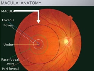
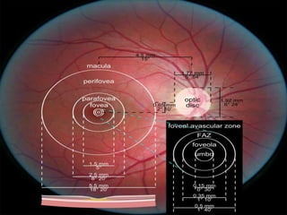
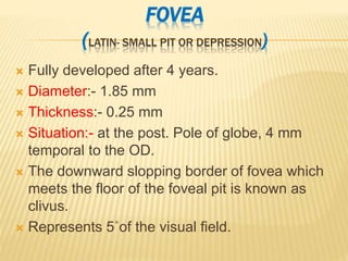
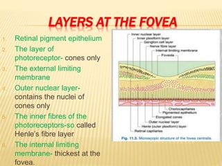
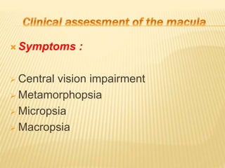
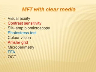
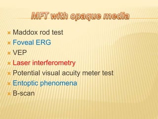
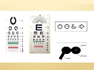
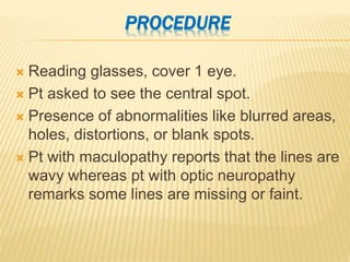
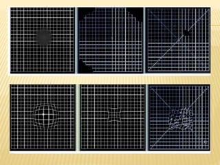
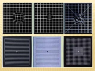
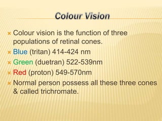
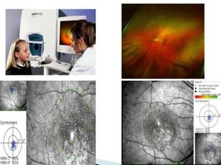
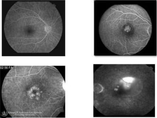
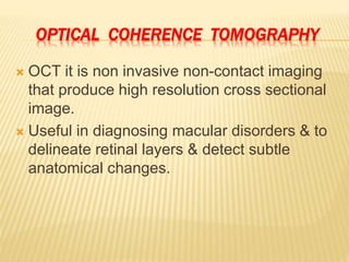
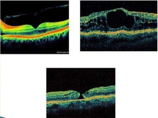
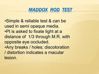
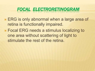
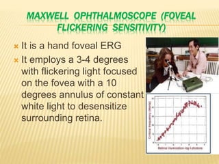
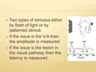
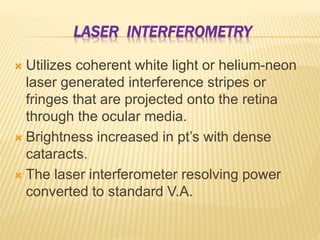
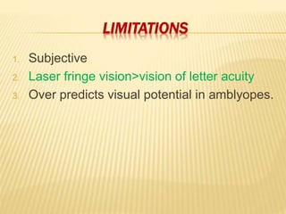
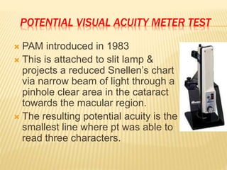
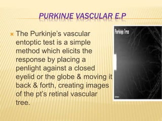
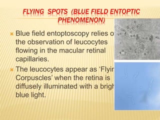
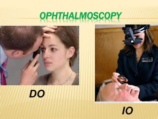
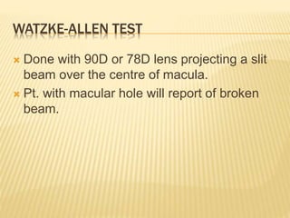
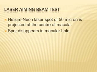
The macular function test is very important test... To understand the maula dis function and amount of disfunction. It is very useful for Central and colour vision.
2. Retina is the innermost Tunic of The Eyeball
& a Transparent Membrane.
Thickness in different region:-
Peripappilary area:- 0.56 mm
Equator:- 0.18 – 0.20 mm
Fovea:- 0.25 mm
Foveola:- 0.13 mm
Ora serrata:- 0.10 mm
3.
4. THREE DISTINCT REGIONS OF RETINA
1. Optic disc :- Diameter – 1.5 mm
2. Peripheral Retina:-
Near periphery (1.5 mm) around macula.
Mid periphery 3 mm wide around near
periphery.
Far periphery.
Ora Serrata – temporally:- 2.1 mm wide
nasally:- 0.7 – 0.8 mm wide
5. 3. Macula Lutea
Site:- temporal to optic disc at the post.
Pole
Shape:- horizontally elliptical
Diameter:- 5.5 mm
Function:- photopic & colour vision
corresponds 15˚of visual field.
6. Yellowish
The coloration probably derives from the
presence of xanthophyll in the ganglion &
bipolar cells
Yellow coloration remains for several hours
to few days in enucleated eyes kept in 4˚C
7.
8. PARTS OF MACULA LUTEA
Umbo:- tiny depression in centre corresponding
to foveal reflex
Foveola:- diameter – 0.35 mm
thickness – 0.13 mm
forms the central floor of fovea.
Devoid of ganglion cell layer & contains cone
photoreceptors only.
Corresponding 1˚of visual field.
Colour:-reddish due to rich choroidal circulation
which shines through it.
9. Fovea:- surrounds the foveola.
Situated 2 DD temporal to the disc margin &
0.8 mm below the horizontal meridian.
The Foveal Avascular zone (FAZ):-
Located within the fovea but extends beyond
the foveola 500μ in diameter.
Parafoveal zone:- 0.55 mm wide around the
fovea centralis
Perifoveal zone:- 1.5 mm wide around the
parafoveal zone.
10.
11. FOVEA
(LATIN- SMALL PIT OR DEPRESSION)
Fully developed after 4 years.
Diameter:- 1.85 mm
Thickness:- 0.25 mm
Situation:- at the post. Pole of globe, 4 mm
temporal to the OD.
The downward slopping border of fovea which
meets the floor of the foveal pit is known as
clivus.
Represents 5˚of the visual field.
12. LAYERS AT THE FOVEA
1. Retinal pigment epithelium
2. The layer of
photoreceptor- cones only
3. The external limiting
membrane
4. Outer nuclear layer-
contains the nuclei of
cones only
5. The inner fibres of the
photoreceptors-so called
Henle’s fibre layer
6. The internal limiting
membrane- thickest at the
fovea.
16. Maddox rod test
Foveal ERG
VEP
Laser interferometry
Potential visual acuity meter test
Entoptic phenomena
B-scan
17. VA is measured by the visual resolution of a
letter, symbol or a pattern under conditions of
maximal contrast.
In pt’s with macular disease VA is frequently
worse when the pt looks through a pin-hole
18.
19. PINHOLE TESTING
Vision improves with
good macular function
Acuity frequently worse
in macular disorder.
20. Contrast sensitivity is a measure of the
minimum amount of contrast needed to
distinguish a test object.
Indirectly assesses the quality of vision.
Can detect early visual loss when VA is
normal.
21.
22. To detect retinal conditions like DR, ARMD &
other macular & optic nerve diseases.
Optical conditions like refractive error,
refractive surgery, cataract & IOL
implantation & normal aging of the eye.
23. •Spatial frequency is the
number of dark light cycles
per visual angle.
•In macular diseases, there is
a marked impairment for the
intermediate & higher spatial
frequencies.
24. Is an effective test for
monitoring potential
decreases in contrast
sensitivity function over
time.
25. With a strong convex
lens affords excellent
vizualizaton of the
macula. (78D & 90D)
26. Principle :
The test involves exposing the macula to a
light source bright enough to bleach a
significant proportion of the visual pigments.
Return of normal retinal function & sensitivity
depends on the regeneration of visual
pigments.
27. Pathological states that affect the
photoreceptors, Bruch's membrane, chorio-
capillaries or can prolong visual recovery
time.
No such prolonged is observed in diseases
affecting the neutral conducting pathways.
28. •Evaluates the 10˚ of visual field
centered on fixation.
•Used in screening and
monitoring macular diseases.
•Square 10*10 cm divided into400
5*5 mm squares to be held at
30cm.
29.
30. PROCEDURE
Reading glasses, cover 1 eye.
Pt asked to see the central spot.
Presence of abnormalities like blurred areas,
holes, distortions, or blank spots.
Pt with maculopathy reports that the lines are
wavy whereas pt with optic neuropathy
remarks some lines are missing or faint.
31.
32.
33. Colour vision is the function of three
populations of retinal cones.
Blue (tritan) 414-424 nm
Green (duetran) 522-539nm
Red (proton) 549-570nm
Normal person possess all these three cones
& called trichromate.
34. Acquired macular diseases tends to produce
blue yellow defects & optic nerve lesions red
green defects.
Deutran anomaly is the most commonly &
those subjects can not differentiate b/t red &
green colours.
37. The principle of microperimetry rests on the
possibility to see –in real time- the retina
under examination (by infrared light) & to
project a defined light stimulus over an
individual, selected location.
38. SCANNING LASER OPHTHALMOSCOPE
MICROPERIMETRY
SLO microperimetry was the 1st technique
which allowed to obtain a fundus related
sensitivity map.
SLO uses a near infrared diode laser
(675nm) beam that rapidly scan the posterior
pole.
The reflected light is detected by a confocal
photodiode & the digitized image is stored in
a computer.
39.
40. FLUORESCEIN ANGIOGRAPHY
Dark appearance of the fovea on FFA is
caused by FAZ & blockage of the choroidal
background by xanthophyll & dense RPE.
FFA is a very useful tool in diagnosing
macular disorders e.g., diabetic maculopathy,
CSR & can reveals the functionality of the
lesion e.g. Ischemic maculopathy.
41.
42. OPTICAL COHERENCE TOMOGRAPHY
OCT it is non invasive non-contact imaging
that produce high resolution cross sectional
image.
Useful in diagnosing macular disorders & to
delineate retinal layers & detect subtle
anatomical changes.
43.
44. Maddox rod
Focal ERG
VEP
Laser interferometry
Potential visual acuity meter test
Entoptic phenomena
Preferential hyperacuity perimeter (PHP)
B-scan
45. MADDOX ROD TEST
•Simple & reliable test & can be
used in semi opaque media.
•Pt is asked to fixate light at a
distance of 1/3 through M.R. with
opposite eye occluded.
•Any breaks / holes; discoloration
/ distortion indicates a macular
lesion.
46. FOCAL ELECTRORETINOGRAM
ERG is only abnormal when a large area of
retina is functionally impaired.
Focal ERG needs a stimulus localizing to
one area without scattering of light to
stimulate the rest of the retina.
47. MAXWELL OPHTHALMOSCOPE (FOVEAL
FLICKERING SENSITIVITY)
It is a hand foveal ERG
It employs a 3-4 degrees
with flickering light focused
on the fovea with a 10
degrees annulus of constant
white light to desensitize
surrounding retina.
48. VISUALLY EVOKED POTENTIAL
VEP measure of the electrical potential
generated in response to entire visual
pathway
It represents integrity of entire visual pathway
from retina to occipital lobe so cannot
differentiate b/t macula, ON & cortical
pathology.
49. Two types of stimulus either
by flash of light or by
patterned stimuli.
If the issue is the V/A then
the amplitude is measured.
If the issue is the lesion in
the visual pathway then the
latency is measured.
50. LASER INTERFEROMETRY
Utilizes coherent white light or helium-neon
laser generated interference stripes or
fringes that are projected onto the retina
through the ocular media.
Brightness increased in pt’s with dense
cataracts.
The laser interferometer resolving power
converted to standard V.A.
52. POTENTIAL VISUAL ACUITY METER TEST
PAM introduced in 1983
This is attached to slit lamp &
projects a reduced Snellen’s chart
via narrow beam of light through a
pinhole clear area in the cataract
towards the macular region.
The resulting potential acuity is the
smallest line where pt was able to
read three characters.
53. LIMITATION
Subjective
Methods that require an alert & cooperative
pt & skilled compassionate examiner.
But it is easier than laser interferometry.
54. ENTOPTIC PHENOMENA
It is refer to visual perceptions that have their
origin in the structure of an observer’s eye.
Three types are used for testing the macula
in opaque media:
1. Purkinje vascular E.P
2. Flying spot (blue field E.P)
3. Haidinger’s brushes
55. PURKINJE VASCULAR E.P
The Purkinje’s vascular
entoptic test is a simple
method which elicits the
response by placing a
penlight against a closed
eyelid or the globe & moving it
back & forth, creating images
of the pt’s retinal vascular
tree.
56. FLYING SPOTS (BLUE FIELD ENTOPTIC
PHENOMENON)
Blue field entoptoscopy relies on
the observation of leucocytes
flowing in the macular retinal
capillaries.
The leucocytes appear as ‘Flying
Corpuscles’ when the retina is
diffusely illuminated with a bright
blue light.
57. HAIDINGER’S BRUSHES
Subject looks at a surface
illuminated with blue light
through a polarizer.
Hourglass shaped yellowish
brushes seen radiating from
the point of fixation.
On rotating polarizer, brushes
rotate.
58. Phenomenon caused by variations in
absorption of plane polarized light by
oriented molecules of xanthophyll pigment in
foveal retina.
Used to sensitize the fovea in amblyopic
child with eccentric fixation.
59. ULTRASONOGRAPHY (B-SCAN)
This gives a gross idea about anatomic
normalcy of the eye , & rules out pathologies
like vitreous haemorrhage, retinal
detachment, optic nerve anomalies, etc.
Scanning does not offer any information on
macular function.
61. WATZKE-ALLEN TEST
Done with 90D or 78D lens projecting a slit
beam over the centre of macula.
Pt. with macular hole will report of broken
beam.
62. LASER AIMING BEAM TEST
Helium-Neon laser spot of 50 micron is
projected at the centre of macula.
Spot disappears in macular hole.