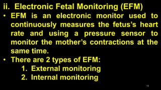
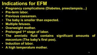
1
Fetal Assessment During Labor
We asses:
1. Amniotic fluid
2. Fetal Heart Rate (FHR):
the primary assessment
2
1. Amniotic fluid analysis:
• Amniotic fluid is a clear, colourless
or pale yellow fluid which is found
within the first 12 days following
conception within the amniotic sac.
• It surrounds the growing baby in the
uterus.
3
4
1. Amniotic fluid analysis:
• Amniotic fluid consists of water and baby's
urine.
• It also contains nutrients, hormones, and
antibodies
• It helps protect the baby from bumps and
injury.
• It maintains constant temperature for the
baby.
• It makes the baby moves easier.
5
1. Amniotic fluid analysis:
• Amniotic fluid volume increases steadily
throughout pregnancy to a maximum of
400–1200 ml at 34–38 weeks.
• If the levels of amniotic fluid levels are
too low or too high, this can pose a
problem.
6
1. Amniotic fluid analysis:
The Amniotic Fluid during Labour:
• Cloudy fluid with foul smell → Infection
• Green Fluid → Fetal Hypoxia due to
meconium (the first stool of the baby)
• Blood streak → Haemorrhage
7
2. Fetal Heart Rate (FHR) Monitoring
• Is the measuring of the fetus’s heart rate
during the labor by using a special
instruments.
• Types and methods of fetal heart
monitoring:
i. Intermittent Auscultation
ii. Electronic Fetal Monitoring (EFM)
8
9
i. Intermittent Auscultation Technique:
• Auscultation is a method of listening
to the fetal heartbeat for about 60
seconds by using a fetal stethoscope
(Fetoscope or Pinard), or a hand-held
Doppler ultrasound device.
10
Fetoscope Pinard Hand-held
Doppler
11
• While listening to the heartbeat, the
doctor also palpates the mother’s uterus
by placing a hand on the abdomen to
measure the contractions.
• Intermittent auscultation should be done:
− Every 15-30 minutes during the active
phase of 1st stage,
− Every 5-15 minutes during the pushing
phase of 2nd stage.
12
Listening
&
Palpation
13
ii. Electronic Fetal Monitoring (EFM)
• EFM is an electronic monitor used to
continuously measures the fetus’s heart
rate and using a pressure sensor to
monitor the mother’s contractions at the
same time.
• There are 2 types of EFM:
1. External monitoring
2. Internal monitoring
14
Indications for EFM
• Pregnancy complications (Diabetes, preeclampsia…)
• Pre-term labor.
• Previous caesarean.
• The baby is smaller than expected.
• Multiple fetuses.
• Overweight mother.
• Prolonged 1st stage of labor.
• The amniotic fluid contains significant amounts of
meconium (The baby's first poo).
• Induction of labor.
• A high temperature mother.
15
1. External monitoring:
• Measuring the heart rate through an
ultrasound device, and measuring the
contractions by using a pressure sensor;
both devises are held against the
mother's belly with a belt.
• The reading of external monitoring is
affecting by changing position.
16
17External Monitoring
18
2. Internal monitoring:
• Measure the heart rate through a wire called
(electrode) contains a needle, inserted
through the vagina and cervix, and placed
under the baby's scalp. And measuring the
contractions with a thin tube inserted into the
uterus.
• Internal monitoring can be done only after the
cervix has dilated to at least 2cm and the
amniotic sac has ruptured.
19
20
Fetal Head Electrode Uterus Tube
21
FHR patterns
• Normal Pattern:
Baseline FHR = 120 – 160 bpm (Beat/min.)
• Tachycardia:
Baseline FHR above 160 bpm
• Bradycardia:
Baseline FHR less than 120 bpm
*Baseline FHR = 10 minutes*
22
Contraction Pattern
• Normal contractions:
5 or fewer contractions in 10 minutes
lasting about 60 seconds in the active
phase.
• Contraction intensity:
30 mmHg in early labor to 70 - 90 mmHg
in the second stage.
23
Normal fetal heart rate with moderate variability
Normal Contraction
24
Causes of Tachycardia
Tachycardia is a FHR above
160 bpm that lasts for at least
10 minutes
• Mother fever
• Mother and fetus infection
• Mother dehydration
• Mother and fetus anemia
• Fetus hypoxemia
• Fetal tachyarrhythmia
• Fetus cardiac abnormalities
Causes of Bradycardia
Fetal bradycardia is a
baseline FHR of less than 110
bpm that lasts for at least 10
minutes
• Mother Dehydration
• Mother Hypotension
• Rupture of uterus or vasa
previa
• Placental abruption
• Medications such as anesthetics
• Fetal hypoxia
• Late or profound hypoxemia
• Umbilical cord occlusion
• Fetal bradyarrythmias
25
Baseline FHR Variability
• The normal fetal heart rate baseline is
from 120 to 160 BPM and has variability
usually with a range of 3-5 bpm from the
baseline.
• The heartbeat will normally go up when
the baby moves or during contractions.
26
Baseline FHR Variability
• Characteristics:
1. Undetectable (less than 5 bpm)
2. Minimal (up to 5 bpm)
3. Moderate (6 to 25 bpm)
4. Marked (more than 25 bpm)
Abnormal if lasts
over 60 min.
27
28
Periodic changes
Periodic changes are accelerations or
decelerations in the FHR that are in
relation to uterine contractions and persist
over time.
29
Accelerations
• Accelerations are transient increases in the FHR
about (15 bpm) above baseline for about (15 sec. to
less than 2 min.) and then return normally to the
base line.
• Prolonged acceleration: Increase in heart rate lasts
for 2 to 10 minutes.
• The presence of accelerations is a sign of normal
fetus, because they are usually associated with
fetus stimulation, such as fetal movement, vaginal
examinations, and contractions.
30
Fetal heart rate accelerations
31
Decelerations
Transitory decrease in the FHR from the
baseline.
1. Early decelerations: the onset and return of a
deceleration is match to the contraction
2. Variable decelerations: variable in the time,
intensity, and duration of a deceleration
3. Late decelerations: the fetal heart rate return
to the baseline after the end of the
contraction
32
Management
1. Early decelerations: No intervention is necessary,
just keep watching. It happen due to uterine
contractions.
2. Variable decelerations: Reposition of the mother,
oxygen mask, and stop oxytocin. It happen due to
Umbilical cord occlusion and problems.
3. Late decelerations: Reposition of the mother,
oxygen mask, stop oxytocin, give IV. It happen
due to reduction in O2
33
Early decelerations
34
Variable decelerations
35
Late decelerations
36
What happens if the fetal heart rate pattern
is abnormal?
• An abnormal fetal heart rate may mean that
the fetus is not getting enough oxygen or that
there are other problems.
• We should first try to find the cause.
• Steps can be taken to help the baby get more
oxygen such as; change the position of the
mother, and giving her an oxygen mask.
37
What happens if the fetal heart rate pattern
is abnormal?
• If the procedures do not work, and the
fetus still has a problem, we should
deliver the baby immediately.
• In this case, the delivery of the baby is
more likely to be by cesarean birth or with
forceps or vacuum delivery.
38
Forceps delivery
Vacuum delivery

Fetal assessment

  • 1.
  • 2.
    Fetal Assessment DuringLabor We asses: 1. Amniotic fluid 2. Fetal Heart Rate (FHR): the primary assessment 2
  • 3.
    1. Amniotic fluidanalysis: • Amniotic fluid is a clear, colourless or pale yellow fluid which is found within the first 12 days following conception within the amniotic sac. • It surrounds the growing baby in the uterus. 3
  • 4.
  • 5.
    1. Amniotic fluidanalysis: • Amniotic fluid consists of water and baby's urine. • It also contains nutrients, hormones, and antibodies • It helps protect the baby from bumps and injury. • It maintains constant temperature for the baby. • It makes the baby moves easier. 5
  • 6.
    1. Amniotic fluidanalysis: • Amniotic fluid volume increases steadily throughout pregnancy to a maximum of 400–1200 ml at 34–38 weeks. • If the levels of amniotic fluid levels are too low or too high, this can pose a problem. 6
  • 7.
    1. Amniotic fluidanalysis: The Amniotic Fluid during Labour: • Cloudy fluid with foul smell → Infection • Green Fluid → Fetal Hypoxia due to meconium (the first stool of the baby) • Blood streak → Haemorrhage 7
  • 8.
    2. Fetal HeartRate (FHR) Monitoring • Is the measuring of the fetus’s heart rate during the labor by using a special instruments. • Types and methods of fetal heart monitoring: i. Intermittent Auscultation ii. Electronic Fetal Monitoring (EFM) 8
  • 9.
    9 i. Intermittent AuscultationTechnique: • Auscultation is a method of listening to the fetal heartbeat for about 60 seconds by using a fetal stethoscope (Fetoscope or Pinard), or a hand-held Doppler ultrasound device.
  • 10.
  • 11.
    11 • While listeningto the heartbeat, the doctor also palpates the mother’s uterus by placing a hand on the abdomen to measure the contractions. • Intermittent auscultation should be done: − Every 15-30 minutes during the active phase of 1st stage, − Every 5-15 minutes during the pushing phase of 2nd stage.
  • 12.
  • 13.
    13 ii. Electronic FetalMonitoring (EFM) • EFM is an electronic monitor used to continuously measures the fetus’s heart rate and using a pressure sensor to monitor the mother’s contractions at the same time. • There are 2 types of EFM: 1. External monitoring 2. Internal monitoring
  • 14.
    14 Indications for EFM •Pregnancy complications (Diabetes, preeclampsia…) • Pre-term labor. • Previous caesarean. • The baby is smaller than expected. • Multiple fetuses. • Overweight mother. • Prolonged 1st stage of labor. • The amniotic fluid contains significant amounts of meconium (The baby's first poo). • Induction of labor. • A high temperature mother.
  • 15.
    15 1. External monitoring: •Measuring the heart rate through an ultrasound device, and measuring the contractions by using a pressure sensor; both devises are held against the mother's belly with a belt. • The reading of external monitoring is affecting by changing position.
  • 16.
  • 17.
  • 18.
    18 2. Internal monitoring: •Measure the heart rate through a wire called (electrode) contains a needle, inserted through the vagina and cervix, and placed under the baby's scalp. And measuring the contractions with a thin tube inserted into the uterus. • Internal monitoring can be done only after the cervix has dilated to at least 2cm and the amniotic sac has ruptured.
  • 19.
  • 20.
  • 21.
    21 FHR patterns • NormalPattern: Baseline FHR = 120 – 160 bpm (Beat/min.) • Tachycardia: Baseline FHR above 160 bpm • Bradycardia: Baseline FHR less than 120 bpm *Baseline FHR = 10 minutes*
  • 22.
    22 Contraction Pattern • Normalcontractions: 5 or fewer contractions in 10 minutes lasting about 60 seconds in the active phase. • Contraction intensity: 30 mmHg in early labor to 70 - 90 mmHg in the second stage.
  • 23.
    23 Normal fetal heartrate with moderate variability Normal Contraction
  • 24.
    24 Causes of Tachycardia Tachycardiais a FHR above 160 bpm that lasts for at least 10 minutes • Mother fever • Mother and fetus infection • Mother dehydration • Mother and fetus anemia • Fetus hypoxemia • Fetal tachyarrhythmia • Fetus cardiac abnormalities Causes of Bradycardia Fetal bradycardia is a baseline FHR of less than 110 bpm that lasts for at least 10 minutes • Mother Dehydration • Mother Hypotension • Rupture of uterus or vasa previa • Placental abruption • Medications such as anesthetics • Fetal hypoxia • Late or profound hypoxemia • Umbilical cord occlusion • Fetal bradyarrythmias
  • 25.
    25 Baseline FHR Variability •The normal fetal heart rate baseline is from 120 to 160 BPM and has variability usually with a range of 3-5 bpm from the baseline. • The heartbeat will normally go up when the baby moves or during contractions.
  • 26.
    26 Baseline FHR Variability •Characteristics: 1. Undetectable (less than 5 bpm) 2. Minimal (up to 5 bpm) 3. Moderate (6 to 25 bpm) 4. Marked (more than 25 bpm) Abnormal if lasts over 60 min.
  • 27.
  • 28.
    28 Periodic changes Periodic changesare accelerations or decelerations in the FHR that are in relation to uterine contractions and persist over time.
  • 29.
    29 Accelerations • Accelerations aretransient increases in the FHR about (15 bpm) above baseline for about (15 sec. to less than 2 min.) and then return normally to the base line. • Prolonged acceleration: Increase in heart rate lasts for 2 to 10 minutes. • The presence of accelerations is a sign of normal fetus, because they are usually associated with fetus stimulation, such as fetal movement, vaginal examinations, and contractions.
  • 30.
    30 Fetal heart rateaccelerations
  • 31.
    31 Decelerations Transitory decrease inthe FHR from the baseline. 1. Early decelerations: the onset and return of a deceleration is match to the contraction 2. Variable decelerations: variable in the time, intensity, and duration of a deceleration 3. Late decelerations: the fetal heart rate return to the baseline after the end of the contraction
  • 32.
    32 Management 1. Early decelerations:No intervention is necessary, just keep watching. It happen due to uterine contractions. 2. Variable decelerations: Reposition of the mother, oxygen mask, and stop oxytocin. It happen due to Umbilical cord occlusion and problems. 3. Late decelerations: Reposition of the mother, oxygen mask, stop oxytocin, give IV. It happen due to reduction in O2
  • 33.
  • 34.
  • 35.
  • 36.
    36 What happens ifthe fetal heart rate pattern is abnormal? • An abnormal fetal heart rate may mean that the fetus is not getting enough oxygen or that there are other problems. • We should first try to find the cause. • Steps can be taken to help the baby get more oxygen such as; change the position of the mother, and giving her an oxygen mask.
  • 37.
    37 What happens ifthe fetal heart rate pattern is abnormal? • If the procedures do not work, and the fetus still has a problem, we should deliver the baby immediately. • In this case, the delivery of the baby is more likely to be by cesarean birth or with forceps or vacuum delivery.
  • 38.