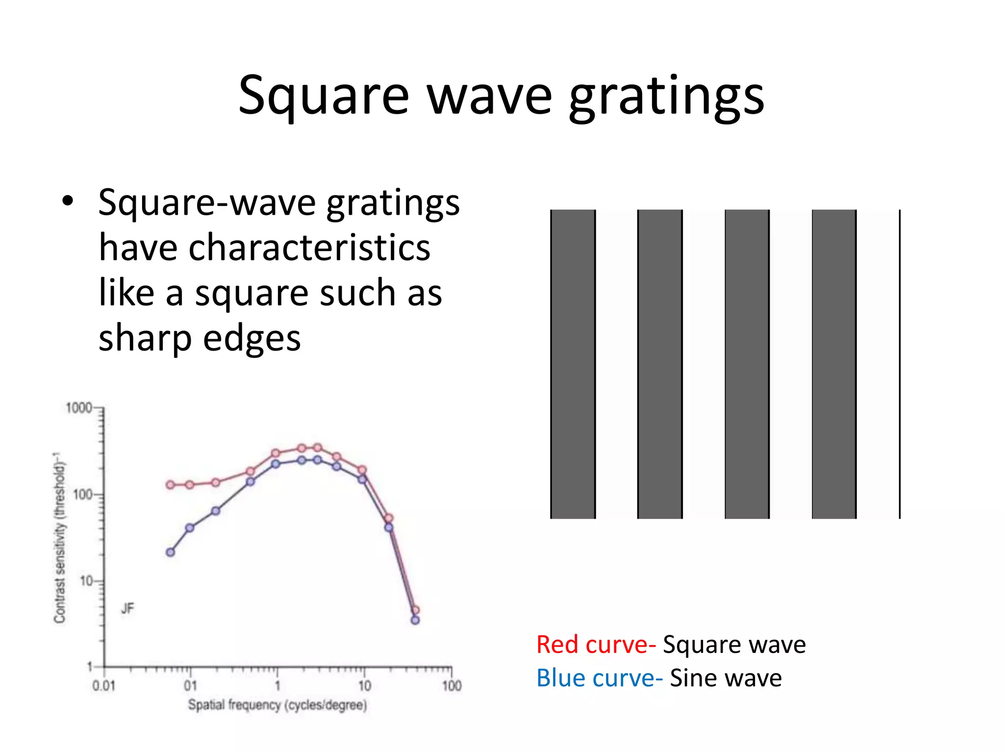
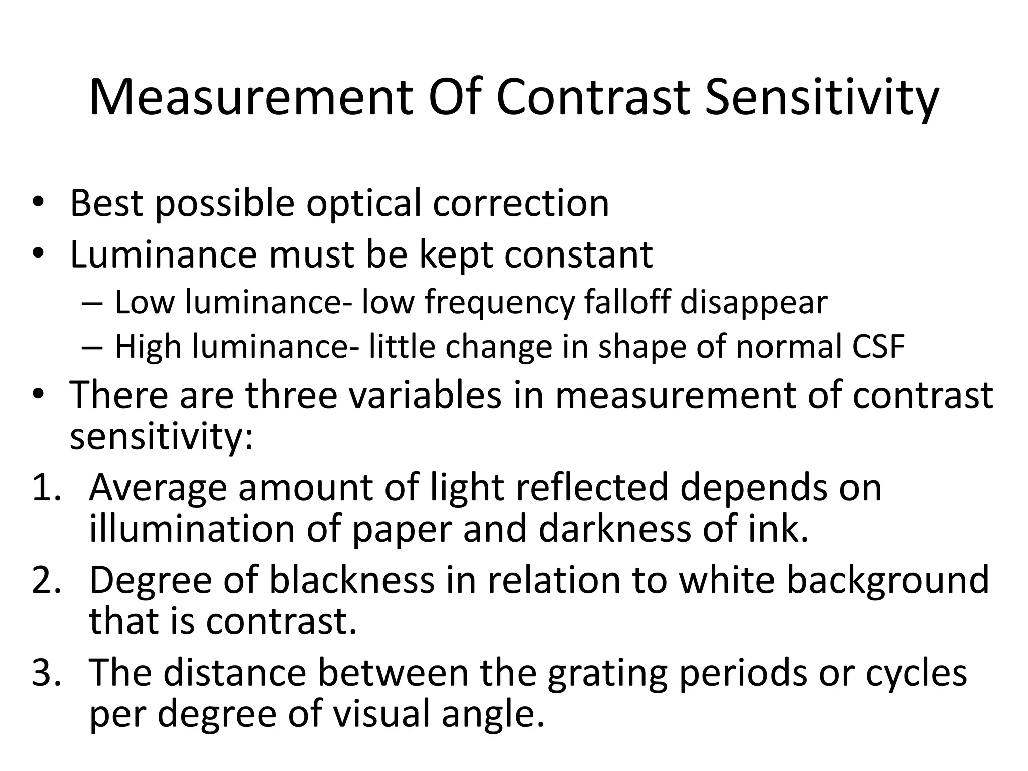
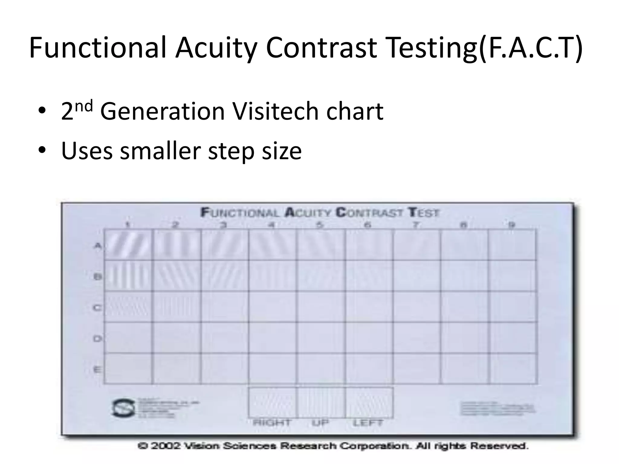
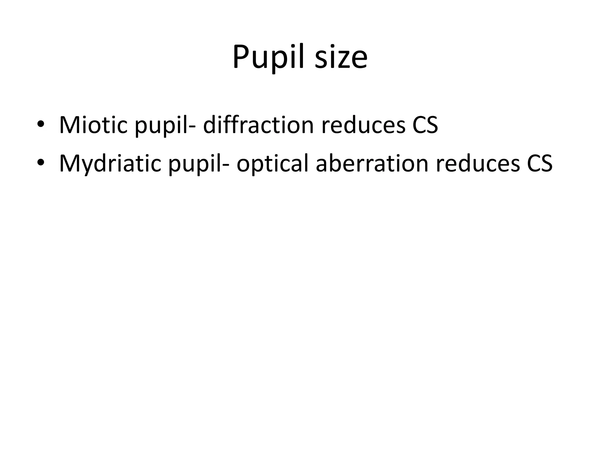
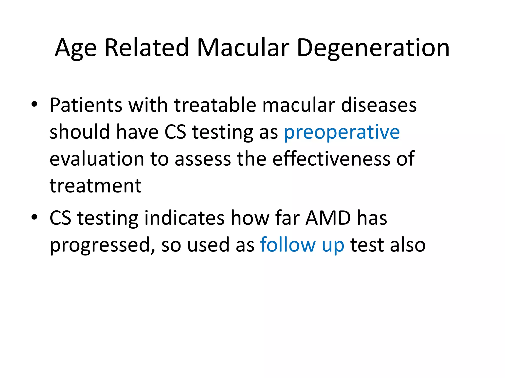
CONTRAST SENSITIVITY
Presenter- Dr. Sharmistha Mukherjee
PGT CNMC&H
Introduction
• Contrast:- is the degree of blackness of target
to whiteness of background.
• Contrast threshold:-is the smallest amount of
contrast required to be able to see the target.
• Contrast sensitivity:- is the reciprocal of the
contrast threshold.
What is Contrast Sensitivity?
• The ability to perceive slight changes in
luminance between regions which are not
separated by definite borders and is just as
important as the ability to perceive sharp
outlines of relatively small objects.
Snellen’s visual acuity versus
contrast sensitivity
• Person may have good VA in Snellen’s chart
due to very high contrast
• But vision drops in the situation of low
contrast in day to day work
• Hence contrast sensitivity is the better
predictor of vision
Gratings
• Equidistant & parallel lines or bars
• One adjacent pair of light and dark
bars makes up one cycle
• The thickness of the grating is
described by their spatial
frequency which expresses the
number of pairs of dark and light
bars subtending an angle of one
degree at the eye
• Units of SF is cycles per degree
(cpd)
ONE CYCLE
Sine wave gratings
• These gratings are lacking in
sharp borders
• Sinusoidal gratings are used
because they are not subject
to defocus aberration,
diffraction, light scatter
Square wave gratings
• Square-wave gratings
have characteristics
like a square such as
sharp edges
Red curve- Square wave
Blue curve- Sine wave
Contrast Sensitivity curve
CONTRAST
SPATIAL FREQUENCYLOW HIGH
LOW
HIGH
Human vision- intermediate spatial frequency range
Cat - low spatial frequencies
Falcon - high spatial frequencies
Contrast Sensitivity Function(CSF)
• The manner in which CS
changes as function of
spatial frequency of the
target is called CSF
• CSF has a peak value of
approximately 1 to 8
cycles/ degree.
• Distinction of high
frequencies not possible
because of limited number
of photoreceptors
Calculation of contrast sensitivity
1. MICHELSON FORMULA:(Lmax - Lmin)/(Lmax + Lmin)
Lmax = Luminance on the lighter surface
Lmin = Luminance on the darker surface
2. WEBERS FORMULA: ( Lb – Lt) /Lb
Lb-Luminance of the background
Lt- Luminance of the target
Weber contrast used in calculating the contrast of letters
Types of contrast sensitivity:
1. Spatial contrast sensitivity
2. Temporal contrast sensitivity
Spatial contrast sensitivity
• Detection of striped pattern at
various levels of contrast and spatial
frequency
• Sine wave gratings of parallel light
and dark bands
• Asked to tell minimum contrast at
which bars can be seen at each
frequency
• Width of the bar is defined as
spatial frequency
• High spatial frequency- narrow bars
• Low spatial frequency- wide bars
Temporal contrast sensitivity
• Temporal contrast sensitivity is measured with
gratings that reverse contrast at various rates over
time.
• Here contrast sensitivity function is generated for
the time related processing in the visual system by
presenting a uniform target field modulated
sinusoidal in time.
Both the system provides more complete and systemic
data on the status of visual performance.
Neural mechanism of contrast
sensitivity
• Campbell and Green gave the concepts that the
retina is not uniform.
• Fovea is specialized for high acuity and is
responsible for high spatial frequencies.
• In the retinal periphery, only low frequency
channels are represented.
• For coarse grating ,central and peripheral retina
have equal CS, but larger the retina is stimulated
greater is the CS. Thus CS is reduced in peripheral
retinal diseases & use of low frequency grating
provide rapid check of peripheral retinal function.
Contribution of M and P pathways
Ganglion cells divided into
– P- Cells :- small , slow conducting axons that input
into parvocellular layers of LGB
– M-Cells :- large , fast conducting axons that input
into magnocellular layers of LGB
• The M-cell information is biased to lower
spatial and mid-temporal frequencies. P-cell
information is biased to mid-higher spatial
and lower temporal frequencies.
Merigan and colleagues represent how the spatial and temporal
contrast sensitivity function is affected by lesions to parvo- and
magno-cellular regions of the LGN of the monkey.
Measurement Of Contrast Sensitivity
• Best possible optical correction
• Luminance must be kept constant
– Low luminance- low frequency falloff disappear
– High luminance- little change in shape of normal CSF
• There are three variables in measurement of contrast
sensitivity:
1. Average amount of light reflected depends on
illumination of paper and darkness of ink.
2. Degree of blackness in relation to white background
that is contrast.
3. The distance between the grating periods or cycles
per degree of visual angle.
• The various methods available to measure CS
include-
Pelli-Robson contrast sensitivity chart
Vision contrast test system (VCTS) by Visitech
Cambridge low contrast gratings
Arden gratings
FACT charts
Vector vision charts (CSV 1000)
Regan charts
Pelli-Robson contrast sensitivity charts
Visitech Contrast Sensitivity Test
• Sine wave
gratings
• Three
orientations:
vertical 90
degrees, 15
degrees
clockwise or
anticlockwise
• VCTS-6500 for
distance testing
and VCTS- 6000
for near testing
• 3m for distance,
40cm for near
Functional Acuity Contrast Testing(F.A.C.T)
• 2nd Generation Visitech chart
• Uses smaller step size
Cambridge Low Contrast Grating
• Performed at a distance of 6m
• Set of 10 plates & 1 page in each pair
contains gratings other is blank
• Shown in order of descending
contrast, stop when first error made
• Then a new series is begun starting 4
plates prior to where the patient
failed to respond.
• Four such series are completed and
the score of each series is noted
(numbered as per the number of
plate read) and added.
• The final total value is converted into
CS from conversion table.
The Arden Plate Test
• Introduced in 1978
• Studied at 57 cm
• Consists of a booklet containing 7 plates(1
screening, 6 diagnostic) with spatial frequency
increasing from 0.2 cpd to 6.4 cpd (each being
double the previous one)
• The contrast changes from top to bottom
• Score 1-20 for each plate
Factors Affecting Contrast Sensitivity
1. Refractive errors
2. Age
3. Cataract
4. Contact lenses
5. Refractive surgery
6. Glaucoma
7. Diabetic retinopathy
8. Optic neuropathies
9. Pituitary adenoma
10. Drugs
11. Toxic chemicals
12. Amblyopia
13. Corneal pathology (edema,
distortion)
Refractive errors
• Decline in contrast sensitivity first at higher
spatial frequencies for mild refractive
disorders
• As the degree of refractive disorder increases,
contrast sensitivity declines at middle to lower
spatial frequencies
Age
• There occurs a definite decrease in contrast
sensitivity with increasing age.
• The pathogenesis of this decline in vision is
likely involves changes in the spherical
aberration of the crystalline lens
Pupil size
• Miotic pupil- diffraction reduces CS
• Mydriatic pupil- optical aberration reduces CS
Cataract
• Early cataracts cause contrast sensitivity losses
similar to refractive disorders at higher spatial
frequencies
• The addition of glare source will exacerbate
the results
• Contrast sensitivity scores may offer
additional information over standard VA tests
in patients with early age-related cataracts
Glaucoma
• Reduce contrast sensitivity for all grating sizes,
mostly at the middle spatial frequencies
• Early progression of Glaucoma can be
measured with CS testing
Diabetic retinopathy
• Before developing DR, diabetic patient may
show lowering of CS
• PRP in PDR & macular laser in DME reduce CS
hence it is important to undergo CS testing
before photocoagulation
Age Related Macular Degeneration
• Patients with treatable macular diseases
should have CS testing as preoperative
evaluation to assess the effectiveness of
treatment
• CS testing indicates how far AMD has
progressed, so used as follow up test also
Amblyopia
•The reciprocal of the
contrast threshold is plotted
against the stimulus spatial
frequency
•The higher the spatial
frequency, the greater the
loss of sensitivity of the
amblyopic (red symbols)
compared with the fellow
fixing eye (blue symbols)
IOLs
CONTRAST
SENSITIVITY
MULTIFOCAL MONOFOCAL
CONTRAST SENSITIVITY AT
DISTANCE(1 ,3,6,12 AND 18
MONTHS)
CONTRAST SENSITIVITY AT
NEAR
L Cataract refractive surgery.2003apr;29(4):703-11
Contrast sensitivity and LASIK
Contrast sensitivity

Contrast sensitivity

  • 1.
    CONTRAST SENSITIVITY Presenter- Dr.Sharmistha Mukherjee PGT CNMC&H
  • 2.
    Introduction • Contrast:- isthe degree of blackness of target to whiteness of background. • Contrast threshold:-is the smallest amount of contrast required to be able to see the target. • Contrast sensitivity:- is the reciprocal of the contrast threshold.
  • 3.
    What is ContrastSensitivity? • The ability to perceive slight changes in luminance between regions which are not separated by definite borders and is just as important as the ability to perceive sharp outlines of relatively small objects.
  • 4.
    Snellen’s visual acuityversus contrast sensitivity • Person may have good VA in Snellen’s chart due to very high contrast • But vision drops in the situation of low contrast in day to day work • Hence contrast sensitivity is the better predictor of vision
  • 5.
    Gratings • Equidistant &parallel lines or bars • One adjacent pair of light and dark bars makes up one cycle • The thickness of the grating is described by their spatial frequency which expresses the number of pairs of dark and light bars subtending an angle of one degree at the eye • Units of SF is cycles per degree (cpd) ONE CYCLE
  • 6.
    Sine wave gratings •These gratings are lacking in sharp borders • Sinusoidal gratings are used because they are not subject to defocus aberration, diffraction, light scatter
  • 7.
    Square wave gratings •Square-wave gratings have characteristics like a square such as sharp edges Red curve- Square wave Blue curve- Sine wave
  • 8.
    Contrast Sensitivity curve CONTRAST SPATIALFREQUENCYLOW HIGH LOW HIGH Human vision- intermediate spatial frequency range Cat - low spatial frequencies Falcon - high spatial frequencies
  • 9.
    Contrast Sensitivity Function(CSF) •The manner in which CS changes as function of spatial frequency of the target is called CSF • CSF has a peak value of approximately 1 to 8 cycles/ degree. • Distinction of high frequencies not possible because of limited number of photoreceptors
  • 10.
    Calculation of contrastsensitivity 1. MICHELSON FORMULA:(Lmax - Lmin)/(Lmax + Lmin) Lmax = Luminance on the lighter surface Lmin = Luminance on the darker surface 2. WEBERS FORMULA: ( Lb – Lt) /Lb Lb-Luminance of the background Lt- Luminance of the target Weber contrast used in calculating the contrast of letters
  • 11.
    Types of contrastsensitivity: 1. Spatial contrast sensitivity 2. Temporal contrast sensitivity
  • 12.
    Spatial contrast sensitivity •Detection of striped pattern at various levels of contrast and spatial frequency • Sine wave gratings of parallel light and dark bands • Asked to tell minimum contrast at which bars can be seen at each frequency • Width of the bar is defined as spatial frequency • High spatial frequency- narrow bars • Low spatial frequency- wide bars
  • 13.
    Temporal contrast sensitivity •Temporal contrast sensitivity is measured with gratings that reverse contrast at various rates over time. • Here contrast sensitivity function is generated for the time related processing in the visual system by presenting a uniform target field modulated sinusoidal in time. Both the system provides more complete and systemic data on the status of visual performance.
  • 14.
    Neural mechanism ofcontrast sensitivity • Campbell and Green gave the concepts that the retina is not uniform. • Fovea is specialized for high acuity and is responsible for high spatial frequencies. • In the retinal periphery, only low frequency channels are represented. • For coarse grating ,central and peripheral retina have equal CS, but larger the retina is stimulated greater is the CS. Thus CS is reduced in peripheral retinal diseases & use of low frequency grating provide rapid check of peripheral retinal function.
  • 15.
    Contribution of Mand P pathways Ganglion cells divided into – P- Cells :- small , slow conducting axons that input into parvocellular layers of LGB – M-Cells :- large , fast conducting axons that input into magnocellular layers of LGB • The M-cell information is biased to lower spatial and mid-temporal frequencies. P-cell information is biased to mid-higher spatial and lower temporal frequencies.
  • 16.
    Merigan and colleaguesrepresent how the spatial and temporal contrast sensitivity function is affected by lesions to parvo- and magno-cellular regions of the LGN of the monkey.
  • 17.
    Measurement Of ContrastSensitivity • Best possible optical correction • Luminance must be kept constant – Low luminance- low frequency falloff disappear – High luminance- little change in shape of normal CSF • There are three variables in measurement of contrast sensitivity: 1. Average amount of light reflected depends on illumination of paper and darkness of ink. 2. Degree of blackness in relation to white background that is contrast. 3. The distance between the grating periods or cycles per degree of visual angle.
  • 18.
    • The variousmethods available to measure CS include- Pelli-Robson contrast sensitivity chart Vision contrast test system (VCTS) by Visitech Cambridge low contrast gratings Arden gratings FACT charts Vector vision charts (CSV 1000) Regan charts
  • 19.
  • 20.
    Visitech Contrast SensitivityTest • Sine wave gratings • Three orientations: vertical 90 degrees, 15 degrees clockwise or anticlockwise • VCTS-6500 for distance testing and VCTS- 6000 for near testing • 3m for distance, 40cm for near
  • 21.
    Functional Acuity ContrastTesting(F.A.C.T) • 2nd Generation Visitech chart • Uses smaller step size
  • 22.
    Cambridge Low ContrastGrating • Performed at a distance of 6m • Set of 10 plates & 1 page in each pair contains gratings other is blank • Shown in order of descending contrast, stop when first error made • Then a new series is begun starting 4 plates prior to where the patient failed to respond. • Four such series are completed and the score of each series is noted (numbered as per the number of plate read) and added. • The final total value is converted into CS from conversion table.
  • 23.
    The Arden PlateTest • Introduced in 1978 • Studied at 57 cm • Consists of a booklet containing 7 plates(1 screening, 6 diagnostic) with spatial frequency increasing from 0.2 cpd to 6.4 cpd (each being double the previous one) • The contrast changes from top to bottom • Score 1-20 for each plate
  • 24.
    Factors Affecting ContrastSensitivity 1. Refractive errors 2. Age 3. Cataract 4. Contact lenses 5. Refractive surgery 6. Glaucoma 7. Diabetic retinopathy 8. Optic neuropathies 9. Pituitary adenoma 10. Drugs 11. Toxic chemicals 12. Amblyopia 13. Corneal pathology (edema, distortion)
  • 25.
    Refractive errors • Declinein contrast sensitivity first at higher spatial frequencies for mild refractive disorders • As the degree of refractive disorder increases, contrast sensitivity declines at middle to lower spatial frequencies
  • 26.
    Age • There occursa definite decrease in contrast sensitivity with increasing age. • The pathogenesis of this decline in vision is likely involves changes in the spherical aberration of the crystalline lens
  • 27.
    Pupil size • Mioticpupil- diffraction reduces CS • Mydriatic pupil- optical aberration reduces CS
  • 28.
    Cataract • Early cataractscause contrast sensitivity losses similar to refractive disorders at higher spatial frequencies • The addition of glare source will exacerbate the results • Contrast sensitivity scores may offer additional information over standard VA tests in patients with early age-related cataracts
  • 29.
    Glaucoma • Reduce contrastsensitivity for all grating sizes, mostly at the middle spatial frequencies • Early progression of Glaucoma can be measured with CS testing
  • 30.
    Diabetic retinopathy • Beforedeveloping DR, diabetic patient may show lowering of CS • PRP in PDR & macular laser in DME reduce CS hence it is important to undergo CS testing before photocoagulation
  • 31.
    Age Related MacularDegeneration • Patients with treatable macular diseases should have CS testing as preoperative evaluation to assess the effectiveness of treatment • CS testing indicates how far AMD has progressed, so used as follow up test also
  • 32.
    Amblyopia •The reciprocal ofthe contrast threshold is plotted against the stimulus spatial frequency •The higher the spatial frequency, the greater the loss of sensitivity of the amblyopic (red symbols) compared with the fellow fixing eye (blue symbols)
  • 33.
    IOLs CONTRAST SENSITIVITY MULTIFOCAL MONOFOCAL CONTRAST SENSITIVITYAT DISTANCE(1 ,3,6,12 AND 18 MONTHS) CONTRAST SENSITIVITY AT NEAR L Cataract refractive surgery.2003apr;29(4):703-11
  • 34.