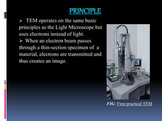
Seminar
on
“ELECTRON MICROSCOPE AND ITS
FORENSIC APPLICATION”
Submitted to- Submitted by-
“Department of Name: Pallavi Kumari
Forensic Science”
Class: M. sc. Forensic science
Sam Higginbottom University of Agriculture, Technology and Sciences
CONTENTS
 Electron microscope
-Introduction
-Types
 Differences between Light Microscope and Electron Microscope.
 A) Transmission Electron Microscope
-Principle, Instrumentation, Working, Sample preparation,
Application, Advantages, Disadvantages.
 B) Scanning Electron Microscope
-Principle, Instrumentation, Working, Sample preparation,
Application, Advantages, Disadvantages.
 Differences between TEM and SEM.
 Some Modifications of TEM.
 Forensic Applications of electron microscope.
 Case report
ELECTRON MICROSCOPE
 An Electron Microscope is a type of
microscope that uses a beam of accelerated
electrons to illuminate a specimen and
creates an enlarged image.
 The wavelength of an electron can be upto
1,00,000 times shorter than that of visible
light photons.
 Thus, Electron microscopes have much
greater resolving power than light
microscopes and can obtain much higher
magnification.
FIG: First Electron microscope
 Some electron microscopes can magnify specimens upto 2
million times, while the best light microscopes are limited
to magnification of 2,000 times.
 The first electron microscope prototype was built in 1931
by German engineers Ernst Ruska and Max Knoll.
 It offers unique possibilities to gain insight into structure,
topology, morphology, composition of materials, etc.
DIFFERENCES BETWEEN LIGHTMICROSCOPEAND
ELECTRON MICROSCOPE
LIGHT MICROSCOPE ELECTRON MICROSCOPE
Illuminating source is the light. Illuminating source is the beam of
electrons.
Specimen preparation takes usually
few minutes to hours.
Specimen preparation takes usually
few days.
Live or dead specimens may be
seen.
Only dead or dried specimens are
seen.
All lenses are made up of glasses. All lenses are electromagnetic.
Has low resolving power. Has high resolving power, about
250 times higher than light
microscope.
It has a magnification of 500X to
1500X.
It has a magnification of 1,00,000X
to 3,00,000X.
Vacuum is not required. Vacuum is essential for its
operation.
 TYPES:
There are 2 basic types of Electron
Microscope-
1. Transmission Electron Microscope
(TEM)- allows only the study of internal
structures.
2. Scanning Electron Microscope
(SEM)- used to visualize the surface of
objects.
 The TEM was the first type of Electron
Microscope to be developed and is patterned
exactly on the Light Transmission Microscope,
except that a focused beam of electrons is used
instead of light to “see through” the specimen.
 It was developed by Max Knoll and Ernst Ruska
in Germany in 1931.
 Transmitted electrons focused by lens system to
form a 2- Dimensional magnified image.
 TEM operates on the same basic
principles as the Light Microscope but
uses electrons instead of light.
 When an electron beam passes
through a thin-section specimen of a
material, electrons are transmitted and
thus creates an image.
FIG: First practical TEM
FIG: Main Components of TEM
The TEM can be broken down into a few components, these are:
1) Electron source;
a. Electron gun- It produces the electron beam.
b. Condenser system- Focuses the beam onto the object.
2) The Image- Producing System:
a. Movable specimen stage.
b. The Objective lens.
c. The Intermediate and projector lenses, which focus the
electrons passing through the specimen to form a real, highly
magnified image.
3) The Image- Recording System:
a. A Fluorescent Screen, for viewing and focusing the image.
b. A Digital Camera, for permanent records.
FIG: Diagram to represent TEM’s working
1. Fixation 2. Rinsing
3. Secondary
Fixation
4.
Dehydration
5. Infiltration
6. Polishing
7. Cutting 8. Staining
 A Transmission Electron Microscopy (TEM) is ideal for a
number of different fields such as life sciences,
nanotechnology, medical, biological and material research,
forensic analysis, gemology and metallurgy as well as
industry and education.
 The main application of Transmission Electron
Microscopy (TEM) is to provide high magnification
images of the internal structure of a sample.
 A TEM operator can investigate the crystalline structure of
an object, see the stress or internal fractures of a sample,
or even view contamination within a sample through the
use of diffraction patterns.
i) High-quality images can be obtained with TEMs.
ii) TEMs have various applications at high spatial resolutions in different
fields such as Scientific, educational and industrial fields.
iii) TEMs provide the highest magnification in microscope field.
iv) TEMs can provide information about surface features, shape, size and
structure.
i) The instruments are very large and expensive.
ii) TEMs require special housing and maintenance because they are
sensitive to mechanical vibration, fluctuation of electromagnetic
fields, and variation of cooling water .
iii) Sample preparations from bulk materials are normally very time-
consuming.
iv) Special training is needed for tool operation and data analysis.
 A Scanning Electron Microscope (SEM) is a type of electron
microscope that images a sample by scanning it with a high-
energy beam of electrons in a raster scan pattern. The electrons
interact with the atoms that make up the sample producing
signals that contain information about the sample’s surface .
 The first Scanning Electron Microscope (SEM) debuted in
1938 (Manfred Von Ardenne) with the first commercial
instruments around 1965.
 The high spatial resolution of a SEM makes it a powerful tool
to characterize a wide range of specimens at the nanometer to
micrometer length scale.
 The basic principle is that a beam
of electrons is generated by a suitable
source, typically a tungsten filament
or a field emission gun.
 The electron beam is accelerated
through a high voltage and pass through
a system of apertures and electromagnetic
lenses to produce a thin beam of electrons.
 Then the beam scans the surface of the
specimen. Electrons are emitted from the
specimen by the action of scanning beam FIG: First practical SEM
and collected by a suitably positioned detector.
FIG: Main Components of SEM
A. Electron optical system (to produce electrons):
 Electron gun
 Condenser lens
 Objective lens
B. Specimen stage (to place the specimen).
C. Secondary electron detector (to collect
secondary electron).
D. Image display and recording unit.
E. Vacuum system (The electron optical system
and a space surrounding the specimen are kept in
vacuum).
FIG: Diagram to represent SEM’s working
1.
• Sample cleaning.
2.
• Sample Fixation and Dehydration.
3.
• Drying.
4.
• Sample preparation of tissue sections.
5.
• Sample stubs, adhesives, and mounting approach.
6. • Sample storage.
 Criminal and other forensic investigations utilize SEMs to
uncover evidence and gain further forensic insight. Uses
include: analysis of gunshot residue, jewellery
examination, bullet marking comparison, handwriting and
print analysis, examination of banknote authenticity, paint
particle and fiber analysis, filament bulb analysis in traffic
incidents.
 In biological sciences, SEMs can be used on anything
from insects and animal tissue to bacteria and viruses.
Uses include: Measuring the effect of climate change of
species, identifying new bacteria and virulent strains,
vaccination testing, uncovering new species, work within
the field of genetics.
 It gives detailed 3D and topological imaging and the versatile
information garnered from different detectors.
 This instrument works very fast.
 Modern SEMs allow for the generation of data in digital form.
 Most SEM samples require minimal preparation actions.
 SEMs are expensive and large.
 Special training is required to operate an SEM.
 SEMs are limited to solid samples.
 SEMs carry a small risk of radiation exposure associated with
the electrons that scatter from beneath the sample surface.
TEM SEM
TEM is based on transmitted
electrons .
SEM is based on scattered
electrons.
TEM has much higher resolution
than SEM. It can resolve objects
as close as 1 nm .
SEM can resolve objects as close
as 20 nm.
TEM provides a 2-dimensional
picture.
SEM provides a 3-dimensional
image
The magnifying power of TEM is
up to 2 million times.
The magnifying power of SEM is
up to 50,000X.
With TEM only small amount of
sample can be analyzed at a time.
SEM allows for large amount of
sample to be analyzed at a time.
The capabilities of the TEM can be further extended
by additional stages and detectors, sometimes
incorporated in the same microscope. These are
certain modifications of Transmission Electron
Microscope:
1. Scanning Transmission Electron Microscope
(STEM).
2. Analytical Electron Microscope (AEM).
3. Reflection Electron Microscope (REM).
4. Low-Voltage Electron Microscope (LVEM).
Since SEMs offer the ability to examine a wide
range of materials at high and low magnification
without sacrificing depth of focus, their use in
forensic sciences makes it possible to draw
conclusions, identify material origins and
contribute to a body of evidence in criminal and
legal matters. Various applications includes:
 Gunshot residue analysis.
 Firearms identification (bullet markings
comparison).
 Investigation of gemstones and jewellery.
Contd……
 Examination of paint particles and fibres.
 Filament bulb investigations at traffic accidents.
 Handwriting and print examination / forgery.
 Counterfeit bank notes.
 Trace comparison.
 Examination of non-conducting materials.
Note: The Phenom desktop SEM GSR
instrument is specifically designed for
automated gunshot residue analysis.
 "The objective lens of aTEM, the heart of the electron
microscope". rodenburg.org
 Alberts, Bruce (2008). Molecular biology of the cell (5th ed.).
NewYork: Garland Science. ISBN 978-0815341116.
 Champness, P. E. (2001). Electron Diffraction in theTransmission
Electron Microscope. Garland Science. ISBN 978-1859961476
 Egerton, R (2005). Physical principles of electron microscopy.
Springer. ISBN 978-0-387-25800-3.
 Fultz, B & Howe, J (2007).Transmission Electron Microscopy
and Diffractometry of Materials. Springer. ISBN 978-3-540-
73885-5.
 Haque, M. A. & Saif, M.T. A. (2001). "In-situ tensile testing of
nano-scale specimens in SEM andTEM". Experimental
Mechanics. 42: 123. doi:10.1007/BF02411059.
 Hawkes, P. (Ed.) (1985).The beginnings of Electron
Microscopy. Academic Press. ISBN 978-0120145782.
ELECTRON MICROSCOPE AND ITS APPLICATION.pptx

ELECTRON MICROSCOPE AND ITS APPLICATION.pptx

  • 1.
    Seminar on “ELECTRON MICROSCOPE ANDITS FORENSIC APPLICATION” Submitted to- Submitted by- “Department of Name: Pallavi Kumari Forensic Science” Class: M. sc. Forensic science Sam Higginbottom University of Agriculture, Technology and Sciences
  • 2.
    CONTENTS  Electron microscope -Introduction -Types Differences between Light Microscope and Electron Microscope.  A) Transmission Electron Microscope -Principle, Instrumentation, Working, Sample preparation, Application, Advantages, Disadvantages.  B) Scanning Electron Microscope -Principle, Instrumentation, Working, Sample preparation, Application, Advantages, Disadvantages.  Differences between TEM and SEM.  Some Modifications of TEM.  Forensic Applications of electron microscope.  Case report
  • 3.
    ELECTRON MICROSCOPE  AnElectron Microscope is a type of microscope that uses a beam of accelerated electrons to illuminate a specimen and creates an enlarged image.  The wavelength of an electron can be upto 1,00,000 times shorter than that of visible light photons.  Thus, Electron microscopes have much greater resolving power than light microscopes and can obtain much higher magnification. FIG: First Electron microscope
  • 4.
     Some electronmicroscopes can magnify specimens upto 2 million times, while the best light microscopes are limited to magnification of 2,000 times.  The first electron microscope prototype was built in 1931 by German engineers Ernst Ruska and Max Knoll.  It offers unique possibilities to gain insight into structure, topology, morphology, composition of materials, etc.
  • 5.
    DIFFERENCES BETWEEN LIGHTMICROSCOPEAND ELECTRONMICROSCOPE LIGHT MICROSCOPE ELECTRON MICROSCOPE Illuminating source is the light. Illuminating source is the beam of electrons. Specimen preparation takes usually few minutes to hours. Specimen preparation takes usually few days. Live or dead specimens may be seen. Only dead or dried specimens are seen. All lenses are made up of glasses. All lenses are electromagnetic. Has low resolving power. Has high resolving power, about 250 times higher than light microscope. It has a magnification of 500X to 1500X. It has a magnification of 1,00,000X to 3,00,000X. Vacuum is not required. Vacuum is essential for its operation.
  • 6.
     TYPES: There are2 basic types of Electron Microscope- 1. Transmission Electron Microscope (TEM)- allows only the study of internal structures. 2. Scanning Electron Microscope (SEM)- used to visualize the surface of objects.
  • 7.
     The TEMwas the first type of Electron Microscope to be developed and is patterned exactly on the Light Transmission Microscope, except that a focused beam of electrons is used instead of light to “see through” the specimen.  It was developed by Max Knoll and Ernst Ruska in Germany in 1931.  Transmitted electrons focused by lens system to form a 2- Dimensional magnified image.
  • 8.
     TEM operateson the same basic principles as the Light Microscope but uses electrons instead of light.  When an electron beam passes through a thin-section specimen of a material, electrons are transmitted and thus creates an image. FIG: First practical TEM
  • 9.
  • 10.
    The TEM canbe broken down into a few components, these are: 1) Electron source; a. Electron gun- It produces the electron beam. b. Condenser system- Focuses the beam onto the object. 2) The Image- Producing System: a. Movable specimen stage. b. The Objective lens. c. The Intermediate and projector lenses, which focus the electrons passing through the specimen to form a real, highly magnified image. 3) The Image- Recording System: a. A Fluorescent Screen, for viewing and focusing the image. b. A Digital Camera, for permanent records.
  • 11.
    FIG: Diagram torepresent TEM’s working
  • 12.
    1. Fixation 2.Rinsing 3. Secondary Fixation 4. Dehydration 5. Infiltration 6. Polishing 7. Cutting 8. Staining
  • 13.
     A TransmissionElectron Microscopy (TEM) is ideal for a number of different fields such as life sciences, nanotechnology, medical, biological and material research, forensic analysis, gemology and metallurgy as well as industry and education.  The main application of Transmission Electron Microscopy (TEM) is to provide high magnification images of the internal structure of a sample.  A TEM operator can investigate the crystalline structure of an object, see the stress or internal fractures of a sample, or even view contamination within a sample through the use of diffraction patterns.
  • 14.
    i) High-quality imagescan be obtained with TEMs. ii) TEMs have various applications at high spatial resolutions in different fields such as Scientific, educational and industrial fields. iii) TEMs provide the highest magnification in microscope field. iv) TEMs can provide information about surface features, shape, size and structure. i) The instruments are very large and expensive. ii) TEMs require special housing and maintenance because they are sensitive to mechanical vibration, fluctuation of electromagnetic fields, and variation of cooling water . iii) Sample preparations from bulk materials are normally very time- consuming. iv) Special training is needed for tool operation and data analysis.
  • 15.
     A ScanningElectron Microscope (SEM) is a type of electron microscope that images a sample by scanning it with a high- energy beam of electrons in a raster scan pattern. The electrons interact with the atoms that make up the sample producing signals that contain information about the sample’s surface .  The first Scanning Electron Microscope (SEM) debuted in 1938 (Manfred Von Ardenne) with the first commercial instruments around 1965.  The high spatial resolution of a SEM makes it a powerful tool to characterize a wide range of specimens at the nanometer to micrometer length scale.
  • 16.
     The basicprinciple is that a beam of electrons is generated by a suitable source, typically a tungsten filament or a field emission gun.  The electron beam is accelerated through a high voltage and pass through a system of apertures and electromagnetic lenses to produce a thin beam of electrons.  Then the beam scans the surface of the specimen. Electrons are emitted from the specimen by the action of scanning beam FIG: First practical SEM and collected by a suitably positioned detector.
  • 17.
  • 18.
    A. Electron opticalsystem (to produce electrons):  Electron gun  Condenser lens  Objective lens B. Specimen stage (to place the specimen). C. Secondary electron detector (to collect secondary electron). D. Image display and recording unit. E. Vacuum system (The electron optical system and a space surrounding the specimen are kept in vacuum).
  • 19.
    FIG: Diagram torepresent SEM’s working
  • 20.
    1. • Sample cleaning. 2. •Sample Fixation and Dehydration. 3. • Drying. 4. • Sample preparation of tissue sections. 5. • Sample stubs, adhesives, and mounting approach. 6. • Sample storage.
  • 21.
     Criminal andother forensic investigations utilize SEMs to uncover evidence and gain further forensic insight. Uses include: analysis of gunshot residue, jewellery examination, bullet marking comparison, handwriting and print analysis, examination of banknote authenticity, paint particle and fiber analysis, filament bulb analysis in traffic incidents.  In biological sciences, SEMs can be used on anything from insects and animal tissue to bacteria and viruses. Uses include: Measuring the effect of climate change of species, identifying new bacteria and virulent strains, vaccination testing, uncovering new species, work within the field of genetics.
  • 22.
     It givesdetailed 3D and topological imaging and the versatile information garnered from different detectors.  This instrument works very fast.  Modern SEMs allow for the generation of data in digital form.  Most SEM samples require minimal preparation actions.  SEMs are expensive and large.  Special training is required to operate an SEM.  SEMs are limited to solid samples.  SEMs carry a small risk of radiation exposure associated with the electrons that scatter from beneath the sample surface.
  • 23.
    TEM SEM TEM isbased on transmitted electrons . SEM is based on scattered electrons. TEM has much higher resolution than SEM. It can resolve objects as close as 1 nm . SEM can resolve objects as close as 20 nm. TEM provides a 2-dimensional picture. SEM provides a 3-dimensional image The magnifying power of TEM is up to 2 million times. The magnifying power of SEM is up to 50,000X. With TEM only small amount of sample can be analyzed at a time. SEM allows for large amount of sample to be analyzed at a time.
  • 24.
    The capabilities ofthe TEM can be further extended by additional stages and detectors, sometimes incorporated in the same microscope. These are certain modifications of Transmission Electron Microscope: 1. Scanning Transmission Electron Microscope (STEM). 2. Analytical Electron Microscope (AEM). 3. Reflection Electron Microscope (REM). 4. Low-Voltage Electron Microscope (LVEM).
  • 25.
    Since SEMs offerthe ability to examine a wide range of materials at high and low magnification without sacrificing depth of focus, their use in forensic sciences makes it possible to draw conclusions, identify material origins and contribute to a body of evidence in criminal and legal matters. Various applications includes:  Gunshot residue analysis.  Firearms identification (bullet markings comparison).  Investigation of gemstones and jewellery.
  • 26.
    Contd……  Examination ofpaint particles and fibres.  Filament bulb investigations at traffic accidents.  Handwriting and print examination / forgery.  Counterfeit bank notes.  Trace comparison.  Examination of non-conducting materials. Note: The Phenom desktop SEM GSR instrument is specifically designed for automated gunshot residue analysis.
  • 27.
     "The objectivelens of aTEM, the heart of the electron microscope". rodenburg.org  Alberts, Bruce (2008). Molecular biology of the cell (5th ed.). NewYork: Garland Science. ISBN 978-0815341116.  Champness, P. E. (2001). Electron Diffraction in theTransmission Electron Microscope. Garland Science. ISBN 978-1859961476  Egerton, R (2005). Physical principles of electron microscopy. Springer. ISBN 978-0-387-25800-3.  Fultz, B & Howe, J (2007).Transmission Electron Microscopy and Diffractometry of Materials. Springer. ISBN 978-3-540- 73885-5.  Haque, M. A. & Saif, M.T. A. (2001). "In-situ tensile testing of nano-scale specimens in SEM andTEM". Experimental Mechanics. 42: 123. doi:10.1007/BF02411059.  Hawkes, P. (Ed.) (1985).The beginnings of Electron Microscopy. Academic Press. ISBN 978-0120145782.