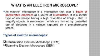
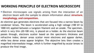
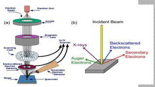
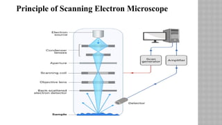
Electron
Microscopy
prepared by:
Emad Adnan
WHAT IS AN ELECTRON MICROSCOPE?
 An electron microscope is a microscope that uses a beam of
accelerated electrons as a source of illumination. It is a special
type of microscope having a high resolution of images, able to
magnify objects in nanometers, which are formed by controlled
use of electrons in a vacuum captured on a phosphorescent
screen.
•Types of electron microscopes:
Transmission Electron Microscope (TEM)
Scanning Electron Microscope (SEM)
WORKING PRINCIPLE OF ELECTRON MICROSCOPE
 Electron microscopes use signals arising from the interaction of an
electron beam with the sample to obtain information about structure,
morphology, and composition.
An electron gun generates electrons that are focused into a narrow beam by
condenser lenses. The beam is accelerated using a high voltage (100 kV to
1000 kV) applied between a tungsten filament and an anode. The specimen,
which is very thin (20-100 nm), is placed on a holder. As the electron beam
passes through, electrons scatter based on the specimen's thickness and
refractive index; denser areas scatter more electrons and appear darker,
while less dense areas appear brighter. The objective lens then creates a
magnified intermediate image, which is further magnified by ocular lenses to
produce the final image.
PARTS OF ELECTRON MICROSCOPE
 Electron Microscope is in the form of a tall vacuum column that is vertically mounted. It has the following components:
1. Electron gun
• The electron gun is a heated tungsten filament, which generates electrons.
2. Electromagnetic lenses
• The condenser lens focuses the electron beam on the specimen. A second condenser lens forms the electrons into a
thin tight beam.
• The electron beam coming out of the specimen passes down the second of magnetic coils called the objective lens,
which has high power and forms the intermediate magnified image.
• The third set of magnetic lenses called projector (ocular) lenses produce the final further magnified image.
• Each of these lenses acts as an image magnifier all the while maintaining an incredible level of detail and resolution.
3. Specimen Holder
• The specimen holder is an extremely thin film of carbon or collodion held by a metal grid.
4. Image viewing and Recording System
• The final image is projected on a fluorescent screen.
• Below the fluorescent screen is a camera for recording the image.
PARTS OF ELECTRON MICROSCOPE
TRANSMISSION ELECTRON MICROSCOPE
(TEM)
 This is a powerful electron microscope that uses a beam of
electrons to focus on a specimen producing a highly magnified
and detailed image of the specimen.
 The working principle of the Transmission Electron Microscope
(TEM) is similar to the light microscope. The major difference is
that light microscopes use light rays to focus and produce an
image while the TEM uses a beam of electrons to focus on the
specimen, to produce an image.
LIMITATIONS OF TRANSMISSION ELECTRON
MICROSCOPE (TEM)
1.Generally, the TEMs are very expensive to purchase
2.They are very big to handle.
3.The preparation of specimens to be viewed under the
TEM is very tedious.
4.The use of chemical fixations, dehydrators, and
embedment's can cause the dangers of artifacts.
5.They are laborious to maintain.
6.It requires a constant inflow of voltage to operate.
SCANNING ELECTRON MICROSCOPE (SEM)
is a type of electron microscope that scans surfaces of
microorganisms that uses a beam of electrons moving
at low energy to focus and scan specimens. The
development of electron microscopes was due to the
inefficiency of the wavelength of light microscopes.
electron microscopes have very short wavelengths in
comparison to the light microscope which enables
better resolution power.
PRINCIPLE OF SCANNING ELECTRON MICROSCOPE (SEM)
 Unlike the Transmission Electron Microscope which uses transmitted electrons,
the scanning electron Microscope uses emitted electrons. The Scanning electron
microscope works on the principle of applying kinetic energy to produce signals
on the interaction of the electrons. These electrons are secondary electrons,
backscattered electrons, and diffracted backscattered electrons which are used to
view crystallized elements and photons. Secondary and backscattered electrons
are used to produce an image. The secondary electrons are emitted from the
specimen play the primary role of detecting the morphology and topography of
the specimen while the backscattered electrons show contrast in the composition
of the elements of the specimen.
Principle of Scanning Electron Microscope
ADVANTAGES OF ELECTRON MICROSCOPY
 Electron microscopy has several main advantages. These include:
• Magnification and higher resolution – as electrons rather than light waves are used, it can be
used to analyze structures which cannot otherwise be seen. The resolution of electron
microscopy images is in the range of up to 0.2 nm, which is 1000x more detailed than light
microscopy.
• Diverse applications – Electron microscopy has a diverse range of applications in many
different fields of research including technology, industry, biomedical science and chemistry.
Examples of applications include semiconductor inspection, computer chip manufacture,
quality control and assurance, analysis of atomic structures, and drug development.
• High-quality images – With proper training, an electron microscope operator can use the
system to produce highly detailed images of structures which are of a high quality, revealing
complex and delicate structures that other techniques may struggle to reproduce
DISADVANTAGES OF ELECTRON MICROSCOPY
• Inability to analyze live specimens – As electrons are
easily scattered by other molecules in the air, samples
must be analyzed in a vacuum. This means that live
specimens cannot be studied by this technique. This
means that biological interactions cannot be properly
observed, which limits the applications of electron
microscopy in biological research.
• Black and white images – Only black and white
images can be produced by an electron microscope.
Images must be falsely colorized.
• Artefacts – These may be present in the image produced.
Artefacts are left over from sample preparation and require
specialized knowledge of sample preparation techniques to
avoid.
• Cost – Electron microscopes are expensive pieces of highly
specialized equipment. As most projects have limited budgets, it
may prove detrimental to use an electron microscope in the
research. However, running costs can be similar to alternatives
such as confocal light microscopes, so the investment in a basic
electron microscope is still worth considering even if budgetary
concerns are a major factor in decisions against utilizing the
technology.
DISADVANTAGES OF ELECTRON
MICROSCOPY
• Size – Despite the advantages in technology over the years,
electron microscopes are still large, bulky pieces of
equipment which require plenty of space in a laboratory. Also,
as electron microscopes are highly sensitive, magnetic fields
and vibrations caused by other lab equipment may interfere
with their operation. Consideration must be given to this if
the researcher is looking to install an electron microscope in
their laboratory.
• Training – Specialist operators are required to operate
electron microscopes, and these can undergo years of
training to properly use this technology.
DISADVANTAGES OF ELECTRON
MICROSCOPY
THANK YOU

Presentation of Electron Microscopy.pptx

  • 1.
  • 2.
    WHAT IS ANELECTRON MICROSCOPE?  An electron microscope is a microscope that uses a beam of accelerated electrons as a source of illumination. It is a special type of microscope having a high resolution of images, able to magnify objects in nanometers, which are formed by controlled use of electrons in a vacuum captured on a phosphorescent screen. •Types of electron microscopes: Transmission Electron Microscope (TEM) Scanning Electron Microscope (SEM)
  • 3.
    WORKING PRINCIPLE OFELECTRON MICROSCOPE  Electron microscopes use signals arising from the interaction of an electron beam with the sample to obtain information about structure, morphology, and composition. An electron gun generates electrons that are focused into a narrow beam by condenser lenses. The beam is accelerated using a high voltage (100 kV to 1000 kV) applied between a tungsten filament and an anode. The specimen, which is very thin (20-100 nm), is placed on a holder. As the electron beam passes through, electrons scatter based on the specimen's thickness and refractive index; denser areas scatter more electrons and appear darker, while less dense areas appear brighter. The objective lens then creates a magnified intermediate image, which is further magnified by ocular lenses to produce the final image.
  • 5.
    PARTS OF ELECTRONMICROSCOPE  Electron Microscope is in the form of a tall vacuum column that is vertically mounted. It has the following components: 1. Electron gun • The electron gun is a heated tungsten filament, which generates electrons. 2. Electromagnetic lenses • The condenser lens focuses the electron beam on the specimen. A second condenser lens forms the electrons into a thin tight beam. • The electron beam coming out of the specimen passes down the second of magnetic coils called the objective lens, which has high power and forms the intermediate magnified image. • The third set of magnetic lenses called projector (ocular) lenses produce the final further magnified image. • Each of these lenses acts as an image magnifier all the while maintaining an incredible level of detail and resolution. 3. Specimen Holder • The specimen holder is an extremely thin film of carbon or collodion held by a metal grid. 4. Image viewing and Recording System • The final image is projected on a fluorescent screen. • Below the fluorescent screen is a camera for recording the image.
  • 6.
  • 7.
    TRANSMISSION ELECTRON MICROSCOPE (TEM) This is a powerful electron microscope that uses a beam of electrons to focus on a specimen producing a highly magnified and detailed image of the specimen.  The working principle of the Transmission Electron Microscope (TEM) is similar to the light microscope. The major difference is that light microscopes use light rays to focus and produce an image while the TEM uses a beam of electrons to focus on the specimen, to produce an image.
  • 9.
    LIMITATIONS OF TRANSMISSIONELECTRON MICROSCOPE (TEM) 1.Generally, the TEMs are very expensive to purchase 2.They are very big to handle. 3.The preparation of specimens to be viewed under the TEM is very tedious. 4.The use of chemical fixations, dehydrators, and embedment's can cause the dangers of artifacts. 5.They are laborious to maintain. 6.It requires a constant inflow of voltage to operate.
  • 10.
    SCANNING ELECTRON MICROSCOPE(SEM) is a type of electron microscope that scans surfaces of microorganisms that uses a beam of electrons moving at low energy to focus and scan specimens. The development of electron microscopes was due to the inefficiency of the wavelength of light microscopes. electron microscopes have very short wavelengths in comparison to the light microscope which enables better resolution power.
  • 12.
    PRINCIPLE OF SCANNINGELECTRON MICROSCOPE (SEM)  Unlike the Transmission Electron Microscope which uses transmitted electrons, the scanning electron Microscope uses emitted electrons. The Scanning electron microscope works on the principle of applying kinetic energy to produce signals on the interaction of the electrons. These electrons are secondary electrons, backscattered electrons, and diffracted backscattered electrons which are used to view crystallized elements and photons. Secondary and backscattered electrons are used to produce an image. The secondary electrons are emitted from the specimen play the primary role of detecting the morphology and topography of the specimen while the backscattered electrons show contrast in the composition of the elements of the specimen.
  • 13.
    Principle of ScanningElectron Microscope
  • 14.
    ADVANTAGES OF ELECTRONMICROSCOPY  Electron microscopy has several main advantages. These include: • Magnification and higher resolution – as electrons rather than light waves are used, it can be used to analyze structures which cannot otherwise be seen. The resolution of electron microscopy images is in the range of up to 0.2 nm, which is 1000x more detailed than light microscopy. • Diverse applications – Electron microscopy has a diverse range of applications in many different fields of research including technology, industry, biomedical science and chemistry. Examples of applications include semiconductor inspection, computer chip manufacture, quality control and assurance, analysis of atomic structures, and drug development. • High-quality images – With proper training, an electron microscope operator can use the system to produce highly detailed images of structures which are of a high quality, revealing complex and delicate structures that other techniques may struggle to reproduce
  • 15.
    DISADVANTAGES OF ELECTRONMICROSCOPY • Inability to analyze live specimens – As electrons are easily scattered by other molecules in the air, samples must be analyzed in a vacuum. This means that live specimens cannot be studied by this technique. This means that biological interactions cannot be properly observed, which limits the applications of electron microscopy in biological research. • Black and white images – Only black and white images can be produced by an electron microscope. Images must be falsely colorized.
  • 16.
    • Artefacts –These may be present in the image produced. Artefacts are left over from sample preparation and require specialized knowledge of sample preparation techniques to avoid. • Cost – Electron microscopes are expensive pieces of highly specialized equipment. As most projects have limited budgets, it may prove detrimental to use an electron microscope in the research. However, running costs can be similar to alternatives such as confocal light microscopes, so the investment in a basic electron microscope is still worth considering even if budgetary concerns are a major factor in decisions against utilizing the technology. DISADVANTAGES OF ELECTRON MICROSCOPY
  • 17.
    • Size –Despite the advantages in technology over the years, electron microscopes are still large, bulky pieces of equipment which require plenty of space in a laboratory. Also, as electron microscopes are highly sensitive, magnetic fields and vibrations caused by other lab equipment may interfere with their operation. Consideration must be given to this if the researcher is looking to install an electron microscope in their laboratory. • Training – Specialist operators are required to operate electron microscopes, and these can undergo years of training to properly use this technology. DISADVANTAGES OF ELECTRON MICROSCOPY
  • 18.

Editor's Notes

  • #2 المجهر الإلكتروني هو مجهر يستخدم شعاعًا من الإلكترونات المتسارعة كمصدر للإضاءة. وهو نوع خاص من المجاهر يتميز بدقة عالية للصور، وقادر على تكبير الأشياء بالنانومتر، والتي يتم تشكيلها عن طريق الاستخدام المتحكم فيه للإلكترونات في الفراغ الملتقطة على شاشة فسفورية. أنواع المجاهر الإلكترونية: (TEMالمجهر الإلكتروني النافذ ( المجهر الإلكتروني الماسح ((SEM
  • #3 تستخدم المجاهر الإلكترونية الإشارات الناتجة عن تفاعل شعاع الإلكترون مع العينة للحصول على معلومات حول البنية والشكل والتكوين. تنتج مدفعة الإلكترونات الإلكترونات التي يتم تركيزها في شعاع ضيق بواسطة عدسات مكثفة. يتم تسريع الشعاع باستخدام جهد عالي (100 كيلو فولت إلى 1000 كيلو فولت) يتم تطبيقه بين خيط التنغستن والأنود. يتم وضع العينة، التي تكون رقيقة جدًا (20-100 نانومتر)، على حامل. عندما يمر شعاع الإلكترون، تتشتت الإلكترونات بناءً على سمك العينة ومؤشر الانكسار؛ المناطق الأكثر كثافة تبعثر المزيد من الإلكترونات وتبدو أغمق، في حين تبدو المناطق الأقل كثافة أكثر سطوعًا. ثم تخلق العدسة الموضوعية صورة وسيطة مكبرة، والتي يتم تكبيرها بشكل أكبر بواسطة العدسات العينية لإنتاج الصورة النهائية.
  • #5 المجهر الإلكتروني على شكل عمود فراغ طويل مثبت رأسياً. يحتوي على المكونات التالية: 1. مسدس الإلكترون مسدس الإلكترون عبارة عن خيط تنجستن ساخن، يولد الإلكترونات. 2. العدسات الكهرومغناطيسية تركز عدسة المكثف شعاع الإلكترون على العينة. تشكل عدسة مكثف ثانية الإلكترونات في شعاع ضيق رفيع. يمر شعاع الإلكترون الخارج من العينة عبر الملف المغناطيسي الثاني المسمى عدسة الهدف، والتي تتمتع بقوة عالية وتشكل الصورة المكبرة المتوسطة. تنتج المجموعة الثالثة من العدسات المغناطيسية المسماة عدسات العرض (العينية) الصورة المكبرة النهائية. تعمل كل من هذه العدسات كمكبر للصورة مع الحفاظ على مستوى لا يصدق من التفاصيل والدقة. 3. حامل العينة حامل العينة عبارة عن طبقة رقيقة للغاية من الكربون أو الكولوديون مثبتة بشبكة معدنية. 4. نظام عرض الصور والتسجيل يتم عرض الصورة النهائية على شاشة فلورية. توجد كاميرا أسفل الشاشة الفلورية لتسجيل الصورة.
  • #7 وهو عبارة عن مجهر إلكتروني قوي يستخدم شعاعًا من الإلكترونات للتركيز على العينة مما ينتج صورة مكبرة ومفصلة للغاية للعينة. مبدأ عمل المجهر الإلكتروني النافذ يشبه مبدأ عمل المجهر الضوئي. والفرق الرئيسي بينهما هو أن المجاهر الضوئية تستخدم أشعة الضوء للتركيز وإنتاج صورة، بينما يستخدم المجهر الإلكتروني النافذ شعاعًا من الإلكترونات للتركيز على العينة لإنتاج صورة.
  • #9 بشكل عام، فإن شراء المجهر الإلكتروني باهظ الثمن للغاية كما أنه كبير الحجم للغاية بحيث يصعب التعامل معه. إن تحضير العينات المراد عرضها تحت المجهر الإلكتروني أمر شاق للغاية. إن استخدام التثبيتات الكيميائية والمجففات والضمادات قد يتسبب في مخاطر حدوث آثار. إن صيانتها شاقة. وتتطلب تدفقًا مستمرًا من الجهد الكهربي للعمل.
  • #10 هو نوع من المجاهر الإلكترونية التي تفحص أسطح الكائنات الحية الدقيقة باستخدام شعاع من الإلكترونات يتحرك بطاقة منخفضة للتركيز وفحص العينات. كان تطوير المجاهر الإلكترونية بسبب عدم كفاءة الطول الموجي للمجاهر الضوئية. المجاهر الإلكترونية لها أطوال موجية قصيرة جدًا مقارنة بالمجهر الضوئي مما يتيح قوة دقة أفضل.
  • #12 على عكس المجهر الإلكتروني النافذ الذي يستخدم الإلكترونات المنقولة، يستخدم المجهر الإلكتروني الماسح الإلكترونات المنبعثة. يعمل المجهر الإلكتروني الماسح على مبدأ تطبيق الطاقة الحركية لإنتاج إشارات حول تفاعل الإلكترونات. هذه الإلكترونات هي إلكترونات ثانوية وإلكترونات مشتتة للخلف وإلكترونات مشتتة للخلف حيود والتي تستخدم لعرض العناصر المتبلورة والفوتونات. تُستخدم الإلكترونات الثانوية والمشتتة للخلف لإنتاج صورة. تلعب الإلكترونات الثانوية المنبعثة من العينة الدور الأساسي في الكشف عن مورفولوجيا وتضاريس العينة بينما تُظهر الإلكترونات المشتتة للخلف تباينًا في تكوين عناصر العينة.
  • #14 تتمتع المجهر الإلكتروني بالعديد من المزايا الرئيسية. وتشمل هذه: التكبير والدقة العالية - حيث يتم استخدام الإلكترونات بدلاً من موجات الضوء، يمكن استخدامه لتحليل الهياكل التي لا يمكن رؤيتها بطريقة أخرى. تصل دقة صور المجهر الإلكتروني إلى 0.2 نانومتر، وهو أكثر تفصيلاً بمقدار 1000 مرة من المجهر الضوئي. تطبيقات متنوعة - يتمتع المجهر الإلكتروني بمجموعة متنوعة من التطبيقات في العديد من مجالات البحث المختلفة بما في ذلك التكنولوجيا والصناعة والعلوم الطبية الحيوية والكيمياء. تشمل أمثلة التطبيقات فحص أشباه الموصلات وتصنيع شرائح الكمبيوتر ومراقبة الجودة وضمانها وتحليل الهياكل الذرية وتطوير الأدوية. صور عالية الجودة - مع التدريب المناسب، يمكن لمشغل المجهر الإلكتروني استخدام النظام لإنتاج صور مفصلة للغاية للهياكل ذات جودة عالية، مما يكشف عن هياكل معقدة ودقيقة قد تكافح التقنيات الأخرى لإعادة إنتاجها
  • #15 عدم القدرة على تحليل العينات الحية – نظرًا لأن الإلكترونات تتشتت بسهولة بواسطة جزيئات أخرى في الهواء، فيجب تحليل العينات في الفراغ. وهذا يعني أنه لا يمكن دراسة العينات الحية بهذه التقنية. وهذا يعني أنه لا يمكن ملاحظة التفاعلات البيولوجية بشكل صحيح، مما يحد من تطبيقات المجهر الإلكتروني في البحث البيولوجي. الصور بالأبيض والأسود – لا يمكن إنتاج سوى صور بالأبيض والأسود بواسطة المجهر الإلكتروني. يجب تلوين الصور بشكل خاطئ.
  • #16 الآثار - قد تكون موجودة في الصورة الناتجة. الآثار هي بقايا من تحضير العينة وتتطلب معرفة متخصصة بتقنيات تحضير العينة لتجنبها. التكلفة - المجاهر الإلكترونية هي قطع باهظة الثمن من المعدات المتخصصة للغاية. نظرًا لأن معظم المشاريع لها ميزانيات محدودة، فقد يكون من الضار استخدام المجهر الإلكتروني في البحث. ومع ذلك، يمكن أن تكون تكاليف التشغيل مماثلة للبدائل مثل المجاهر الضوئية البؤرية، لذلك فإن الاستثمار في المجهر الإلكتروني الأساسي لا يزال يستحق النظر فيه حتى لو كانت المخاوف المتعلقة بالميزانية عاملاً رئيسيًا في القرارات ضد استخدام التكنولوجيا.
  • #17 الحجم – على الرغم من المزايا التي اكتسبتها التكنولوجيا على مر السنين، لا تزال المجاهر الإلكترونية عبارة عن معدات ضخمة وكبيرة الحجم تتطلب مساحة كبيرة في المختبر. كما أن المجاهر الإلكترونية شديدة الحساسية، وقد تتداخل المجالات المغناطيسية والاهتزازات الناتجة عن معدات المختبر الأخرى مع تشغيلها. يجب مراعاة هذا الأمر إذا كان الباحث يتطلع إلى تركيب مجهر إلكتروني في مختبره. التدريب – يلزم وجود مشغلين متخصصين لتشغيل المجاهر الإلكترونية، ويمكن أن يخضع هؤلاء لسنوات من التدريب لاستخدام هذه التكنولوجيا بشكل صحيح.