SlideShare a Scribd company logo
ELECTRON MICROSCOPE
Presenter: Kinza Haroon
1
History of Electron Microscope:
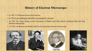
• In 1897 J.J Thomson discovered electron.
• In 1924 Louis deBorglie identifies wavelength for electron.
• In 1931 Dr. Ernst Ruska at the University of Berlin and Max Knoll combined built the first
electron microscope.
• 1n 1938 von Borries and Ruska built the first practical electron microscope.
2
Principle of Electron Microscope:
• In electron microscope, tungsten is heated by applying a high voltage current,
electron from a continues stream, which is used like a light beam. The lenses used
in EM are magnetic coils capable of focusing the electron beam on the specimen
and illuminating it. The strength of the magnetic lens depends upon the current
that flows through it. Greater the flow of the current, greater will be strength of the
magnetic field. The electron beam cannot pass through the glass lens.
3
Components of Electron
Microscope:
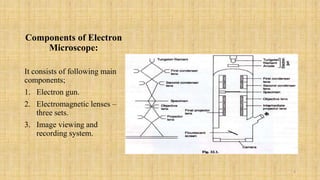
It consists of following main
components;
1. Electron gun.
2. Electromagnetic lenses –
three sets.
3. Image viewing and
recording system.
4
Preparation of Specimen:
1. Fixation and Dehydration
 Specimens are fixed in glutaraldehyde to stabilize the cell structure. After
fixation, dehydration is carried out slowly with organic solvents.
2. Embedding:
 Resins such as araldite and epoxy are used for this purpose. Microbes are
embedded in plastic resin.
3. Ultra Sectioning:
 to obtain extremely thin sections Ultra-microtomes with diamond knife or glass
knives are used.
4. Staining:
 Specimens are stained with heavy metals such as lead, uranium.
5
Working on Electron Microscope:
• The specimen to be observed is prepared as extremely thin dry film on slide.
• They are then introduced into the instrument at a point between the magnetic
condenser and the magnetic objective.
• The magnified image is viewed on a fluorescent screen through an airtight
window.
• The image can be recorded on a photographic plate by a camera built into the
instrument.
6
Types of Electron Microscope:
• There are two basic types of electron microscope:
1. Transmission Electron Microscope (TEM);
 Allows one to study of the inner structures.
2. Scanning Electron Microscope (SEM);
 Used to visualize the surface of object.
7
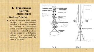
1. Transmission
Electron
Microscope:
• Working Principle:
 When an electron beam passes
through a thin-sectioned
specimen of a material, electrons
are scattered. A sophisticated
system of electromagnetic lenses
focuses the scattered electrons
into an image or a diffraction
pattern, or a nano-analytical
spectrum, depending upon the
mode of operation.
8
Biological Applications of TEM:
• Its applications in biological sciences are enormous in which some of them are
mentioned below;
Cancer research
Cellular tomography,
Toxicology,
Chemical identity,
Pollution nanotechnology.
9
Limitations of TEM:
• TEMs are large and very expensive.
• It is difficult to produce thin sample.
• Relatively time consuming process with a low throughput of samples.
• The structure of the sample may be changed during the preparation process.
• Require special housing and maintenance.
• Operation and analysis requires special training.
• Images are black and white.
10
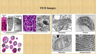
TEM Images:
11
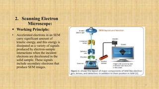
2. Scanning Electron
Microscope:
 Working Principle:
• Accelerated electrons in an SEM
carry significant amount of
kinetic energy, and this energy is
dissipated as a variety of signals
produced by electron-sample
interactions when the incident
electrons are decelerated in the
solid sample. These signals
include secondary electrons that
produce SEM images.
12
Biological Applications of SEM:
• For investigation of viral structure,
• Cryo-electron microscopy,
• 3D tissue imaging,
• Forensic sciences,
• For anatomical study of certain biological molecules,
• For visualization of intracellular changes,
• Used in ultra high vacuum, air, water and various liquid environment.
13
Limitations of SEM:
• SEMs are expensive and large.
• Special training is required to operate the SEM.
• SEMs are limited to solid sample.
• Couldn’t detect elements with atomic number less than 11.
14
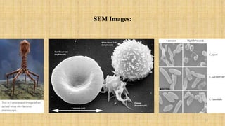
SEM Images:
15
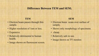
Difference Between TEM and SEM;
TEM
• Electron beam passes through thin
sample.
• Higher resolution of 1nm or less.
• Expensive.
• Relatively detrimental to human
health.
• Image shown on fluorescent screen.
SEM
• Electron beam scans over surface of
sample.
• Shows only morphology of specimens.
• cheap.
• Relatively safe to use.
• Image shown on TV monitor.
16
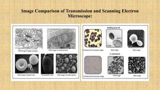
Image Comparison of Transmission and Scanning Electron
Microscope:
17
Conclusion;
• By the time electron microscope been invented it becomes a valuable tool in
development of scientific theories.
• It has contributed greatly in biological sciences, medicines, physical and chemical
sciences, its wide spread use is because, it has the ability to observe the material
up to nanometer and micrometer scale.
• Its fact that they are highly expensive but they are still widely used in research
field because they produce a detailed image of working specimen.
• In a nut shell, we can say that electron microscope invention made a revolution in
every field and brought a whole new universe.
18
19

More Related Content

What's hot

Electron microscope
Electron microscopeElectron microscope
Electron microscope
microbiology Notes
 
Simple microscope
Simple microscopeSimple microscope
Simple microscope
SuganyaPaulraj
 
Electron microscope, principle and application
Electron microscope, principle and applicationElectron microscope, principle and application
Electron microscope, principle and application
KAUSHAL SAHU
 
Recent advancement in microscopic techniques cryoem and super
Recent advancement in microscopic techniques  cryoem and superRecent advancement in microscopic techniques  cryoem and super
Recent advancement in microscopic techniques cryoem and super
DanishNigar
 
Light microscope
Light microscopeLight microscope
Light microscope
Rasika Deshmukh
 
Electron microscope
Electron microscopeElectron microscope
Electron microscope
sanjaysingh1256
 
Electron microscope
Electron microscopeElectron microscope
Electron microscope
Nithiya Pappuraj
 
Fluorescence Microscopy
Fluorescence MicroscopyFluorescence Microscopy
Fluorescence Microscopy
University of Allahabad
 
Electron microscope (TEM & SEM)
Electron microscope (TEM & SEM)Electron microscope (TEM & SEM)
Electron microscope (TEM & SEM)
Subhankar Das
 
Electron microscope - SEM and TEM
Electron microscope - SEM and TEMElectron microscope - SEM and TEM
Electron microscope - SEM and TEM
gaurav raja
 
Electron microscopy
Electron microscopyElectron microscopy
Electron microscopy
Najam ul Sehar Aiman
 
Fluoresence microscope
Fluoresence microscopeFluoresence microscope
Fluoresence microscope
SuganyaPaulraj
 
Fluorescence microscope
Fluorescence microscopeFluorescence microscope
Fluorescence microscope
gohil sanjay bhagvanji
 
Transmission electron
Transmission        electronTransmission        electron
Transmission electron
Takeen Khurshid
 
ELECTRON MICROSCOPE AND ITS APPLICATION.pptx
ELECTRON MICROSCOPE AND ITS APPLICATION.pptxELECTRON MICROSCOPE AND ITS APPLICATION.pptx
ELECTRON MICROSCOPE AND ITS APPLICATION.pptx
PallaviKumari112
 
Electron microscope
Electron microscopeElectron microscope
Electron microscope
Reena Rai
 
Fluorescence microscopy
Fluorescence microscopyFluorescence microscopy
Fluorescence microscopy
Jyotsna Verma
 
Light Microscopy
Light MicroscopyLight Microscopy
Types of Microscopes with their applications
Types of Microscopes with their applicationsTypes of Microscopes with their applications
Types of Microscopes with their applications
Microbiology Note
 
Bright field microscope and its working principle
Bright field microscope and its working principleBright field microscope and its working principle
Bright field microscope and its working principle
KavimugarajaM
 

What's hot (20)

Electron microscope
Electron microscopeElectron microscope
Electron microscope
 
Simple microscope
Simple microscopeSimple microscope
Simple microscope
 
Electron microscope, principle and application
Electron microscope, principle and applicationElectron microscope, principle and application
Electron microscope, principle and application
 
Recent advancement in microscopic techniques cryoem and super
Recent advancement in microscopic techniques  cryoem and superRecent advancement in microscopic techniques  cryoem and super
Recent advancement in microscopic techniques cryoem and super
 
Light microscope
Light microscopeLight microscope
Light microscope
 
Electron microscope
Electron microscopeElectron microscope
Electron microscope
 
Electron microscope
Electron microscopeElectron microscope
Electron microscope
 
Fluorescence Microscopy
Fluorescence MicroscopyFluorescence Microscopy
Fluorescence Microscopy
 
Electron microscope (TEM & SEM)
Electron microscope (TEM & SEM)Electron microscope (TEM & SEM)
Electron microscope (TEM & SEM)
 
Electron microscope - SEM and TEM
Electron microscope - SEM and TEMElectron microscope - SEM and TEM
Electron microscope - SEM and TEM
 
Electron microscopy
Electron microscopyElectron microscopy
Electron microscopy
 
Fluoresence microscope
Fluoresence microscopeFluoresence microscope
Fluoresence microscope
 
Fluorescence microscope
Fluorescence microscopeFluorescence microscope
Fluorescence microscope
 
Transmission electron
Transmission        electronTransmission        electron
Transmission electron
 
ELECTRON MICROSCOPE AND ITS APPLICATION.pptx
ELECTRON MICROSCOPE AND ITS APPLICATION.pptxELECTRON MICROSCOPE AND ITS APPLICATION.pptx
ELECTRON MICROSCOPE AND ITS APPLICATION.pptx
 
Electron microscope
Electron microscopeElectron microscope
Electron microscope
 
Fluorescence microscopy
Fluorescence microscopyFluorescence microscopy
Fluorescence microscopy
 
Light Microscopy
Light MicroscopyLight Microscopy
Light Microscopy
 
Types of Microscopes with their applications
Types of Microscopes with their applicationsTypes of Microscopes with their applications
Types of Microscopes with their applications
 
Bright field microscope and its working principle
Bright field microscope and its working principleBright field microscope and its working principle
Bright field microscope and its working principle
 

Similar to Electron microscope 1

Electron microscope
Electron microscopeElectron microscope
Electron microscope
ISHRAK MAHADI
 
scanning electron microscope
scanning electron microscopescanning electron microscope
scanning electron microscope
DrAkhilaCNV
 
Electron microscope
Electron microscopeElectron microscope
Electron microscope
SuganyaPaulraj
 
Tem or transmitted electron microscopy
Tem or transmitted electron microscopyTem or transmitted electron microscopy
Tem or transmitted electron microscopy
vijayakumars66
 
electron microscope by Priyanka patel..pptx
electron microscope by Priyanka patel..pptxelectron microscope by Priyanka patel..pptx
electron microscope by Priyanka patel..pptx
MAMTESHBANJARE
 
Sem n tem
Sem n temSem n tem
Sem n tem
damarisb
 
ect 292 nano electronics
ect 292 nano electronicsect 292 nano electronics
ect 292 nano electronics
RenjithThomas13
 
Powerpoint presentation on electron microscopy
Powerpoint presentation on electron microscopyPowerpoint presentation on electron microscopy
Powerpoint presentation on electron microscopy
kumar virbhadra
 
SCANNING ELECTRON MICROSCOPY (SEM)
SCANNING ELECTRON MICROSCOPY (SEM)SCANNING ELECTRON MICROSCOPY (SEM)
SCANNING ELECTRON MICROSCOPY (SEM)
Hamza Suharwardi
 
Scanning Electron Microscopy
Scanning Electron MicroscopyScanning Electron Microscopy
Scanning Electron Microscopy
MalikNumairZeeshan
 
Electron Microscope
Electron MicroscopeElectron Microscope
Electron Microscope
Ningaraj Belagall
 
Electron Microscopy Principle and Application
Electron Microscopy Principle and ApplicationElectron Microscopy Principle and Application
Electron Microscopy Principle and Application
KARTHIK REDDY C A
 
Electron microscopy ameena
Electron microscopy ameenaElectron microscopy ameena
Electron microscopy ameena
Annakurian9
 
Scanning and transmission electroscopy.pptx
Scanning and transmission electroscopy.pptxScanning and transmission electroscopy.pptx
Scanning and transmission electroscopy.pptx
Nirmal P J
 
Electron microscope
Electron microscopeElectron microscope
Electron microscope
Rasika Deshmukh
 
Confocal and electron microscopy.pptx
Confocal and electron microscopy.pptxConfocal and electron microscopy.pptx
Confocal and electron microscopy.pptx
Sathish SV
 
Scanning Electron Microscopy (SEM)
Scanning Electron Microscopy (SEM)Scanning Electron Microscopy (SEM)
Scanning Electron Microscopy (SEM)
Junaid Ahmad
 
Transmission electron microscopeppt
Transmission electron microscopepptTransmission electron microscopeppt
Transmission electron microscopeppt
Genevia Vincent
 
ELECTRON MICROSCOPE TYPES: SEM & TEM NOTES PPT
ELECTRON MICROSCOPE TYPES: SEM & TEM  NOTES PPTELECTRON MICROSCOPE TYPES: SEM & TEM  NOTES PPT
ELECTRON MICROSCOPE TYPES: SEM & TEM NOTES PPT
SruthyPB3
 
Electron microscope
Electron microscopeElectron microscope
Electron microscope
Akhil s
 

Similar to Electron microscope 1 (20)

Electron microscope
Electron microscopeElectron microscope
Electron microscope
 
scanning electron microscope
scanning electron microscopescanning electron microscope
scanning electron microscope
 
Electron microscope
Electron microscopeElectron microscope
Electron microscope
 
Tem or transmitted electron microscopy
Tem or transmitted electron microscopyTem or transmitted electron microscopy
Tem or transmitted electron microscopy
 
electron microscope by Priyanka patel..pptx
electron microscope by Priyanka patel..pptxelectron microscope by Priyanka patel..pptx
electron microscope by Priyanka patel..pptx
 
Sem n tem
Sem n temSem n tem
Sem n tem
 
ect 292 nano electronics
ect 292 nano electronicsect 292 nano electronics
ect 292 nano electronics
 
Powerpoint presentation on electron microscopy
Powerpoint presentation on electron microscopyPowerpoint presentation on electron microscopy
Powerpoint presentation on electron microscopy
 
SCANNING ELECTRON MICROSCOPY (SEM)
SCANNING ELECTRON MICROSCOPY (SEM)SCANNING ELECTRON MICROSCOPY (SEM)
SCANNING ELECTRON MICROSCOPY (SEM)
 
Scanning Electron Microscopy
Scanning Electron MicroscopyScanning Electron Microscopy
Scanning Electron Microscopy
 
Electron Microscope
Electron MicroscopeElectron Microscope
Electron Microscope
 
Electron Microscopy Principle and Application
Electron Microscopy Principle and ApplicationElectron Microscopy Principle and Application
Electron Microscopy Principle and Application
 
Electron microscopy ameena
Electron microscopy ameenaElectron microscopy ameena
Electron microscopy ameena
 
Scanning and transmission electroscopy.pptx
Scanning and transmission electroscopy.pptxScanning and transmission electroscopy.pptx
Scanning and transmission electroscopy.pptx
 
Electron microscope
Electron microscopeElectron microscope
Electron microscope
 
Confocal and electron microscopy.pptx
Confocal and electron microscopy.pptxConfocal and electron microscopy.pptx
Confocal and electron microscopy.pptx
 
Scanning Electron Microscopy (SEM)
Scanning Electron Microscopy (SEM)Scanning Electron Microscopy (SEM)
Scanning Electron Microscopy (SEM)
 
Transmission electron microscopeppt
Transmission electron microscopepptTransmission electron microscopeppt
Transmission electron microscopeppt
 
ELECTRON MICROSCOPE TYPES: SEM & TEM NOTES PPT
ELECTRON MICROSCOPE TYPES: SEM & TEM  NOTES PPTELECTRON MICROSCOPE TYPES: SEM & TEM  NOTES PPT
ELECTRON MICROSCOPE TYPES: SEM & TEM NOTES PPT
 
Electron microscope
Electron microscopeElectron microscope
Electron microscope
 

Recently uploaded

MYASTHENIA GRAVIS POWER POINT PRESENTATION
MYASTHENIA GRAVIS POWER POINT PRESENTATIONMYASTHENIA GRAVIS POWER POINT PRESENTATION
MYASTHENIA GRAVIS POWER POINT PRESENTATION
blessyjannu21
 
Exploring the Benefits of Binaural Hearing: Why Two Hearing Aids Are Better T...
Exploring the Benefits of Binaural Hearing: Why Two Hearing Aids Are Better T...Exploring the Benefits of Binaural Hearing: Why Two Hearing Aids Are Better T...
Exploring the Benefits of Binaural Hearing: Why Two Hearing Aids Are Better T...
Ear Solutions (ESPL)
 
Surface guided deep inspiration breath hold (SG-DIBH) in ultra-hypofractionat...
Surface guided deep inspiration breath hold (SG-DIBH) in ultra-hypofractionat...Surface guided deep inspiration breath hold (SG-DIBH) in ultra-hypofractionat...
Surface guided deep inspiration breath hold (SG-DIBH) in ultra-hypofractionat...
SGRT Community
 
Top Rated Massage Center In Ajman Chandrima Spa
Top Rated Massage Center In Ajman Chandrima SpaTop Rated Massage Center In Ajman Chandrima Spa
Top Rated Massage Center In Ajman Chandrima Spa
Chandrima Spa Ajman
 
MBC Support Group for Black Women – Insights in Genetic Testing.pdf
MBC Support Group for Black Women – Insights in Genetic Testing.pdfMBC Support Group for Black Women – Insights in Genetic Testing.pdf
MBC Support Group for Black Women – Insights in Genetic Testing.pdf
bkling
 
一比一原版(USF毕业证)旧金山大学毕业证如何办理
一比一原版(USF毕业证)旧金山大学毕业证如何办理一比一原版(USF毕业证)旧金山大学毕业证如何办理
一比一原版(USF毕业证)旧金山大学毕业证如何办理
40fortunate
 
Unlocking the Secrets to Safe Patient Handling.pdf
Unlocking the Secrets to Safe Patient Handling.pdfUnlocking the Secrets to Safe Patient Handling.pdf
Unlocking the Secrets to Safe Patient Handling.pdf
Lift Ability
 
Innovative Minds France's Most Impactful Healthcare Leaders.pdf
Innovative Minds France's Most Impactful Healthcare Leaders.pdfInnovative Minds France's Most Impactful Healthcare Leaders.pdf
Innovative Minds France's Most Impactful Healthcare Leaders.pdf
eurohealthleaders
 
R3 Stem Cell Therapy: A New Hope for Women with Ovarian Failure
R3 Stem Cell Therapy: A New Hope for Women with Ovarian FailureR3 Stem Cell Therapy: A New Hope for Women with Ovarian Failure
R3 Stem Cell Therapy: A New Hope for Women with Ovarian Failure
R3 Stem Cell
 
NKTI Annual Report - Annual Report FY 2022
NKTI Annual Report - Annual Report FY 2022NKTI Annual Report - Annual Report FY 2022
NKTI Annual Report - Annual Report FY 2022
nktiacc3
 
DELIRIUM BY DR JAGMOHAN PRAJAPATI.......
DELIRIUM BY DR JAGMOHAN PRAJAPATI.......DELIRIUM BY DR JAGMOHAN PRAJAPATI.......
DELIRIUM BY DR JAGMOHAN PRAJAPATI.......
DR Jag Mohan Prajapati
 
Friendly Massage in Ajman - Malayali Kerala Spa Ajman
Friendly Massage in Ajman - Malayali Kerala Spa AjmanFriendly Massage in Ajman - Malayali Kerala Spa Ajman
Friendly Massage in Ajman - Malayali Kerala Spa Ajman
Malayali Kerala Spa Ajman
 
Get Covid Testing at Fit to Fly PCR Test
Get Covid Testing at Fit to Fly PCR TestGet Covid Testing at Fit to Fly PCR Test
Get Covid Testing at Fit to Fly PCR Test
NX Healthcare
 
FACIAL NERVE
FACIAL NERVEFACIAL NERVE
FACIAL NERVE
aditigupta1117
 
Champions of Health Spotlight On Leaders Shaping Germany's Healthcare.pdf
Champions of Health Spotlight On Leaders Shaping Germany's Healthcare.pdfChampions of Health Spotlight On Leaders Shaping Germany's Healthcare.pdf
Champions of Health Spotlight On Leaders Shaping Germany's Healthcare.pdf
eurohealthleaders
 
The Power of Superfoods and Exercise.pdf
The Power of Superfoods and Exercise.pdfThe Power of Superfoods and Exercise.pdf
The Power of Superfoods and Exercise.pdf
Dr Rachana Gujar
 
Hypotension and role of physiotherapy in it
Hypotension and role of physiotherapy in itHypotension and role of physiotherapy in it
Hypotension and role of physiotherapy in it
Vishal kr Thakur
 
Top massage center in ajman chandrima Spa
Top massage center in ajman chandrima  SpaTop massage center in ajman chandrima  Spa
Top massage center in ajman chandrima Spa
Chandrima Spa Ajman
 
CANSA support - Caring for Cancer Patients' Caregivers
CANSA support - Caring for Cancer Patients' CaregiversCANSA support - Caring for Cancer Patients' Caregivers
CANSA support - Caring for Cancer Patients' Caregivers
CANSA The Cancer Association of South Africa
 
TEST BANK FOR Health Assessment in Nursing 7th Edition by Weber Chapters 1 - ...
TEST BANK FOR Health Assessment in Nursing 7th Edition by Weber Chapters 1 - ...TEST BANK FOR Health Assessment in Nursing 7th Edition by Weber Chapters 1 - ...
TEST BANK FOR Health Assessment in Nursing 7th Edition by Weber Chapters 1 - ...
rightmanforbloodline
 

Recently uploaded (20)

MYASTHENIA GRAVIS POWER POINT PRESENTATION
MYASTHENIA GRAVIS POWER POINT PRESENTATIONMYASTHENIA GRAVIS POWER POINT PRESENTATION
MYASTHENIA GRAVIS POWER POINT PRESENTATION
 
Exploring the Benefits of Binaural Hearing: Why Two Hearing Aids Are Better T...
Exploring the Benefits of Binaural Hearing: Why Two Hearing Aids Are Better T...Exploring the Benefits of Binaural Hearing: Why Two Hearing Aids Are Better T...
Exploring the Benefits of Binaural Hearing: Why Two Hearing Aids Are Better T...
 
Surface guided deep inspiration breath hold (SG-DIBH) in ultra-hypofractionat...
Surface guided deep inspiration breath hold (SG-DIBH) in ultra-hypofractionat...Surface guided deep inspiration breath hold (SG-DIBH) in ultra-hypofractionat...
Surface guided deep inspiration breath hold (SG-DIBH) in ultra-hypofractionat...
 
Top Rated Massage Center In Ajman Chandrima Spa
Top Rated Massage Center In Ajman Chandrima SpaTop Rated Massage Center In Ajman Chandrima Spa
Top Rated Massage Center In Ajman Chandrima Spa
 
MBC Support Group for Black Women – Insights in Genetic Testing.pdf
MBC Support Group for Black Women – Insights in Genetic Testing.pdfMBC Support Group for Black Women – Insights in Genetic Testing.pdf
MBC Support Group for Black Women – Insights in Genetic Testing.pdf
 
一比一原版(USF毕业证)旧金山大学毕业证如何办理
一比一原版(USF毕业证)旧金山大学毕业证如何办理一比一原版(USF毕业证)旧金山大学毕业证如何办理
一比一原版(USF毕业证)旧金山大学毕业证如何办理
 
Unlocking the Secrets to Safe Patient Handling.pdf
Unlocking the Secrets to Safe Patient Handling.pdfUnlocking the Secrets to Safe Patient Handling.pdf
Unlocking the Secrets to Safe Patient Handling.pdf
 
Innovative Minds France's Most Impactful Healthcare Leaders.pdf
Innovative Minds France's Most Impactful Healthcare Leaders.pdfInnovative Minds France's Most Impactful Healthcare Leaders.pdf
Innovative Minds France's Most Impactful Healthcare Leaders.pdf
 
R3 Stem Cell Therapy: A New Hope for Women with Ovarian Failure
R3 Stem Cell Therapy: A New Hope for Women with Ovarian FailureR3 Stem Cell Therapy: A New Hope for Women with Ovarian Failure
R3 Stem Cell Therapy: A New Hope for Women with Ovarian Failure
 
NKTI Annual Report - Annual Report FY 2022
NKTI Annual Report - Annual Report FY 2022NKTI Annual Report - Annual Report FY 2022
NKTI Annual Report - Annual Report FY 2022
 
DELIRIUM BY DR JAGMOHAN PRAJAPATI.......
DELIRIUM BY DR JAGMOHAN PRAJAPATI.......DELIRIUM BY DR JAGMOHAN PRAJAPATI.......
DELIRIUM BY DR JAGMOHAN PRAJAPATI.......
 
Friendly Massage in Ajman - Malayali Kerala Spa Ajman
Friendly Massage in Ajman - Malayali Kerala Spa AjmanFriendly Massage in Ajman - Malayali Kerala Spa Ajman
Friendly Massage in Ajman - Malayali Kerala Spa Ajman
 
Get Covid Testing at Fit to Fly PCR Test
Get Covid Testing at Fit to Fly PCR TestGet Covid Testing at Fit to Fly PCR Test
Get Covid Testing at Fit to Fly PCR Test
 
FACIAL NERVE
FACIAL NERVEFACIAL NERVE
FACIAL NERVE
 
Champions of Health Spotlight On Leaders Shaping Germany's Healthcare.pdf
Champions of Health Spotlight On Leaders Shaping Germany's Healthcare.pdfChampions of Health Spotlight On Leaders Shaping Germany's Healthcare.pdf
Champions of Health Spotlight On Leaders Shaping Germany's Healthcare.pdf
 
The Power of Superfoods and Exercise.pdf
The Power of Superfoods and Exercise.pdfThe Power of Superfoods and Exercise.pdf
The Power of Superfoods and Exercise.pdf
 
Hypotension and role of physiotherapy in it
Hypotension and role of physiotherapy in itHypotension and role of physiotherapy in it
Hypotension and role of physiotherapy in it
 
Top massage center in ajman chandrima Spa
Top massage center in ajman chandrima  SpaTop massage center in ajman chandrima  Spa
Top massage center in ajman chandrima Spa
 
CANSA support - Caring for Cancer Patients' Caregivers
CANSA support - Caring for Cancer Patients' CaregiversCANSA support - Caring for Cancer Patients' Caregivers
CANSA support - Caring for Cancer Patients' Caregivers
 
TEST BANK FOR Health Assessment in Nursing 7th Edition by Weber Chapters 1 - ...
TEST BANK FOR Health Assessment in Nursing 7th Edition by Weber Chapters 1 - ...TEST BANK FOR Health Assessment in Nursing 7th Edition by Weber Chapters 1 - ...
TEST BANK FOR Health Assessment in Nursing 7th Edition by Weber Chapters 1 - ...
 

Electron microscope 1

  • 2. History of Electron Microscope: • In 1897 J.J Thomson discovered electron. • In 1924 Louis deBorglie identifies wavelength for electron. • In 1931 Dr. Ernst Ruska at the University of Berlin and Max Knoll combined built the first electron microscope. • 1n 1938 von Borries and Ruska built the first practical electron microscope. 2
  • 3. Principle of Electron Microscope: • In electron microscope, tungsten is heated by applying a high voltage current, electron from a continues stream, which is used like a light beam. The lenses used in EM are magnetic coils capable of focusing the electron beam on the specimen and illuminating it. The strength of the magnetic lens depends upon the current that flows through it. Greater the flow of the current, greater will be strength of the magnetic field. The electron beam cannot pass through the glass lens. 3
  • 4. Components of Electron Microscope: It consists of following main components; 1. Electron gun. 2. Electromagnetic lenses – three sets. 3. Image viewing and recording system. 4
  • 5. Preparation of Specimen: 1. Fixation and Dehydration  Specimens are fixed in glutaraldehyde to stabilize the cell structure. After fixation, dehydration is carried out slowly with organic solvents. 2. Embedding:  Resins such as araldite and epoxy are used for this purpose. Microbes are embedded in plastic resin. 3. Ultra Sectioning:  to obtain extremely thin sections Ultra-microtomes with diamond knife or glass knives are used. 4. Staining:  Specimens are stained with heavy metals such as lead, uranium. 5
  • 6. Working on Electron Microscope: • The specimen to be observed is prepared as extremely thin dry film on slide. • They are then introduced into the instrument at a point between the magnetic condenser and the magnetic objective. • The magnified image is viewed on a fluorescent screen through an airtight window. • The image can be recorded on a photographic plate by a camera built into the instrument. 6
  • 7. Types of Electron Microscope: • There are two basic types of electron microscope: 1. Transmission Electron Microscope (TEM);  Allows one to study of the inner structures. 2. Scanning Electron Microscope (SEM);  Used to visualize the surface of object. 7
  • 8. 1. Transmission Electron Microscope: • Working Principle:  When an electron beam passes through a thin-sectioned specimen of a material, electrons are scattered. A sophisticated system of electromagnetic lenses focuses the scattered electrons into an image or a diffraction pattern, or a nano-analytical spectrum, depending upon the mode of operation. 8
  • 9. Biological Applications of TEM: • Its applications in biological sciences are enormous in which some of them are mentioned below; Cancer research Cellular tomography, Toxicology, Chemical identity, Pollution nanotechnology. 9
  • 10. Limitations of TEM: • TEMs are large and very expensive. • It is difficult to produce thin sample. • Relatively time consuming process with a low throughput of samples. • The structure of the sample may be changed during the preparation process. • Require special housing and maintenance. • Operation and analysis requires special training. • Images are black and white. 10
  • 12. 2. Scanning Electron Microscope:  Working Principle: • Accelerated electrons in an SEM carry significant amount of kinetic energy, and this energy is dissipated as a variety of signals produced by electron-sample interactions when the incident electrons are decelerated in the solid sample. These signals include secondary electrons that produce SEM images. 12
  • 13. Biological Applications of SEM: • For investigation of viral structure, • Cryo-electron microscopy, • 3D tissue imaging, • Forensic sciences, • For anatomical study of certain biological molecules, • For visualization of intracellular changes, • Used in ultra high vacuum, air, water and various liquid environment. 13
  • 14. Limitations of SEM: • SEMs are expensive and large. • Special training is required to operate the SEM. • SEMs are limited to solid sample. • Couldn’t detect elements with atomic number less than 11. 14
  • 16. Difference Between TEM and SEM; TEM • Electron beam passes through thin sample. • Higher resolution of 1nm or less. • Expensive. • Relatively detrimental to human health. • Image shown on fluorescent screen. SEM • Electron beam scans over surface of sample. • Shows only morphology of specimens. • cheap. • Relatively safe to use. • Image shown on TV monitor. 16
  • 17. Image Comparison of Transmission and Scanning Electron Microscope: 17
  • 18. Conclusion; • By the time electron microscope been invented it becomes a valuable tool in development of scientific theories. • It has contributed greatly in biological sciences, medicines, physical and chemical sciences, its wide spread use is because, it has the ability to observe the material up to nanometer and micrometer scale. • Its fact that they are highly expensive but they are still widely used in research field because they produce a detailed image of working specimen. • In a nut shell, we can say that electron microscope invention made a revolution in every field and brought a whole new universe. 18
  • 19. 19