Downloaded 715 times
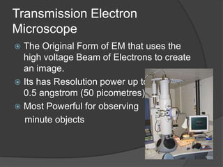
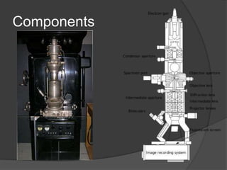
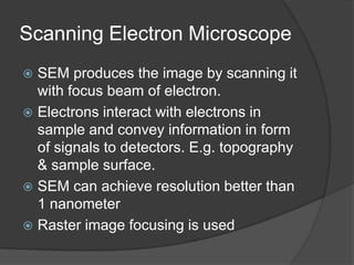

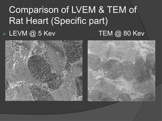


This document discusses four main types of electron microscopes: transmission electron microscope (TEM), scanning electron microscope (SEM), reflection electron microscope (REM), and low-voltage electron microscope (LVEM). It provides details on the components, imaging mechanisms, advantages, limitations, and typical voltages used for each microscope type. The TEM is noted as the original and most powerful microscope for high resolution imaging, but requires thin sample preparation. The SEM can image thicker bulk samples but provides 3D surface images rather than internal structure. The REM uses elastically scattered electrons for raster scanning. The LVEM provides high contrast images at lower voltages than TEMs, with improved thickness limits over conventional TEM.


![Light Microscope and Electron Microscope [Best one]](https://cdn.slidesharecdn.com/ss_thumbnails/presentation-170404212835-thumbnail.jpg?width=640&height=640&fit=bounds)


