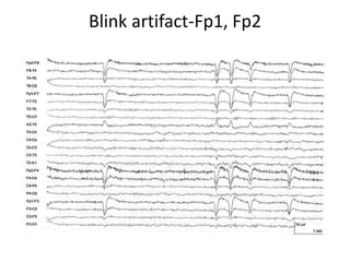
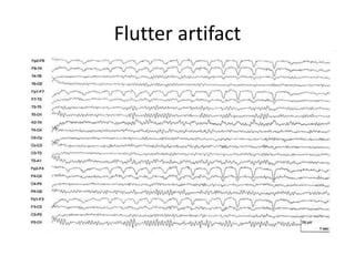
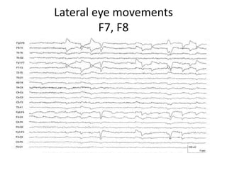
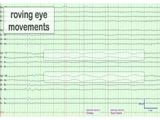
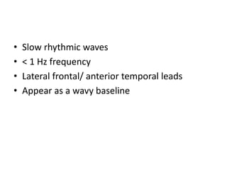
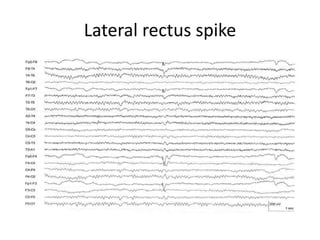
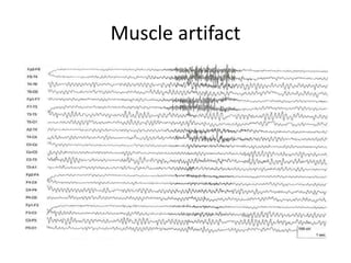
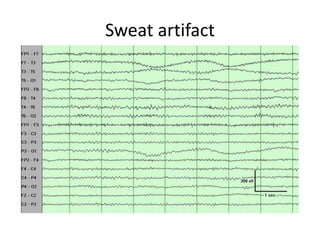
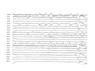
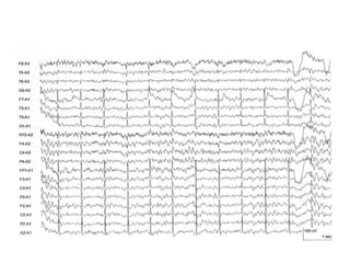
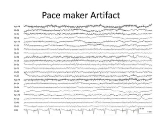
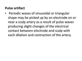
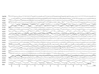
Artifacts in EEG
• EEG artifacts are recorded signals that are
non-cerebral in origin.
• They may be divided into one of two
categories:
• physiological artifacts
• non- physiological artifacts.
Physiological artifacts arise from a variety of body activities
that are due to either:
• Movement of head, body or scalp (e.g. pulsations of the
scalp arteries)- that affect the electrode scalp interface
• Bioelectrical potentials generated within the body from
moving sources (such as eye, tongue, or pharyngeal muscle
movement),
stationary sources such as the scalp muscles, heart or
sweat glands
• Altered volume conduction due to changes in the
conductance of tissues (scalp, bone, muscle) and fluids
(CSF, blood) between the cerebral cortex and the recording
electrodes.
Non- physiological artifacts arise from two main
sources:
• external electrical interference from other power
sources such as power lines or electrical
equipment,
• internal electrical malfunctioning of the recording
system, arising from recording electrodes
(electrode integrity, positioning and application),
cables, amplifiers.
• Artifacts can often be recognized by their
characteristic morphology and distribution.
• The patient and record must both be closely
observed throughout the recording
Blinking and other eye movements.
• These movements cause potential changes which
are picked up mainly by frontal electrodes.
• The electrodes that record the largest potential
change with vertical eye movement are Fp1 and
Fp2 because they are placed directly above the
eyes.
• The electrodes that record the largest potential
change with horizontal (lateral) eye movements
are F7 and F8 because they are closest electrodes
that are approximately lateral to the eyes.
• Eye movement artifacts in the EEG can usually
be identified by their frontal distribution, their
bilateral symmetry and their characteristic
shape.
• Eye movement artifacts can be identified
during the recording by observing the patient
and correlating eye blinks and movements
with pen deflections.
Blink artifact-Fp1, Fp2
Flutter artifact
Flutter artifact
• Medium amplitude, activity confined to
frontal poles
• Compared to blink artifact, flutter artifact
typically has a lower amplitude and a more
rhythmic appearance(more frequency)
Lateral eye movements
F7, F8
• Slow rhythmic waves
• < 1 Hz frequency
• Lateral frontal/ anterior temporal leads
• Appear as a wavy baseline
Lateral rectus spike
Muscle artifact
• Muscle activity causes very short potentials
which usually recur.
• Muscle artifacts from scalp and face muscles
occur mainly in the frontal and temporal regions.
• Easily identified by its shape and repetition.
• It can be reduced and often eliminated by asking
the patient to relax the jaw or open the mouth
slightly, or change position.
Muscle artifact
Muscle artifact
• High amplitude, fast activity.
• Distribution that reflects the locations of the
muscles generating it.
• Typical of muscle artifact, it begins and ends
abruptly.
Perspiration artifacts
• consists of slow waveforms that are usually
greater than 2 sec in duration.
• In addition, sweat gland activity produces slowly
changing electrical potentials that are recorded
by the electrodes.
• Perspiration artifact almost always appear in
more than one channel, but may be lateralized or
asymmetric.
Sweat artifact
Electrocardiogram.
• The artifact may appear in all channels using a
common reference. Or only in one or a few
channels.
• Small amplitude artifacts reflects mainly the
R- wave of the electrocardiogram.
• The R- wave usually appears maximally over
the left posterior head region as a positive
sharply contoured wave form and, with lower
amplitude, over the right temporal head
regions as a negative waveform.
• If there is any doubt whether sharp waves are
due to the ECG artifact or to the cerebral
activity, the technician should record the ECG
in one channel and compare it with the timing
of the suspected sharp waves.
Pace maker Artifact
Pulse artifact
• Periodic waves of sinusoidal or triangular
shape may be picked up by an electrode on or
near a scalp artery as a result of pulse waves
producing slight changes of the electrical
contact between electrode and scalp with
each dilation and contraction of the artery.
• Electrical Interference.
• Artifacts due to electrical interference emanates
from electrical equipment and nearby power
lines.
• Strong interference can cause artifacts even with
good recording electrodes and equipment;
• these artifacts are then likely to appear in all
channels of all recordings made in the same
recording room.
• When recordings are made in an environment
with excessive interference such as an intensive
care unit or an operating room, the patient’s
head and the connections to the EEG machine
should be kept as far from power cables as
possible.
• Electrode wires should be straightened and
bundled together.
• Equipment other than the EEG machine should
be unplugged if feasible.
Mechanical ventilator artifacts
Artifacts arising from electrodes
• A common electrode artifact referred to as an
‘electrode pop’ is due to a sudden change of
electrode contact causing pen deflections
which abruptly deviate from the baseline and
then gradually return.
Electrode pop
Electrode pop
• Nearly vertical rise followed by the slower fall
at the F3 electrode is typical of electrode pop
artifact.
• Amplitude that is much greater than the
surrounding activity, a field that is limited to
one electrode.
Electrode movement artifact
• focal slowing in the T4-T6 and T6-O2 channels
• oscillations typical of rhythmic electrode
movement
• poor contact
• which leads to sharp or slow waves of varying
morphology and amplitude.
Lead movement
Lead movement
• Artifact through activity that is both unusually
high amplitude and low frequency and also
disorganized without a plausible field
• Lead movement has a more disorganized
morphology that does not resemble true EEG
activity in any form
38

Eeg artifacts

  • 1.
  • 2.
    • EEG artifactsare recorded signals that are non-cerebral in origin. • They may be divided into one of two categories: • physiological artifacts • non- physiological artifacts.
  • 3.
    Physiological artifacts arisefrom a variety of body activities that are due to either: • Movement of head, body or scalp (e.g. pulsations of the scalp arteries)- that affect the electrode scalp interface • Bioelectrical potentials generated within the body from moving sources (such as eye, tongue, or pharyngeal muscle movement), stationary sources such as the scalp muscles, heart or sweat glands • Altered volume conduction due to changes in the conductance of tissues (scalp, bone, muscle) and fluids (CSF, blood) between the cerebral cortex and the recording electrodes.
  • 4.
    Non- physiological artifactsarise from two main sources: • external electrical interference from other power sources such as power lines or electrical equipment, • internal electrical malfunctioning of the recording system, arising from recording electrodes (electrode integrity, positioning and application), cables, amplifiers.
  • 5.
    • Artifacts canoften be recognized by their characteristic morphology and distribution. • The patient and record must both be closely observed throughout the recording
  • 6.
    Blinking and othereye movements. • These movements cause potential changes which are picked up mainly by frontal electrodes. • The electrodes that record the largest potential change with vertical eye movement are Fp1 and Fp2 because they are placed directly above the eyes. • The electrodes that record the largest potential change with horizontal (lateral) eye movements are F7 and F8 because they are closest electrodes that are approximately lateral to the eyes.
  • 7.
    • Eye movementartifacts in the EEG can usually be identified by their frontal distribution, their bilateral symmetry and their characteristic shape. • Eye movement artifacts can be identified during the recording by observing the patient and correlating eye blinks and movements with pen deflections.
  • 8.
  • 9.
  • 10.
    Flutter artifact • Mediumamplitude, activity confined to frontal poles • Compared to blink artifact, flutter artifact typically has a lower amplitude and a more rhythmic appearance(more frequency)
  • 11.
  • 13.
    • Slow rhythmicwaves • < 1 Hz frequency • Lateral frontal/ anterior temporal leads • Appear as a wavy baseline
  • 14.
  • 15.
    Muscle artifact • Muscleactivity causes very short potentials which usually recur. • Muscle artifacts from scalp and face muscles occur mainly in the frontal and temporal regions. • Easily identified by its shape and repetition. • It can be reduced and often eliminated by asking the patient to relax the jaw or open the mouth slightly, or change position.
  • 16.
  • 17.
    Muscle artifact • Highamplitude, fast activity. • Distribution that reflects the locations of the muscles generating it. • Typical of muscle artifact, it begins and ends abruptly.
  • 18.
    Perspiration artifacts • consistsof slow waveforms that are usually greater than 2 sec in duration. • In addition, sweat gland activity produces slowly changing electrical potentials that are recorded by the electrodes. • Perspiration artifact almost always appear in more than one channel, but may be lateralized or asymmetric.
  • 19.
  • 20.
    Electrocardiogram. • The artifactmay appear in all channels using a common reference. Or only in one or a few channels. • Small amplitude artifacts reflects mainly the R- wave of the electrocardiogram.
  • 21.
    • The R-wave usually appears maximally over the left posterior head region as a positive sharply contoured wave form and, with lower amplitude, over the right temporal head regions as a negative waveform.
  • 22.
    • If thereis any doubt whether sharp waves are due to the ECG artifact or to the cerebral activity, the technician should record the ECG in one channel and compare it with the timing of the suspected sharp waves.
  • 25.
  • 26.
    Pulse artifact • Periodicwaves of sinusoidal or triangular shape may be picked up by an electrode on or near a scalp artery as a result of pulse waves producing slight changes of the electrical contact between electrode and scalp with each dilation and contraction of the artery.
  • 28.
    • Electrical Interference. •Artifacts due to electrical interference emanates from electrical equipment and nearby power lines. • Strong interference can cause artifacts even with good recording electrodes and equipment; • these artifacts are then likely to appear in all channels of all recordings made in the same recording room.
  • 29.
    • When recordingsare made in an environment with excessive interference such as an intensive care unit or an operating room, the patient’s head and the connections to the EEG machine should be kept as far from power cables as possible. • Electrode wires should be straightened and bundled together. • Equipment other than the EEG machine should be unplugged if feasible.
  • 30.
  • 31.
    Artifacts arising fromelectrodes • A common electrode artifact referred to as an ‘electrode pop’ is due to a sudden change of electrode contact causing pen deflections which abruptly deviate from the baseline and then gradually return.
  • 32.
  • 33.
    Electrode pop • Nearlyvertical rise followed by the slower fall at the F3 electrode is typical of electrode pop artifact. • Amplitude that is much greater than the surrounding activity, a field that is limited to one electrode.
  • 34.
  • 35.
    Electrode movement artifact •focal slowing in the T4-T6 and T6-O2 channels • oscillations typical of rhythmic electrode movement • poor contact • which leads to sharp or slow waves of varying morphology and amplitude.
  • 36.
  • 37.
    Lead movement • Artifactthrough activity that is both unusually high amplitude and low frequency and also disorganized without a plausible field • Lead movement has a more disorganized morphology that does not resemble true EEG activity in any form
  • 38.