Downloaded 268 times
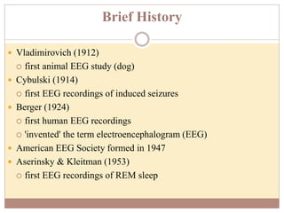
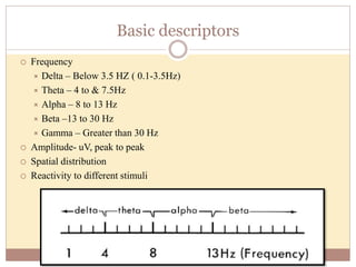
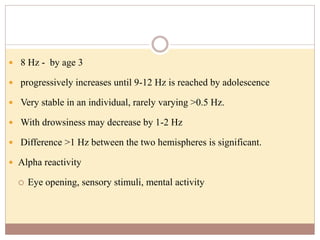
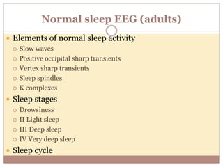
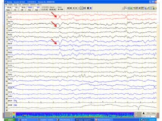
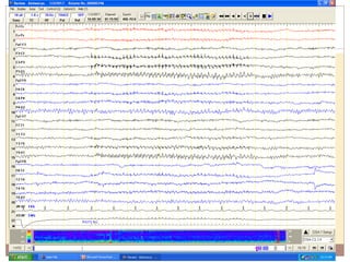

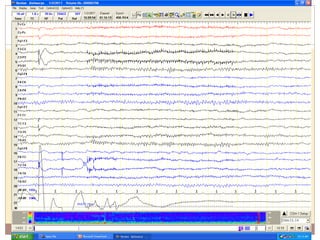
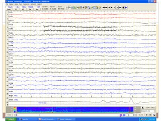


This document provides an overview of normal EEG patterns in adults. It begins with a brief history of EEG and then describes the basic electrical activity generated by the brain and how EEG recordings work. It outlines the normal frequency bands seen in EEG - delta, theta, alpha, beta and gamma. Specific normal EEG patterns like the alpha rhythm, vertex waves, sleep spindles and K-complexes are described. It also discusses benign variants and activation procedures. In summary, the document serves as a reference for the typical EEG patterns seen in healthy, awake and sleeping adults.


![CASE_PRESENTATION_ON_subdural_hematoma(SDH)[1 FINAL PPT]-1.pptx](https://cdn.slidesharecdn.com/ss_thumbnails/casepresentationonsubduralhematomasdh1finalppt-1-260129172522-d405d375-thumbnail.jpg?width=640&height=640&fit=bounds)


