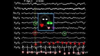
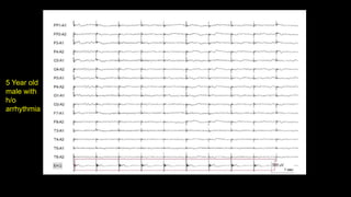
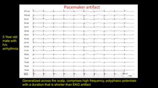
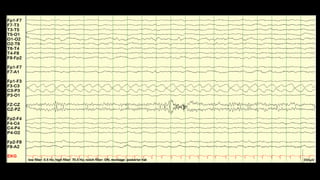
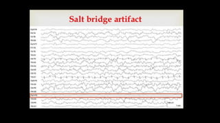
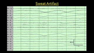
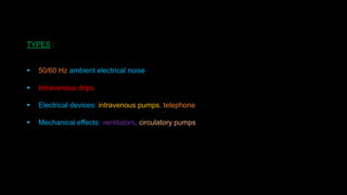
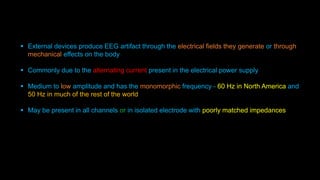
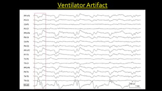
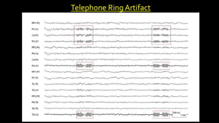
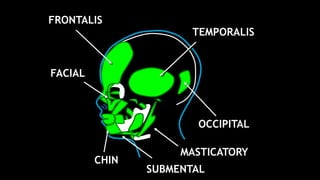
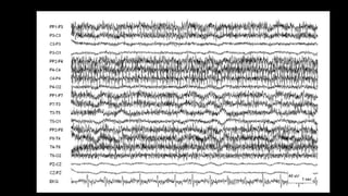
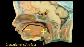
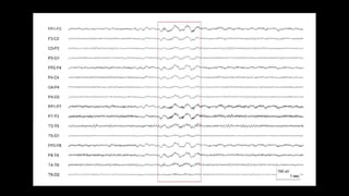
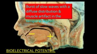
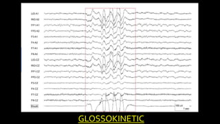
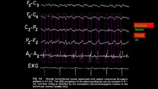
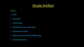
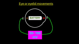
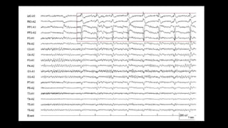
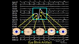
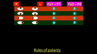
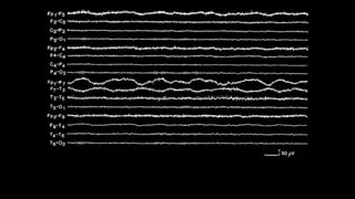
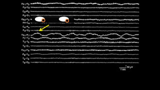
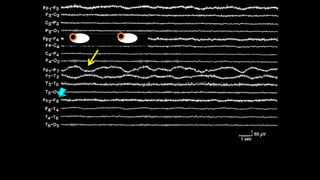
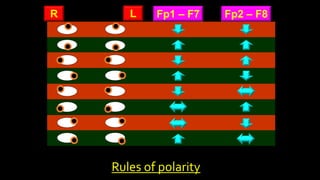
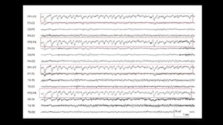
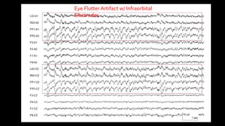
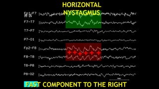
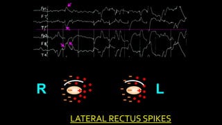
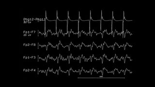
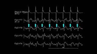
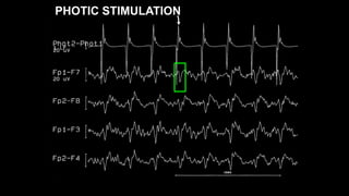
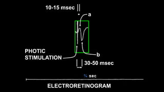
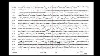
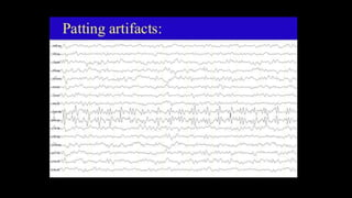
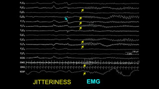
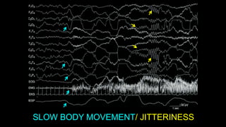
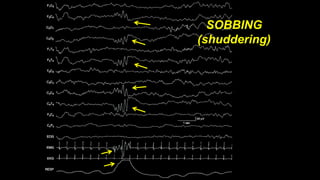
EEG Artifacts
&
How to Resolve
Lalit Bansal M.D.
Director of Epilepsy Surgery Medical Program
Pediatric Epilepsy & Clinical Neurophysiology
Children’s Mercy Hospital
05/15/2018
 EEG records cerebral activity and electrical activities from sites other than the brain
 Anything that is NOT of cerebral origin is termed as ARTIFACT
Types of Artifact:
1. Physiological artifact - generated other than brain ie. body
2. Extraphysiological artifact - arise outside the body, eg: equipment, enviornment
Introduction
 Physiological activity has a logical topographic field of distribution with an
excepted fall of the voltage potentials
 Artifact have an illogical distribution that defies the principles of localization
Principles to discriminate artifacts from EEG signals
 Good, clean prep
 Balanced impedances
 Good hook-up, neatly bundled electrodes
 Place jack-box close to patients head
 Keep patient cool, not cold
 Unplug all electrical items close to patient, i.e. bed, radio, fan, etc.
KEYTO AN ARTIFACT FREE RECORDING
 Cardiac
 Electrode
 External device artifact
 Muscle artifact
 Ocular artifact
Artifacts
Heart produce 2 types of artifact
1. Electrical
2. Mechanical
 Timed lock to cardiac contractions and synchronized with EKG complexes
 P/T wave are usually not visible on EEG (distance from the heart and the suboptimal axis)
 Artifact is a poorly formed QRS complex
 Prominent in patients with short neck
 Most prominent over temporal region
 R wave - A1 Negative and A2 Positive
Cardiac Artifact
5 Year old
male with
h/o
arrhythmia
Generalized across the scalp, comprises high frequency, polyphasic potentials
with a duration that is shorter than EKG artifact
Pacemaker artifact
5 Year old
male with
h/o
arrhythmia
Image of pulse artifact
 Mechanical artifact from the heart arise through the circulatory pulse
 Electrode artifact - occurs when an electrode rests over a vessel
 Periodic slow wave with a regular interval - follows EKG artifact’s peak by about 200msec
 Most common over frontal and temporal, less common over occipital
How to Identify:
 Applying pressure on electrode alters its appearance on the EEG
Pulse Artifact
PULSE
ARTIFACT
PULSE
ARTIFACT
 Mechanical cardiac artifact
 Results from slight movement of the head or body that occurs with cardiac contraction
 Similar to pulse artifact but is more widespread
 May involve one or few electrodes – due to electrode lead movement
 Biposterior electrodes if movement of the head on the pillow
 Occasionally can be generalized
How to resolve:
 Reposition the head
Ballistocardiographic Artifact
BallistocardiographicArtifact
Electrode Artifact
Types:
1. Electrode pop
2. Electrode contact
3. Electrode/lead movement
4. Perspiration
5. Salt bridge
6. Movement artifact
 Usually manifest as one of two disparate waveforms, brief transients that are limited to
one electrode and low frequency rhythms across a scalp region
 Due to spontaneous discharging of electrical potential present between the electrode or
its lead
 Electrode pops - reflect the ability of the electrode and skin interface to function as a
capacitor and store electrical charge across the electrolyte paste or gel that holds the
electrode in place
 With the release of the charge there is a change in impedance, and a sudden potential
appears in all channels that include the electrode
 Sometimes more than one pop occurs within a few seconds
 Characteristic morphology - very steep rise and a more shallow fall
Electrode Pop
F3 electrode
Pop
 Produces artifact with a less conserved morphology than electrode pop
 Poor contact produces instability in the impedance, which leads to sharp or slow waves
of varying morphology and amplitude
 These waves may be rhythmic if the poor contact occurs in the context of rhythmic
movement, such as from a tremor.
Poor electrode contact or lead movement
Reference Montage ipsilateral ear–T5 electrode activity
60 Hz Off – high Impedance inT5
 Lead movement has more disorganized morphology that does not resemble true EEG activity
 Often includes double phase reversal (without consistency in polarity that indicates a cerebrally
generated electrical field)
Lead Movement
Artifact
Electrode Movement
Artifact
 Slowing in T4-T6 and T6-O2 channels
 No field beyond T6
 Oscillations typical of rhythmic electrode movement
 Seen due to smearing of the electrode paste between electrodes or presence of
perspiration across the scalp
 Forms an unwanted electrical connection between the electrodes forming a channel
 Perspiration artifact
- manifests as low amplitude
- undulating (smooth) waves
- duration is typically greater than 2 sec
 Slat bridge artifact
- lower in amplitude
- not wavering with low frequency oscillation
- typically include only one channel
 It may appear flat and close to isoelectric
Salt Bridge and Perspiration
Artifact
Sweat Artifact
External device artifact
TYPES :
 50/60 Hz ambient electrical noise
 Intravenous drips
 Electrical devices: intravenous pumps, telephone
 Mechanical effects: ventilators, circulatory pumps
 External devices produce EEG artifact through the electrical fields they generate or through
mechanical effects on the body
 Commonly due to the alternating current present in the electrical power supply
 Medium to low amplitude and has the monomorphic frequency - 60 Hz in North America and
50 Hz in much of the rest of the world
 May be present in all channels or in isolated electrode with poorly matched impedances
60 Hz Artifact
 Electrical noise may also result from falling electrostatically charged droplets in an IV drip
 In-phase activity
 A spike like EEG potential results, which has the regularity of the drip
EEG
 Ventilators and circulatory pumps produce artifacts with slower components than other
electrical devices
 Resemble ballistocardiographic or other electrode artifact
 Monomorphic frequency with fixed interval
 Slow wave or a complex including a mixture of frequencies superimposed on a slow wave
 Exceptions to typical pattern – High frequency oscillator
Mechanical devices
Ventilator Artifact
Telephone Ring Artifact
Muscle artifact
Types :
 Glossokenetic (chew/swallow)
 Photomyogenic (photomyoclonic)
 Surface Electromyographic (scalp/facial muscle)
 Frequency is higher than that of clinical EEG and too fast to be visually estimated
 Without filtering, EMG artifact usually has a more disorganized appearance because the
individual myogenic potentials overlap with each other
 Occasionally, individual potentials are discernible.
 Duration of EMG artifact varies according to the duration of the muscle activity; a second to an
entire EEG recording.
 Artifact occurs most commonly in frontal and temporal electrodes
Muscle Artifact
FRONTALIS
TEMPORALIS
OCCIPITAL
FACIAL
MASTICATORY
SUBMENTAL
CHIN
Glossokinetic Artifact
BIOELECTRICAL POTENTIAL
Burst of slow waves with a
diffuse distribution &
muscle artifact in the
temporal region!
GLOSSOKINETIC
Distinguishing from Ictal Activity
Ictal epileptic
activity
Continuous glossokinetic
artifact
Sucking Artifact
6 Month old male
Pontine
lesion
Tinnit
us
Types :
 Blink
 Eye flutter
 Lateral gaze
 Slow/Roving eye movements
 Lateral rectus spike
 Rapid eye movements of REM sleep
 Electroretinogram
Ocular Artifact
BATTERY
50- 100
mV
Eye or eyelid movements
Eye Blink Artifact
Fp1 – F7 Fp2 – F8
R L
Rules of polarity
Fp1 – F7 Fp2 – F8
R L
Rules of polarity
Eye Flutter Artifact w/ Infraorbital
Electrodes
1
2
1
1 1
2
Conjugate EM:
Vertical
Horizontal
Out of phase
EEG
In-phase
Out of phase
In-phase
Detect all EM
Out of phase
In-phase
EEG = EM
EEG = vertical EM
Misses low
amplitude EM
Good because:
Bad because:
VERTICAL
FAST COMPONENT TO THE RIGHT
HORIZONTAL
NYSTAGMUS
R L
3
T
LATERAL RECTUS SPIKES
PHOTIC STIMULATION
¾
a
b
PHOTIC
STIMULATION
10-15 msec
30-50 msec
ELECTRORETINOGRAM
JITTERINESS EMG
SLOW BODY MOVEMENT/ JITTERINESS
SOBBING
(shuddering)
Sympathetic Skin Response
(Galvanic Skin Response)
 Skin potential medicated by unmyelinated cholinergic sympathetic fibers
 Changes in electrical properties of skin during sweating, sensory stimulation or
emotional stress
 Appear as long lasting potential of abrupt onset with a monophasic, biphasic or
triphasic morphology.
 Most commonly from frontal but may be diffuse
 Confirmation: extracranial recording from palm of the hand
SYMPATHETIC SKIN
RESPONSE
PALM
DORSUM
EEG Artifacts & How to Resolve (Lalit Bansal M.D.)

EEG Artifacts & How to Resolve (Lalit Bansal M.D.)

  • 1.
    EEG Artifacts & How toResolve Lalit Bansal M.D. Director of Epilepsy Surgery Medical Program Pediatric Epilepsy & Clinical Neurophysiology Children’s Mercy Hospital 05/15/2018
  • 2.
     EEG recordscerebral activity and electrical activities from sites other than the brain  Anything that is NOT of cerebral origin is termed as ARTIFACT Types of Artifact: 1. Physiological artifact - generated other than brain ie. body 2. Extraphysiological artifact - arise outside the body, eg: equipment, enviornment Introduction
  • 3.
     Physiological activityhas a logical topographic field of distribution with an excepted fall of the voltage potentials  Artifact have an illogical distribution that defies the principles of localization Principles to discriminate artifacts from EEG signals
  • 4.
     Good, cleanprep  Balanced impedances  Good hook-up, neatly bundled electrodes  Place jack-box close to patients head  Keep patient cool, not cold  Unplug all electrical items close to patient, i.e. bed, radio, fan, etc. KEYTO AN ARTIFACT FREE RECORDING
  • 5.
     Cardiac  Electrode External device artifact  Muscle artifact  Ocular artifact Artifacts
  • 6.
    Heart produce 2types of artifact 1. Electrical 2. Mechanical  Timed lock to cardiac contractions and synchronized with EKG complexes  P/T wave are usually not visible on EEG (distance from the heart and the suboptimal axis)  Artifact is a poorly formed QRS complex  Prominent in patients with short neck  Most prominent over temporal region  R wave - A1 Negative and A2 Positive Cardiac Artifact
  • 10.
    5 Year old malewith h/o arrhythmia
  • 11.
    Generalized across thescalp, comprises high frequency, polyphasic potentials with a duration that is shorter than EKG artifact Pacemaker artifact 5 Year old male with h/o arrhythmia
  • 12.
  • 13.
     Mechanical artifactfrom the heart arise through the circulatory pulse  Electrode artifact - occurs when an electrode rests over a vessel  Periodic slow wave with a regular interval - follows EKG artifact’s peak by about 200msec  Most common over frontal and temporal, less common over occipital How to Identify:  Applying pressure on electrode alters its appearance on the EEG Pulse Artifact
  • 15.
  • 16.
  • 17.
     Mechanical cardiacartifact  Results from slight movement of the head or body that occurs with cardiac contraction  Similar to pulse artifact but is more widespread  May involve one or few electrodes – due to electrode lead movement  Biposterior electrodes if movement of the head on the pillow  Occasionally can be generalized How to resolve:  Reposition the head Ballistocardiographic Artifact
  • 18.
  • 19.
  • 20.
    Types: 1. Electrode pop 2.Electrode contact 3. Electrode/lead movement 4. Perspiration 5. Salt bridge 6. Movement artifact
  • 21.
     Usually manifestas one of two disparate waveforms, brief transients that are limited to one electrode and low frequency rhythms across a scalp region  Due to spontaneous discharging of electrical potential present between the electrode or its lead  Electrode pops - reflect the ability of the electrode and skin interface to function as a capacitor and store electrical charge across the electrolyte paste or gel that holds the electrode in place  With the release of the charge there is a change in impedance, and a sudden potential appears in all channels that include the electrode  Sometimes more than one pop occurs within a few seconds  Characteristic morphology - very steep rise and a more shallow fall Electrode Pop
  • 22.
  • 23.
     Produces artifactwith a less conserved morphology than electrode pop  Poor contact produces instability in the impedance, which leads to sharp or slow waves of varying morphology and amplitude  These waves may be rhythmic if the poor contact occurs in the context of rhythmic movement, such as from a tremor. Poor electrode contact or lead movement
  • 25.
    Reference Montage ipsilateralear–T5 electrode activity
  • 26.
    60 Hz Off– high Impedance inT5
  • 27.
     Lead movementhas more disorganized morphology that does not resemble true EEG activity  Often includes double phase reversal (without consistency in polarity that indicates a cerebrally generated electrical field) Lead Movement Artifact
  • 28.
    Electrode Movement Artifact  Slowingin T4-T6 and T6-O2 channels  No field beyond T6  Oscillations typical of rhythmic electrode movement
  • 29.
     Seen dueto smearing of the electrode paste between electrodes or presence of perspiration across the scalp  Forms an unwanted electrical connection between the electrodes forming a channel  Perspiration artifact - manifests as low amplitude - undulating (smooth) waves - duration is typically greater than 2 sec  Slat bridge artifact - lower in amplitude - not wavering with low frequency oscillation - typically include only one channel  It may appear flat and close to isoelectric Salt Bridge and Perspiration Artifact
  • 31.
  • 32.
  • 33.
    TYPES :  50/60Hz ambient electrical noise  Intravenous drips  Electrical devices: intravenous pumps, telephone  Mechanical effects: ventilators, circulatory pumps
  • 34.
     External devicesproduce EEG artifact through the electrical fields they generate or through mechanical effects on the body  Commonly due to the alternating current present in the electrical power supply  Medium to low amplitude and has the monomorphic frequency - 60 Hz in North America and 50 Hz in much of the rest of the world  May be present in all channels or in isolated electrode with poorly matched impedances
  • 35.
  • 36.
     Electrical noisemay also result from falling electrostatically charged droplets in an IV drip  In-phase activity  A spike like EEG potential results, which has the regularity of the drip EEG
  • 37.
     Ventilators andcirculatory pumps produce artifacts with slower components than other electrical devices  Resemble ballistocardiographic or other electrode artifact  Monomorphic frequency with fixed interval  Slow wave or a complex including a mixture of frequencies superimposed on a slow wave  Exceptions to typical pattern – High frequency oscillator Mechanical devices
  • 39.
  • 40.
  • 41.
    Muscle artifact Types : Glossokenetic (chew/swallow)  Photomyogenic (photomyoclonic)  Surface Electromyographic (scalp/facial muscle)
  • 42.
     Frequency ishigher than that of clinical EEG and too fast to be visually estimated  Without filtering, EMG artifact usually has a more disorganized appearance because the individual myogenic potentials overlap with each other  Occasionally, individual potentials are discernible.  Duration of EMG artifact varies according to the duration of the muscle activity; a second to an entire EEG recording.  Artifact occurs most commonly in frontal and temporal electrodes Muscle Artifact
  • 43.
  • 45.
  • 47.
    BIOELECTRICAL POTENTIAL Burst ofslow waves with a diffuse distribution & muscle artifact in the temporal region!
  • 48.
  • 50.
    Distinguishing from IctalActivity Ictal epileptic activity Continuous glossokinetic artifact
  • 51.
  • 54.
  • 55.
    Types :  Blink Eye flutter  Lateral gaze  Slow/Roving eye movements  Lateral rectus spike  Rapid eye movements of REM sleep  Electroretinogram Ocular Artifact
  • 56.
    BATTERY 50- 100 mV Eye oreyelid movements
  • 58.
  • 59.
    Fp1 – F7Fp2 – F8 R L Rules of polarity
  • 63.
    Fp1 – F7Fp2 – F8 R L Rules of polarity
  • 65.
    Eye Flutter Artifactw/ Infraorbital Electrodes
  • 66.
    1 2 1 1 1 2 Conjugate EM: Vertical Horizontal Outof phase EEG In-phase Out of phase In-phase Detect all EM Out of phase In-phase EEG = EM EEG = vertical EM Misses low amplitude EM Good because: Bad because:
  • 68.
  • 70.
    FAST COMPONENT TOTHE RIGHT HORIZONTAL NYSTAGMUS
  • 72.
  • 75.
  • 76.
  • 79.
  • 80.
  • 81.
  • 82.
    Sympathetic Skin Response (GalvanicSkin Response)  Skin potential medicated by unmyelinated cholinergic sympathetic fibers  Changes in electrical properties of skin during sweating, sensory stimulation or emotional stress  Appear as long lasting potential of abrupt onset with a monophasic, biphasic or triphasic morphology.  Most commonly from frontal but may be diffuse  Confirmation: extracranial recording from palm of the hand
  • 83.