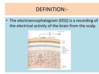
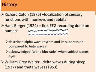
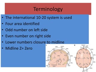
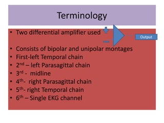
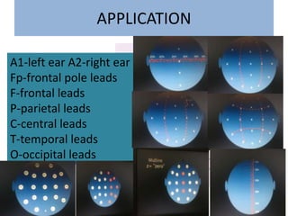
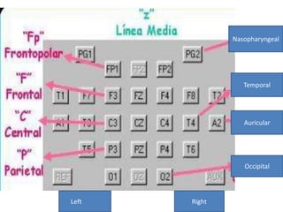
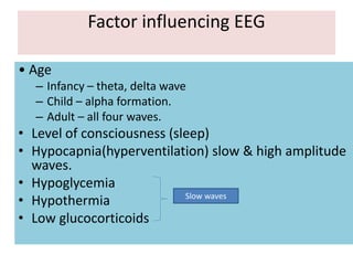
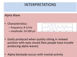
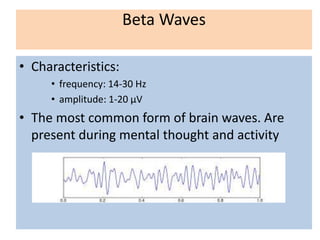
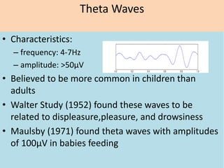
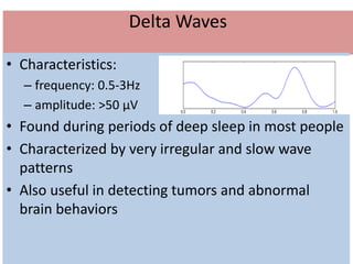
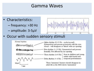
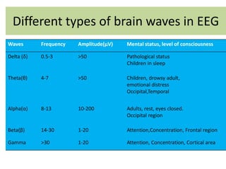
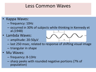
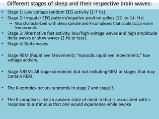
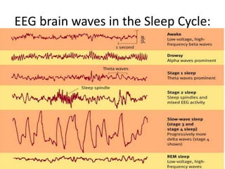
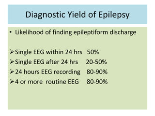
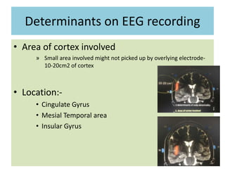
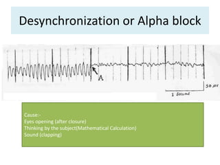
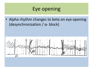
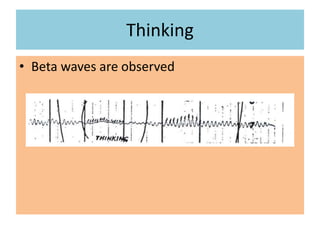
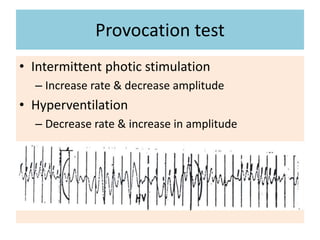
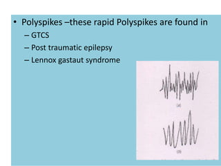
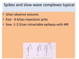
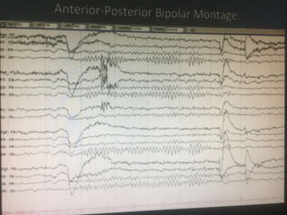
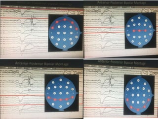
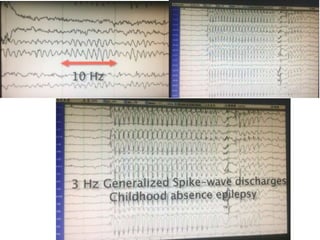
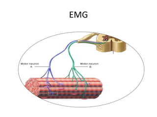
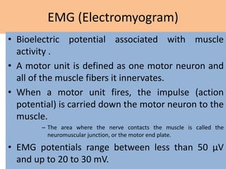
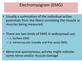
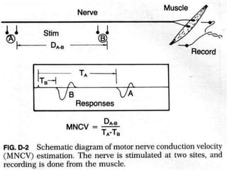
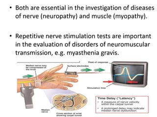
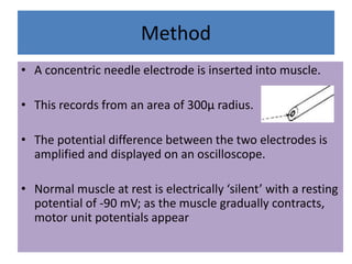
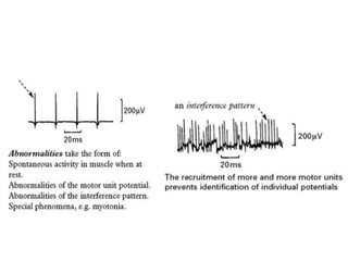
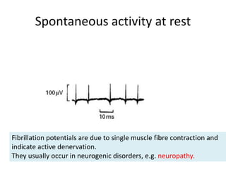
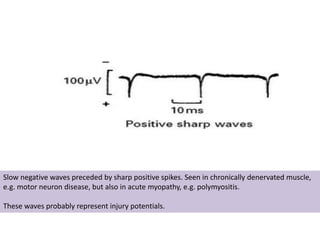
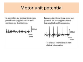
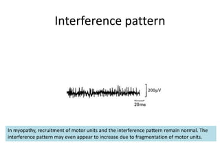
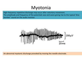
EEG/EMG
DR.RAJEEV BHANDARI
NATIONAL ACADEMY OF MEDICAL
SCIENCES
BIR HOSPITAL
DEFINITION:-
• The electroencephalogram (EEG) is a recording of
the electrical activity of the brain from the scalp.
History
Richard Caton (1875) –localization of sensory
functions with monkeys and rabbits
Hans Berger (1924) – first EEG recording done on
humans
described alpha wave rhythm and its suppression
compared to beta waves
acknowledged “alpha blockade” when subject opens
eyes
William Grey Walter –delta waves during sleep
(1937) and theta waves (1953)
Terminology
• The international 10-20 system is used
• Four area identified
• Odd number on left side
• Even number on right side
• Lower numbers closure to midline
• Midline Z= Zero
Terminology
• Two differential amplifier used
• Consists of bipolar and unipolar montages
• First-left Temporal chain
• 2nd – left Parasagittal chain
• 3rd - midline
• 4th- right Parasagittal chain
• 5th- right Temporal chain
• 6th – Single EKG channel
Output
APPLICATION
A1-left ear A2-right ear
Fp-frontal pole leads
F-frontal leads
P-parietal leads
C-central leads
T-temporal leads
O-occipital leads
Nasopharyngeal
Temporal
Auricular
Occipital
Left Right
Factor influencing EEG
• Age
– Infancy – theta, delta wave
– Child – alpha formation.
– Adult – all four waves.
• Level of consciousness (sleep)
• Hypocapnia(hyperventilation) slow & high amplitude
waves.
• Hypoglycemia
• Hypothermia
• Low glucocorticoids
Slow waves
INTERPRETATIONS
Alpha Wave
• Characteristics:
– frequency: 8-13 Hz
– amplitude: 10-200 μV
• Easily produced when quietly sitting in relaxed
position with eyes closed (few people have trouble
producing alpha waves)
• Alpha blockade occurs with mental activity
Beta Waves
• Characteristics:
• frequency: 14-30 Hz
• amplitude: 1-20 μV
• The most common form of brain waves. Are
present during mental thought and activity
Theta Waves
• Characteristics:
– frequency: 4-7Hz
– amplitude: >50μV
• Believed to be more common in children than
adults
• Walter Study (1952) found these waves to be
related to displeasure,pleasure, and drowsiness
• Maulsby (1971) found theta waves with amplitudes
of 100μV in babies feeding
Delta Waves
• Characteristics:
– frequency: 0.5-3Hz
– amplitude: >50 μV
• Found during periods of deep sleep in most people
• Characterized by very irregular and slow wave
patterns
• Also useful in detecting tumors and abnormal
brain behaviors
Gamma Waves
• Characteristics:
– frequency: >30 Hz
– amplitude: 3-5μV
• Occur with sudden sensory stimuli
Different types of brain waves in EEG
Waves Frequency Amplitude(μV) Mental status, level of consciousness
Delta (δ) 0.5-3 >50 Pathological status
Children in sleep
Theta(θ) 4-7 >50 Children, drowsy adult,
emotional distress
Occipital,Temporal
Alpha(α) 8-13 10-200 Adults, rest, eyes closed.
Occipital region
Beta(β) 14-30 1-20 Attention,Concentration, Frontal region
Gamma >30 1-20 Attention, Concentration, Cortical area
Less Common Waves
• Kappa Waves:
– frequency: 10Hz
– occurred in 30% of subjects while thinking in Kennedy et
al.(1948)
• Lambda Waves:
– amplitude: 20-50μV
– last 250 msec, related to response of shifting visual image
– triangular in shape
• Mu Waves:
– frequency: 8-13Hz
– sharp peeks with rounded negative portions (7% of
population)
Different stages of sleep and their respective brain waves:
• Stage 1: Low voltage random EEG activity (2-7 Hz)
• Stage 2: Irregular EEG pattern/negative-positive spikes (12- to 14- Hz)
– Also characterized with sleep spindle and K-complexes that could occur every
few seconds.
• Stage 3: Alternative fast activity, low/high voltage waves and high amplitude
delta waves or slow waves (2 Hz or less).
• Stage 4: Delta waves
• Stage REM (Rapid eye Movement): “episodic rapid eye movements,” low
voltage activity.
• Stage NREM: All stage combined, but not including REM or stages that may
contain REM.
• The K-complex occurs randomly in stage 2 and stage 3
• The K complex is like an awaken state of mind in that is associated with a
response to a stimulus that one would experience while awake
EEG brain waves in the Sleep Cycle:
Diagnostic Yield of Epilepsy
• Likelihood of finding epileptiform discharge
Single EEG within 24 hrs 50%
Single EEG after 24 hrs 20-50%
24 hours EEG recording 80-90%
4 or more routine EEG 80-90%
Determinants on EEG recording
• Area of cortex involved
» Small area involved might not picked up by overlying electrode-
10-20cm2 of cortex
• Location:-
• Cingulate Gyrus
• Mesial Temporal area
• Insular Gyrus
Desynchronization or Alpha block
Cause:-
Eyes opening (after closure)
Thinking by the subject(Mathematical Calculation)
Sound (clapping)
Eye opening
• Alpha rhythm changes to beta on eye opening
(desynchronization / α- block)
Thinking
• Beta waves are observed
Provocation test
• Intermittent photic stimulation
– Increase rate & decrease amplitude
• Hyperventilation
– Decrease rate & increase in amplitude
• Polyspikes –these rapid Polyspikes are found in
– GTCS
– Post traumatic epilepsy
– Lennox gastaut syndrome
Spikes and slow wave complexes typical
• 3/sec-absence seizures
• Fast - 4-6/sec-myoclonic jerks
• Sow -1-2.5/sec-intractable epilepsy with MR
More advanced methods of telemetry
and foramen ovale recording
– to establish the diagnosis of ‘epilepsy’ if doubt remains
– to determine the exact frequency and site of origin of the attacks
– to aid classification of seizure type.
• Telemetry: utilises a continuous 24–48 hour recording of EEG,
often combined with a videotape recording of the patient.
• Increasing availability of this and ambulatory recording has
greatly improved diagnostic accuracy and reliability of seizure
classification.
• Foramen ovale recording: a needle electrode is passed
percutaneously through the foramen ovale to record activity
from the adjacent temporal lobe.
EMG
EMG (Electromyogram)
• Bioelectric potential associated with muscle
activity .
• A motor unit is defined as one motor neuron and
all of the muscle fibers it innervates.
• When a motor unit fires, the impulse (action
potential) is carried down the motor neuron to the
muscle.
– The area where the nerve contacts the muscle is called the
neuromuscular junction, or the motor end plate.
• EMG potentials range between less than 50 μV
and up to 20 to 30 mV.
Electromyogram (EMG)
• Usually a summation of the individual action
potentials from the fibers consisting the muscle or
muscles being measured.
• There are two kinds of EMG in widespread use:
– 1. Surface EMG
– 2. Intramuscular (needle and fine-wire) EMG.
• Abnormal spontaneous activity might indicate
some nerve and/or muscle damage
• Both are essential in the investigation of diseases
of nerve (neuropathy) and muscle (myopathy).
• Repetitive nerve stimulation tests are important
in the evaluation of disorders of neuromuscular
transmission, e.g. myasthenia gravis.
Method
• A concentric needle electrode is inserted into muscle.
• This records from an area of 300μ radius.
• The potential difference between the two electrodes is
amplified and displayed on an oscilloscope.
• Normal muscle at rest is electrically ‘silent’ with a resting
potential of -90 mV; as the muscle gradually contracts,
motor unit potentials appear
Spontaneous activity at rest
Fibrillation potentials are due to single muscle fibre contraction and
indicate active denervation.
They usually occur in neurogenic disorders, e.g. neuropathy.
Slow negative waves preceded by sharp positive spikes. Seen in chronically denervated muscle,
e.g. motor neuron disease, but also in acute myopathy, e.g. polymyositis.
These waves probably represent injury potentials.
Motor unit potential
Interference pattern
In myopathy, recruitment of motor units and the interference pattern remain normal. The
interference pattern may even appear to increase due to fragmentation of motor units.
Myotonia
High frequency repetitive discharge may occur after voluntary movement.
The amplitude and frequency of the potentials wax and wane giving rise to the typical ‘dive
bomber’ sound on the audio monitor.
An abnormal myotonic discharge provoked by moving the needle electrode.
• Thank you!

EEG INTERPRETATION

  • 1.
    EEG/EMG DR.RAJEEV BHANDARI NATIONAL ACADEMYOF MEDICAL SCIENCES BIR HOSPITAL
  • 2.
    DEFINITION:- • The electroencephalogram(EEG) is a recording of the electrical activity of the brain from the scalp.
  • 3.
    History Richard Caton (1875)–localization of sensory functions with monkeys and rabbits Hans Berger (1924) – first EEG recording done on humans described alpha wave rhythm and its suppression compared to beta waves acknowledged “alpha blockade” when subject opens eyes William Grey Walter –delta waves during sleep (1937) and theta waves (1953)
  • 4.
    Terminology • The international10-20 system is used • Four area identified • Odd number on left side • Even number on right side • Lower numbers closure to midline • Midline Z= Zero
  • 5.
    Terminology • Two differentialamplifier used • Consists of bipolar and unipolar montages • First-left Temporal chain • 2nd – left Parasagittal chain • 3rd - midline • 4th- right Parasagittal chain • 5th- right Temporal chain • 6th – Single EKG channel Output
  • 6.
    APPLICATION A1-left ear A2-rightear Fp-frontal pole leads F-frontal leads P-parietal leads C-central leads T-temporal leads O-occipital leads
  • 7.
  • 8.
    Factor influencing EEG •Age – Infancy – theta, delta wave – Child – alpha formation. – Adult – all four waves. • Level of consciousness (sleep) • Hypocapnia(hyperventilation) slow & high amplitude waves. • Hypoglycemia • Hypothermia • Low glucocorticoids Slow waves
  • 9.
    INTERPRETATIONS Alpha Wave • Characteristics: –frequency: 8-13 Hz – amplitude: 10-200 μV • Easily produced when quietly sitting in relaxed position with eyes closed (few people have trouble producing alpha waves) • Alpha blockade occurs with mental activity
  • 10.
    Beta Waves • Characteristics: •frequency: 14-30 Hz • amplitude: 1-20 μV • The most common form of brain waves. Are present during mental thought and activity
  • 11.
    Theta Waves • Characteristics: –frequency: 4-7Hz – amplitude: >50μV • Believed to be more common in children than adults • Walter Study (1952) found these waves to be related to displeasure,pleasure, and drowsiness • Maulsby (1971) found theta waves with amplitudes of 100μV in babies feeding
  • 12.
    Delta Waves • Characteristics: –frequency: 0.5-3Hz – amplitude: >50 μV • Found during periods of deep sleep in most people • Characterized by very irregular and slow wave patterns • Also useful in detecting tumors and abnormal brain behaviors
  • 13.
    Gamma Waves • Characteristics: –frequency: >30 Hz – amplitude: 3-5μV • Occur with sudden sensory stimuli
  • 14.
    Different types ofbrain waves in EEG Waves Frequency Amplitude(μV) Mental status, level of consciousness Delta (δ) 0.5-3 >50 Pathological status Children in sleep Theta(θ) 4-7 >50 Children, drowsy adult, emotional distress Occipital,Temporal Alpha(α) 8-13 10-200 Adults, rest, eyes closed. Occipital region Beta(β) 14-30 1-20 Attention,Concentration, Frontal region Gamma >30 1-20 Attention, Concentration, Cortical area
  • 15.
    Less Common Waves •Kappa Waves: – frequency: 10Hz – occurred in 30% of subjects while thinking in Kennedy et al.(1948) • Lambda Waves: – amplitude: 20-50μV – last 250 msec, related to response of shifting visual image – triangular in shape • Mu Waves: – frequency: 8-13Hz – sharp peeks with rounded negative portions (7% of population)
  • 16.
    Different stages ofsleep and their respective brain waves: • Stage 1: Low voltage random EEG activity (2-7 Hz) • Stage 2: Irregular EEG pattern/negative-positive spikes (12- to 14- Hz) – Also characterized with sleep spindle and K-complexes that could occur every few seconds. • Stage 3: Alternative fast activity, low/high voltage waves and high amplitude delta waves or slow waves (2 Hz or less). • Stage 4: Delta waves • Stage REM (Rapid eye Movement): “episodic rapid eye movements,” low voltage activity. • Stage NREM: All stage combined, but not including REM or stages that may contain REM. • The K-complex occurs randomly in stage 2 and stage 3 • The K complex is like an awaken state of mind in that is associated with a response to a stimulus that one would experience while awake
  • 17.
    EEG brain wavesin the Sleep Cycle:
  • 20.
    Diagnostic Yield ofEpilepsy • Likelihood of finding epileptiform discharge Single EEG within 24 hrs 50% Single EEG after 24 hrs 20-50% 24 hours EEG recording 80-90% 4 or more routine EEG 80-90%
  • 21.
    Determinants on EEGrecording • Area of cortex involved » Small area involved might not picked up by overlying electrode- 10-20cm2 of cortex • Location:- • Cingulate Gyrus • Mesial Temporal area • Insular Gyrus
  • 22.
    Desynchronization or Alphablock Cause:- Eyes opening (after closure) Thinking by the subject(Mathematical Calculation) Sound (clapping)
  • 23.
    Eye opening • Alpharhythm changes to beta on eye opening (desynchronization / α- block)
  • 24.
  • 25.
    Provocation test • Intermittentphotic stimulation – Increase rate & decrease amplitude • Hyperventilation – Decrease rate & increase in amplitude
  • 26.
    • Polyspikes –theserapid Polyspikes are found in – GTCS – Post traumatic epilepsy – Lennox gastaut syndrome
  • 27.
    Spikes and slowwave complexes typical • 3/sec-absence seizures • Fast - 4-6/sec-myoclonic jerks • Sow -1-2.5/sec-intractable epilepsy with MR
  • 31.
    More advanced methodsof telemetry and foramen ovale recording – to establish the diagnosis of ‘epilepsy’ if doubt remains – to determine the exact frequency and site of origin of the attacks – to aid classification of seizure type. • Telemetry: utilises a continuous 24–48 hour recording of EEG, often combined with a videotape recording of the patient. • Increasing availability of this and ambulatory recording has greatly improved diagnostic accuracy and reliability of seizure classification. • Foramen ovale recording: a needle electrode is passed percutaneously through the foramen ovale to record activity from the adjacent temporal lobe.
  • 32.
  • 33.
    EMG (Electromyogram) • Bioelectricpotential associated with muscle activity . • A motor unit is defined as one motor neuron and all of the muscle fibers it innervates. • When a motor unit fires, the impulse (action potential) is carried down the motor neuron to the muscle. – The area where the nerve contacts the muscle is called the neuromuscular junction, or the motor end plate. • EMG potentials range between less than 50 μV and up to 20 to 30 mV.
  • 34.
    Electromyogram (EMG) • Usuallya summation of the individual action potentials from the fibers consisting the muscle or muscles being measured. • There are two kinds of EMG in widespread use: – 1. Surface EMG – 2. Intramuscular (needle and fine-wire) EMG. • Abnormal spontaneous activity might indicate some nerve and/or muscle damage
  • 36.
    • Both areessential in the investigation of diseases of nerve (neuropathy) and muscle (myopathy). • Repetitive nerve stimulation tests are important in the evaluation of disorders of neuromuscular transmission, e.g. myasthenia gravis.
  • 37.
    Method • A concentricneedle electrode is inserted into muscle. • This records from an area of 300μ radius. • The potential difference between the two electrodes is amplified and displayed on an oscilloscope. • Normal muscle at rest is electrically ‘silent’ with a resting potential of -90 mV; as the muscle gradually contracts, motor unit potentials appear
  • 39.
    Spontaneous activity atrest Fibrillation potentials are due to single muscle fibre contraction and indicate active denervation. They usually occur in neurogenic disorders, e.g. neuropathy.
  • 40.
    Slow negative wavespreceded by sharp positive spikes. Seen in chronically denervated muscle, e.g. motor neuron disease, but also in acute myopathy, e.g. polymyositis. These waves probably represent injury potentials.
  • 41.
  • 42.
    Interference pattern In myopathy,recruitment of motor units and the interference pattern remain normal. The interference pattern may even appear to increase due to fragmentation of motor units.
  • 43.
    Myotonia High frequency repetitivedischarge may occur after voluntary movement. The amplitude and frequency of the potentials wax and wane giving rise to the typical ‘dive bomber’ sound on the audio monitor. An abnormal myotonic discharge provoked by moving the needle electrode.
  • 44.