Downloaded 448 times
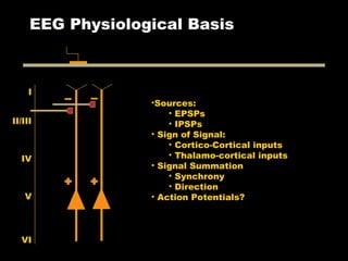


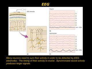
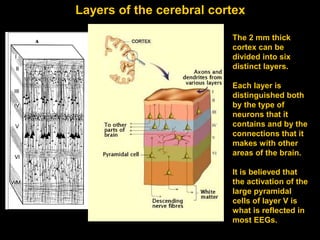
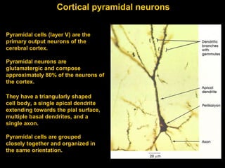
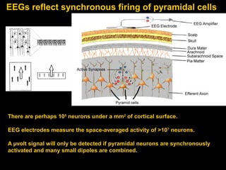
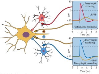
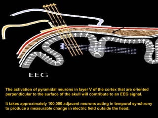

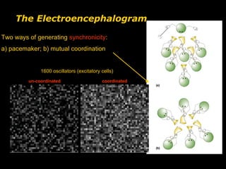
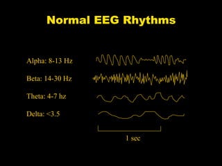

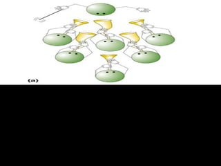
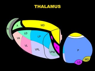
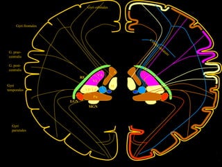
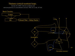
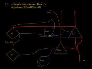
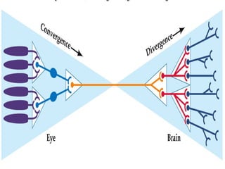
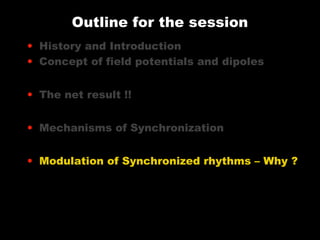

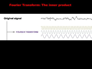
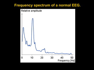
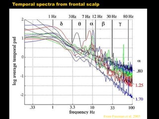


The document discusses the physiological basis of electroencephalograms (EEG), focusing on the synchronization of neural activity in the cortex and the role of pyramidal neurons in generating detectable EEG signals. It outlines the history of EEG discovery, mechanisms of synchronization, and the significance of different rhythmic activities (alpha, beta, theta, and delta) in cognitive processes. Additionally, it emphasizes the importance of neuronal oscillations for memory, attention, and conscious awareness.


![ONFH[AVN HIP] -TRIPLE REGIME -A NOVAL SURGICAL CONCEPT .pptx](https://cdn.slidesharecdn.com/ss_thumbnails/onfhavnhip2026koaconcalicutdrgokuldevdrmashraf-260210064517-213ec005-thumbnail.jpg?width=640&height=640&fit=bounds)


