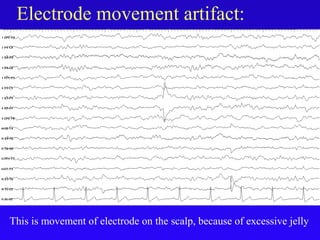
Downloaded 2,245 times
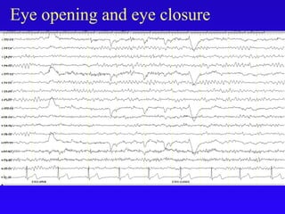
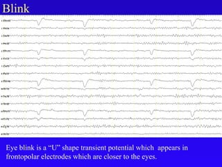
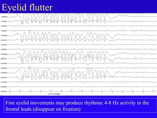
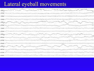
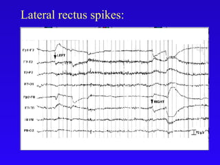
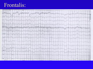
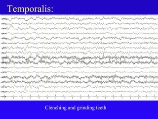
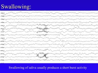
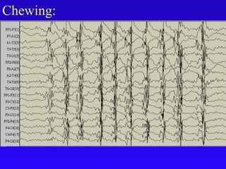
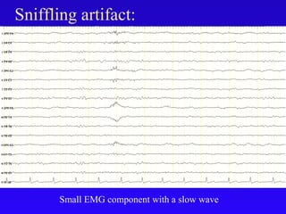
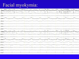
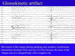
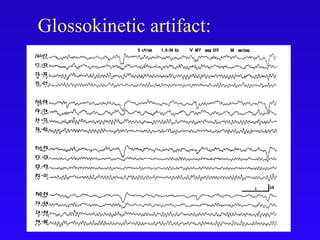
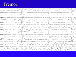
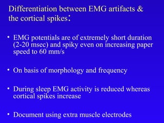
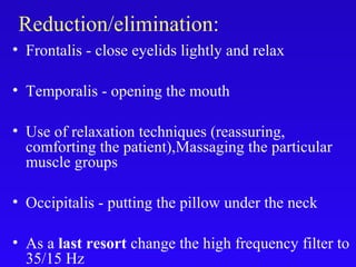

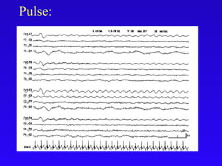

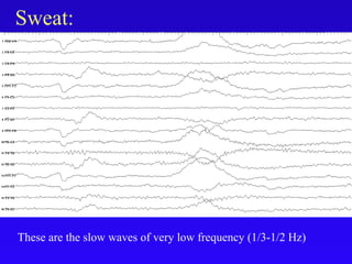
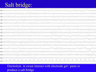

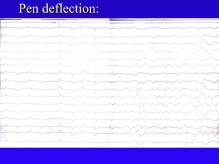
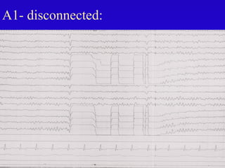
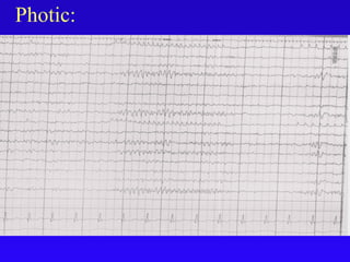
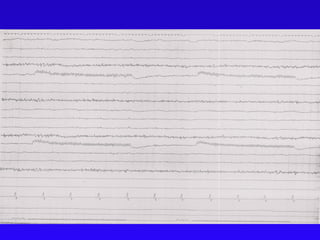
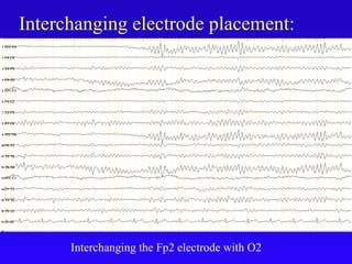
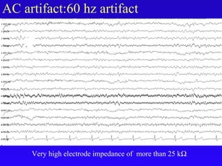
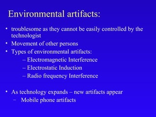
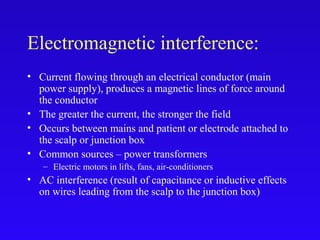
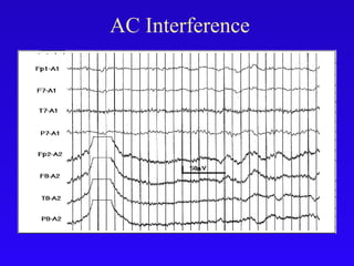
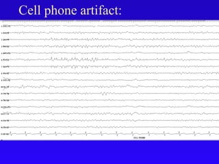
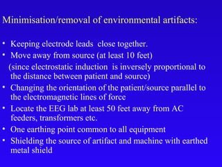
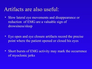


This document discusses different types of artifacts that can appear on an EEG, including how to identify and eliminate them. It separates artifacts into physiological artifacts originating from the patient's body (e.g. eye movements, muscle activity), and extraphysiological artifacts from external sources (e.g. electrodes, equipment, environment). Specific artifact types like blinks, lateral eye movements, muscle activity are described. Guidelines provided on how to reduce artifacts include closing the eyes, relaxing muscles, ensuring good electrode contact, and shielding from environmental interference.


