Downloaded 282 times

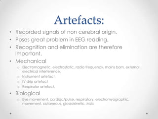
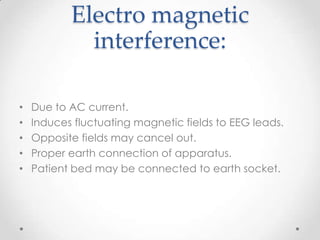


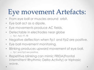
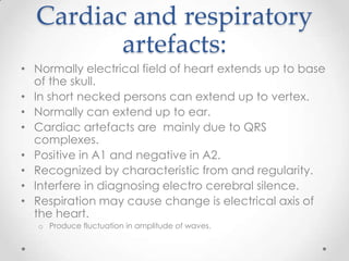
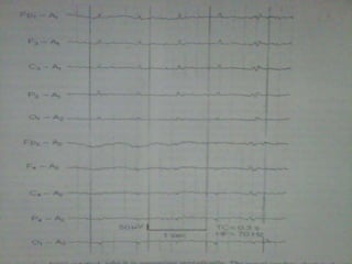
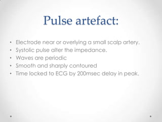
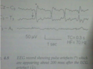
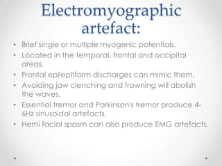


This document discusses various EEG artifacts and benign variants that can appear on an EEG reading. It describes mechanical artifacts such as those caused by electromagnetic interference, electrostatic interference, radio frequency interference, and instrument artifacts. It also discusses biological artifacts including those from eye movements, cardiac/pulse activity, electromyography, movement, skin potentials, and tongue movements. Some benign rhythmic activities and epileptiform variants are also outlined such as frontal arousal rhythm, midline theta rhythm, and 14-16Hz positive bursts. Proper identification and differentiation of artifacts and variants is important for accurate EEG interpretation.