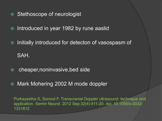
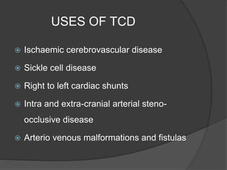
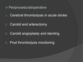
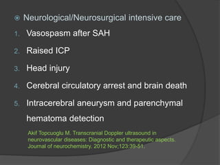
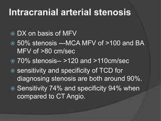
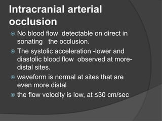
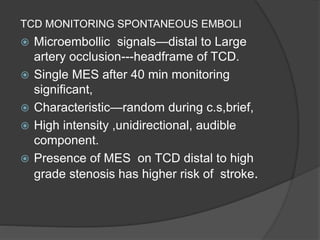
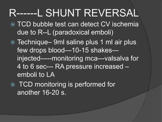
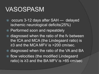
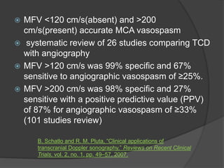
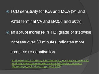
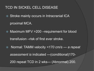
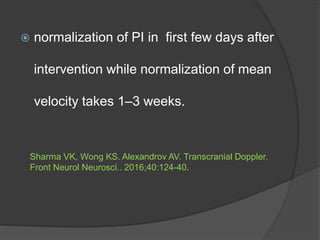
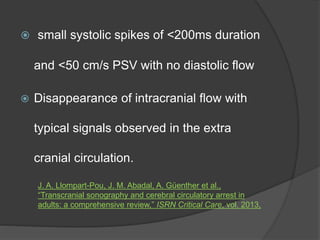
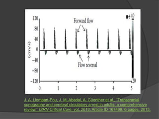
SlideShare a Scribd company logo
Dr ASHISH SHARMA
SR 1 NEUROLOGY
GMC KOTA
 Stethoscope of neurologist
 Introduced in year 1982 by rune aaslid
 Initially introduced for detecton of vasospasm of
SAH.
 cheaper,noninvasive,bed side
 Mark Mohering 2002 M mode doppler
Purkayastha S, Sorond F. Transcranial Doppler ultrasound: technique and
application. Semin Neurol. 2012 Sep;32(4):411-20. doi: 10.1055/s-0032-
1331812
American academy of neurology guidelines
Bathala L, Mehndiratta MM, Sharma VK. Transcranial doppler: Technique and
common findings (Part 1). Annals of Indian Academy of Neurology. 2013
Apr;16(2):174.
TERMS IN TCD
 PULSALITY INDEX---The difference between PSV and EDV divided by
the mean flow velocity (MFV). PI values greater than 1.2 represent high
resistance blood flow.
 PEAK SYSTOLIC VELOCITY --This is the initial peak on each cardiac
cycle’s TCD waveform
 END DIASTOLIC VELOCITY---indicates a low resistance cerebral
artery flow pattern in all main intracranial arteries, ranging from 20 to
50% of the PSV
 MEAN FLOW VELOCITY --calculated as EDV plus one-third of the
difference between PSV and EDV
 RESISTANCE INDEX--The RI is determined by subtracting EDV from
PSV and dividing the value by PSV. If the RI value is less than 0.75, it
is considered normal.
Alexandrov AV, Demchuk AM, Wein TH, Grotta JC. Yield of transcranial Doppler in
acute cerebral ischemia. Stroke. 1999 Aug;30(8):1604-9.
FREQUENTLY USED TERMINOLOGY IN
TCD
PRINCIPALS OF TCD
 The insonation is performed from the
proximal to the distal artery.
 start closest to the heart and proceed
distally
 located, find and record the best signal
 inspect at least two locations for each
artery
 Find location where the waveforms
exhibit the greatest abnormalities
USES OF TCD
 Ischaemic cerebrovascular disease
 Sickle cell disease
 Right to left cardiac shunts
 Intra and extra-cranial arterial steno-
occlusive disease
 Arterio venous malformations and fistulas
 Periprocedural/operative
1. Cerebral thrombolysis in acute stroke
2. Carotid end arterectomy
3. Carotid angioplasty and stenting
4. Post thrombolysis monitoring
 Neurological/Neurosurgical intensive care
1. Vasospasm after SAH
2. Raised ICP
3. Head injury
4. Cerebral circulatory arrest and brain death
5. Intracerebral aneurysm and parenchymal
hematoma detection
Akif Topcuoglu M. Transcranial Doppler ultrasound in
neurovascular diseases: Diagnostic and therapeutic aspects.
Journal of neurochemistry. 2012 Nov;123:39-51.
Intracranial arterial stenosis
 DX on basis of MFV
 50% stenosis ---MCA MFV of >100 and BA
MFV of >80 cm/sec
 70% stenosis-- >120 and >110cm/sec
 sensitivity and specificity of TCD for
diagnosing stenosis are both around 90%.
 Sensitivity 74% and specificity 94% when
compared to CT Angio.
Intracranial arterial
occlusion
 No blood flow detectable on direct in
sonating the occlusion.
 The systolic acceleration -lower and
diastolic blood flow observed at more-
distal sites.
 waveform is normal at sites that are
even more distal
 the flow velocity is low, at ≤30 cm/sec
TCD MONITORING SPONTANEOUS EMBOLI
 Microembollic signals—distal to Large
artery occlusion---headframe of TCD.
 Single MES after 40 min monitoring
significant,
 Characteristic—random during c.s,brief,
 High intensity ,unidirectional, audible
component.
 Presence of MES on TCD distal to high
grade stenosis has higher risk of stroke.
R------L SHUNT REVERSAL
 TCD bubble test can detect CV ischemia
due to R--L (paradoxical emboli)
 Technique– 9ml saline plus 1 ml air plus
few drops blood—10-15 shakes—
injected-----monitoring mca---valsalva for
4 to 6 sec--- RA pressure increased –
emboli to LA
 TCD monitoring is performed for
another 16-20 s.
VASOSPASM
 occurs 3-12 days after SAH --- delayed
ischemic neurological deficits(25%)
 Performed soon and repeatdely
 diagnosed when the ratio of the fv between
the ICA and MCA (the Lindegaard ratio) is
≥3 and the MCA MFV is >200 cm/sec.
 diagnosed when the ratio of the VA and BA
 flow velocities (the modified Lindegaard
ratio) is ≥3 and the BA MFV is >85 cm/sec
 MFV <120 cm/s(absent) and >200
cm/s(present) accurate MCA vasospasm
 systematic review of 26 studies comparing TCD
with angiography
 MFV >120 cm/s was 99% specific and 67%
sensitive to angiographic vasospasm of ≥25%.
 MFV >200 cm/s was 98% specific and 27%
sensitive with a positive predictive value (PPV)
of 87% for angiographic vasospasm of ≥33%
(101 studies review)
B. Schatlo and R. M. Pluta, “Clinical applications of
transcranial Doppler sonography,” Reviews on Recent Clinical
Trials, vol. 2, no. 1, pp. 49–57, 2007.
TCD IN ACUTE STROKE
 Prognostication value in acute stroke.
 Sensitivity 90% in MCA acute occlusion
detection
 Occlusion –6 hrs – in M1 segment detected---
high chance of hemorrhagic transformation
 Effectiveness ,duration, dose of re canalisation
 TCD sensitivity for ICA and MCA (94 and
93%) terminal VA and BA(56 and 60%).
 an abrupt increase in TIBI grade or stepwise
increase over 30 minutes indicates more
complete re canalisation
A. M. Demchuk, I. Christou, T. H. Wein et al., “Accuracy and criteria for
localizing arterial occlusion with transcranial Doppler,” Journal of
Neuroimaging, vol. 10, no. 1, pp. 1–12, 2000
TCD IN SICKEL CELL DISEASE
 Stroke mainly occurs in Intracranial ICA
proximal MCA.
 Maximum MFV >200 –requirement for blood
transfusion –risk of first ever stroke.
 Normal: TAMM velocity <170 cm/s — a repeat
assessment is indicated ---(conditional)170-
200 repeat TCD in 2 wks--- (Abnormal) 200.
TCD IN AVM
 AVM –supplied by high flow shunts with absent
vasomotor reactivity easily detected by TCD
 Sensitivity in detecting middle and large AVM>
small (2.5 cm)
 2 parameters –velocity and PI
 AVM (high velocity and low PI) as size increases
 normalization of PI in first few days after
intervention while normalization of mean
velocity takes 1–3 weeks.
Sharma VK, Wong KS. Alexandrov AV. Transcranial Doppler.
Front Neurol Neurosci.. 2016;40:124-40.
TCD IN BRAIN STEM DEATH
 Criteria for the diagnosis on TCD state
that
 one of the following waveforms must be
observed in the BA, B/L ICA, and B/L
MCA on two examinations at least 30
minutes apart
 an oscillating waveform
 small systolic spikes of <200ms duration
and <50 cm/s PSV with no diastolic flow
 Disappearance of intracranial flow with
typical signals observed in the extra
cranial circulation.
J. A. Llompart-Pou, J. M. Abadal, A. Güenther et al.,
“Transcranial sonography and cerebral circulatory arrest in
adults: a comprehensive review,” ISRN Critical Care, vol. 2013,
J. A. Llompart-Pou, J. M. Abadal, A. Güenther et al., “Transcranial
sonography and cerebral circulatory arrest in adults: a comprehensive
review,” ISRN Critical Care, vol. 2013, Article ID 167468, 6 pages, 2013.
REFERENCES
 Pan Y, Wan W, Xiang M and Guan Y (2022) Transcranial Doppler
ultrasonography as a Diagnostic Tool for Cerebrovascular Disorders
Front. Hum. Neurosci. 16:841809
 Transcranial Doppler Ultrasound: A Review of the Physical Principles
and Major Applications in Critical Care (Review Article)
 Transcranial Doppler: examination techniques and interpretation
Youngrok Do1, Yong-Jae Kim2, and Jun Hong Lee3
 Role of Transcranial Doppler Ultrasonography in Evaluation of Patients
with Cerebrovascular Disease Vijay K. Sharma, MD, Georgios
Tsivgoulis, MD, Annabelle Y. Lao, MD, and Andrei V. Alexandrov, MD
 Transcranial doppler: Technique and common findings (Part
1) Lokesh Bathala, Man Mohan Mehndiratta1, Vijay K.
Sharma2
 Transcranial Doppler Ultrasound: Technique and
Application Sushmita Purkayastha, Farzaneh Sorond, MD
 Role of transcranial Doppler ultrasonography in stroke
Sanjukta Sarkar, Sujoy Ghosh, Sandip Kumar Ghosh,
Andrew Collier
 Transcranial Doppler: Techniques and advanced
applications: Part 2 Arvind K. Sharma, Lokesh Bathala1,
Amit Batra2, Man Mohan Mehndiratta3, Vijay K. Sharma4
TRANSCRANIAL DOPPLER (1).pptx

More Related Content

What's hot

Benign variants of eeg
Benign variants of eegBenign variants of eeg
Benign variants of eeg
NeurologyKota
 
Sympathetic Skin Response (SSR) Testing
Sympathetic Skin Response (SSR) TestingSympathetic Skin Response (SSR) Testing
Sympathetic Skin Response (SSR) Testing
Murtaza Syed
 
Imaging in SAH
Imaging  in  SAHImaging  in  SAH
Imaging in SAH
Sarath Menon
 
Vagal Nerve stimulation
Vagal Nerve stimulationVagal Nerve stimulation
Vagal Nerve stimulation
Amr Hassan
 
CSF Production, Dynamics and Physiology
CSF Production, Dynamics and PhysiologyCSF Production, Dynamics and Physiology
CSF Production, Dynamics and Physiology
Dr Fakir Mohan Sahu
 
Neurosonology
NeurosonologyNeurosonology
Neurosonology
NeurologyKota
 
Periodic Lateralizing Epileptiform Discharges
Periodic Lateralizing Epileptiform DischargesPeriodic Lateralizing Epileptiform Discharges
Periodic Lateralizing Epileptiform Discharges
Roopchand Ps
 
Epilepsy surgery
Epilepsy surgeryEpilepsy surgery
Epilepsy surgery
Dr. Chirag Solanki
 
Neurosonology
NeurosonologyNeurosonology
Neurosonology
dahmed hamed
 
Mca anatomy
Mca anatomyMca anatomy
Mca anatomy
Dikpal Singh
 
Cv junction
Cv junctionCv junction
Cerebral Amyloid Angiopathy
Cerebral Amyloid Angiopathy Cerebral Amyloid Angiopathy
Cerebral Amyloid Angiopathy
Ade Wijaya
 
Digital Subtraction Neuroangiography: What a Resident Should Know
Digital Subtraction Neuroangiography: What a Resident Should Know Digital Subtraction Neuroangiography: What a Resident Should Know
Digital Subtraction Neuroangiography: What a Resident Should Know
Dr. Shahnawaz Alam
 
Brain AVM (ArterioVenous Malformation) Managment.pptx
Brain AVM (ArterioVenous Malformation) Managment.pptxBrain AVM (ArterioVenous Malformation) Managment.pptx
Brain AVM (ArterioVenous Malformation) Managment.pptx
Dr. Rahul Jain
 
Anatomy of cerebral veins
Anatomy of cerebral veinsAnatomy of cerebral veins
Anatomy of cerebral veins
Amar Patil
 
Cerebral Vasospasm
Cerebral Vasospasm Cerebral Vasospasm
Cerebral Vasospasm
Dhaval Shukla
 
Arterial Spin Labelling in stroke
Arterial Spin Labelling in strokeArterial Spin Labelling in stroke
Arterial Spin Labelling in stroke
Dr. Yash Kumar Achantani
 
The Role of Intraoperative Neuromonitoring (IONM)
The Role of Intraoperative Neuromonitoring (IONM)The Role of Intraoperative Neuromonitoring (IONM)
The Role of Intraoperative Neuromonitoring (IONM)
David Barnkow, AuD, DABNM, CNIM, CCC/A
 
An Update on Blood Biomarkers for Stroke
An Update on Blood Biomarkers for StrokeAn Update on Blood Biomarkers for Stroke
An Update on Blood Biomarkers for Stroke
Prisma Health Upstate
 

What's hot (20)

Benign variants of eeg
Benign variants of eegBenign variants of eeg
Benign variants of eeg
 
Sympathetic Skin Response (SSR) Testing
Sympathetic Skin Response (SSR) TestingSympathetic Skin Response (SSR) Testing
Sympathetic Skin Response (SSR) Testing
 
Imaging in SAH
Imaging  in  SAHImaging  in  SAH
Imaging in SAH
 
Vagal Nerve stimulation
Vagal Nerve stimulationVagal Nerve stimulation
Vagal Nerve stimulation
 
Moyamoya disease
Moyamoya diseaseMoyamoya disease
Moyamoya disease
 
CSF Production, Dynamics and Physiology
CSF Production, Dynamics and PhysiologyCSF Production, Dynamics and Physiology
CSF Production, Dynamics and Physiology
 
Neurosonology
NeurosonologyNeurosonology
Neurosonology
 
Periodic Lateralizing Epileptiform Discharges
Periodic Lateralizing Epileptiform DischargesPeriodic Lateralizing Epileptiform Discharges
Periodic Lateralizing Epileptiform Discharges
 
Epilepsy surgery
Epilepsy surgeryEpilepsy surgery
Epilepsy surgery
 
Neurosonology
NeurosonologyNeurosonology
Neurosonology
 
Mca anatomy
Mca anatomyMca anatomy
Mca anatomy
 
Cv junction
Cv junctionCv junction
Cv junction
 
Cerebral Amyloid Angiopathy
Cerebral Amyloid Angiopathy Cerebral Amyloid Angiopathy
Cerebral Amyloid Angiopathy
 
Digital Subtraction Neuroangiography: What a Resident Should Know
Digital Subtraction Neuroangiography: What a Resident Should Know Digital Subtraction Neuroangiography: What a Resident Should Know
Digital Subtraction Neuroangiography: What a Resident Should Know
 
Brain AVM (ArterioVenous Malformation) Managment.pptx
Brain AVM (ArterioVenous Malformation) Managment.pptxBrain AVM (ArterioVenous Malformation) Managment.pptx
Brain AVM (ArterioVenous Malformation) Managment.pptx
 
Anatomy of cerebral veins
Anatomy of cerebral veinsAnatomy of cerebral veins
Anatomy of cerebral veins
 
Cerebral Vasospasm
Cerebral Vasospasm Cerebral Vasospasm
Cerebral Vasospasm
 
Arterial Spin Labelling in stroke
Arterial Spin Labelling in strokeArterial Spin Labelling in stroke
Arterial Spin Labelling in stroke
 
The Role of Intraoperative Neuromonitoring (IONM)
The Role of Intraoperative Neuromonitoring (IONM)The Role of Intraoperative Neuromonitoring (IONM)
The Role of Intraoperative Neuromonitoring (IONM)
 
An Update on Blood Biomarkers for Stroke
An Update on Blood Biomarkers for StrokeAn Update on Blood Biomarkers for Stroke
An Update on Blood Biomarkers for Stroke
 

Similar to TRANSCRANIAL DOPPLER (1).pptx

Imaging in stroke
Imaging in strokeImaging in stroke
Imaging in stroke
Navni Garg
 
Magnetic resonance angiography
Magnetic resonance angiographyMagnetic resonance angiography
Magnetic resonance angiography
Rahman Ud Din
 
magnetic resonance angiography
magnetic resonance angiographymagnetic resonance angiography
magnetic resonance angiography
qavi786
 
Magnetic Resonance Angiography and Venography
Magnetic Resonance Angiography and VenographyMagnetic Resonance Angiography and Venography
Magnetic Resonance Angiography and Venography
Anjan Dangal
 
CEREBRAL VENOUS THROMBOSIS
CEREBRAL VENOUS THROMBOSISCEREBRAL VENOUS THROMBOSIS
CEREBRAL VENOUS THROMBOSIS
Srirama Anjaneyulu
 
Imaging in stroke
Imaging in stroke Imaging in stroke
Imaging in stroke
Deepak Garg
 
Cardiac CT Angiography to detect Myocardial Bridging
Cardiac CT Angiography to detect Myocardial Bridging Cardiac CT Angiography to detect Myocardial Bridging
Cardiac CT Angiography to detect Myocardial Bridging
Han Naung Tun
 
Stroke PPT.pptx
Stroke PPT.pptxStroke PPT.pptx
Stroke PPT.pptx
SwarupaDeulkar
 
Tcd sandeep.ppt.oct 2009.ppt
Tcd sandeep.ppt.oct 2009.pptTcd sandeep.ppt.oct 2009.ppt
Tcd sandeep.ppt.oct 2009.ppt
sandeepsandeepkundra
 
Thrombotic Occlusion Of The Common Carotid Artery
Thrombotic Occlusion Of The Common Carotid ArteryThrombotic Occlusion Of The Common Carotid Artery
Thrombotic Occlusion Of The Common Carotid ArteryMedicineAndHealthNeurolog
 
Laser 1470nm , low leed,radial fiber and non
Laser 1470nm , low leed,radial fiber and  nonLaser 1470nm , low leed,radial fiber and  non
Laser 1470nm , low leed,radial fiber and non
Enrique Luis Ferracani
 
Imaging in stroke
Imaging in strokeImaging in stroke
Imaging in strokeRaju Soni
 
Journal Reading TCD.pptx
Journal Reading TCD.pptxJournal Reading TCD.pptx
Journal Reading TCD.pptx
lailatulfadhila2
 
Carotid doppler anamika
Carotid doppler anamikaCarotid doppler anamika
Carotid doppler anamika
aenagupta
 
Brain MRI
Brain MRIBrain MRI
Brain MRI
Alireza Bahmani
 

Similar to TRANSCRANIAL DOPPLER (1).pptx (20)

Imaging in stroke
Imaging in strokeImaging in stroke
Imaging in stroke
 
Magnetic resonance angiography
Magnetic resonance angiographyMagnetic resonance angiography
Magnetic resonance angiography
 
magnetic resonance angiography
magnetic resonance angiographymagnetic resonance angiography
magnetic resonance angiography
 
Cardiac dyssynchrony ppt by dr awadhesh
Cardiac dyssynchrony ppt   by dr awadheshCardiac dyssynchrony ppt   by dr awadhesh
Cardiac dyssynchrony ppt by dr awadhesh
 
Magnetic Resonance Angiography and Venography
Magnetic Resonance Angiography and VenographyMagnetic Resonance Angiography and Venography
Magnetic Resonance Angiography and Venography
 
CEREBRAL VENOUS THROMBOSIS
CEREBRAL VENOUS THROMBOSISCEREBRAL VENOUS THROMBOSIS
CEREBRAL VENOUS THROMBOSIS
 
Imaging in stroke
Imaging in stroke Imaging in stroke
Imaging in stroke
 
Acute brain attack 911
Acute brain attack  911Acute brain attack  911
Acute brain attack 911
 
Cardiac CT Angiography to detect Myocardial Bridging
Cardiac CT Angiography to detect Myocardial Bridging Cardiac CT Angiography to detect Myocardial Bridging
Cardiac CT Angiography to detect Myocardial Bridging
 
Stroke PPT.pptx
Stroke PPT.pptxStroke PPT.pptx
Stroke PPT.pptx
 
Tcd sandeep.ppt.oct 2009.ppt
Tcd sandeep.ppt.oct 2009.pptTcd sandeep.ppt.oct 2009.ppt
Tcd sandeep.ppt.oct 2009.ppt
 
Thrombotic Occlusion Of The Common Carotid Artery
Thrombotic Occlusion Of The Common Carotid ArteryThrombotic Occlusion Of The Common Carotid Artery
Thrombotic Occlusion Of The Common Carotid Artery
 
Laser 1470nm , low leed,radial fiber and non
Laser 1470nm , low leed,radial fiber and  nonLaser 1470nm , low leed,radial fiber and  non
Laser 1470nm , low leed,radial fiber and non
 
Imaging in stroke
Imaging in strokeImaging in stroke
Imaging in stroke
 
12 B. CT Brain.ppt
12 B. CT Brain.ppt12 B. CT Brain.ppt
12 B. CT Brain.ppt
 
Journal Reading TCD.pptx
Journal Reading TCD.pptxJournal Reading TCD.pptx
Journal Reading TCD.pptx
 
12 b. ct brain
12 b. ct brain12 b. ct brain
12 b. ct brain
 
Ec ic bypass
Ec ic bypassEc ic bypass
Ec ic bypass
 
Carotid doppler anamika
Carotid doppler anamikaCarotid doppler anamika
Carotid doppler anamika
 
Brain MRI
Brain MRIBrain MRI
Brain MRI
 

More from NeurologyKota

CONCEPT OF NODOPATHIES AND PARANODOPATHIES.pptx
CONCEPT OF NODOPATHIES AND PARANODOPATHIES.pptxCONCEPT OF NODOPATHIES AND PARANODOPATHIES.pptx
CONCEPT OF NODOPATHIES AND PARANODOPATHIES.pptx
NeurologyKota
 
NEUROLOGICAL SCALES FOR ASSESSMENT OF CONSCIOUSNESS.pptx
NEUROLOGICAL SCALES FOR ASSESSMENT OF CONSCIOUSNESS.pptxNEUROLOGICAL SCALES FOR ASSESSMENT OF CONSCIOUSNESS.pptx
NEUROLOGICAL SCALES FOR ASSESSMENT OF CONSCIOUSNESS.pptx
NeurologyKota
 
LOCALISATION OF LESION CAUSING COMA.pptx
LOCALISATION OF LESION CAUSING COMA.pptxLOCALISATION OF LESION CAUSING COMA.pptx
LOCALISATION OF LESION CAUSING COMA.pptx
NeurologyKota
 
TREADMILL For_BRAIN_Dr Bharat Bhushan sir.pptx
TREADMILL For_BRAIN_Dr Bharat Bhushan sir.pptxTREADMILL For_BRAIN_Dr Bharat Bhushan sir.pptx
TREADMILL For_BRAIN_Dr Bharat Bhushan sir.pptx
NeurologyKota
 
REMOTE ROBOTIC.pptx
REMOTE ROBOTIC.pptxREMOTE ROBOTIC.pptx
REMOTE ROBOTIC.pptx
NeurologyKota
 
DUAL AND TRIPLE ANTITHROMBOTIC THERAPY FOR SECONDARY STROKE [Autosaved].pptx
DUAL AND TRIPLE ANTITHROMBOTIC THERAPY FOR SECONDARY STROKE [Autosaved].pptxDUAL AND TRIPLE ANTITHROMBOTIC THERAPY FOR SECONDARY STROKE [Autosaved].pptx
DUAL AND TRIPLE ANTITHROMBOTIC THERAPY FOR SECONDARY STROKE [Autosaved].pptx
NeurologyKota
 
SMART WEARABLE DEVICES IN NEUROLOGY new.pptx
SMART WEARABLE DEVICES IN NEUROLOGY new.pptxSMART WEARABLE DEVICES IN NEUROLOGY new.pptx
SMART WEARABLE DEVICES IN NEUROLOGY new.pptx
NeurologyKota
 
ASSESSMENT OF AUTONOMIC FUNCTION TEST.pptx
ASSESSMENT OF AUTONOMIC FUNCTION TEST.pptxASSESSMENT OF AUTONOMIC FUNCTION TEST.pptx
ASSESSMENT OF AUTONOMIC FUNCTION TEST.pptx
NeurologyKota
 
INTRACEREBRAL HEMORRHAGE IN YOUNG ADULTS.pptx
INTRACEREBRAL HEMORRHAGE IN YOUNG ADULTS.pptxINTRACEREBRAL HEMORRHAGE IN YOUNG ADULTS.pptx
INTRACEREBRAL HEMORRHAGE IN YOUNG ADULTS.pptx
NeurologyKota
 
CAROTID WEB.pptx
CAROTID WEB.pptxCAROTID WEB.pptx
CAROTID WEB.pptx
NeurologyKota
 
CNS IRIS.pptx
CNS IRIS.pptxCNS IRIS.pptx
CNS IRIS.pptx
NeurologyKota
 
EPILEPTIC ENCEPHALOPATHY
 EPILEPTIC ENCEPHALOPATHY  EPILEPTIC ENCEPHALOPATHY
EPILEPTIC ENCEPHALOPATHY
NeurologyKota
 
Domain Assessment in Dementia.pptx
Domain Assessment in Dementia.pptxDomain Assessment in Dementia.pptx
Domain Assessment in Dementia.pptx
NeurologyKota
 
Young Onset Dementia.pptx
Young Onset Dementia.pptxYoung Onset Dementia.pptx
Young Onset Dementia.pptx
NeurologyKota
 
ENCEPHALOPATHY
ENCEPHALOPATHY ENCEPHALOPATHY
ENCEPHALOPATHY
NeurologyKota
 
NEWER INSIGHT IN FUNCTIONAL NEUROLOGICAL DISORDER
NEWER INSIGHT IN FUNCTIONAL NEUROLOGICAL DISORDER NEWER INSIGHT IN FUNCTIONAL NEUROLOGICAL DISORDER
NEWER INSIGHT IN FUNCTIONAL NEUROLOGICAL DISORDER
NeurologyKota
 
Hyperthermic syndrome in ICU and their management.pptx
Hyperthermic syndrome in ICU and their management.pptxHyperthermic syndrome in ICU and their management.pptx
Hyperthermic syndrome in ICU and their management.pptx
NeurologyKota
 
Entrapment Syndromes of Lower Limb.pptx
Entrapment Syndromes of Lower Limb.pptxEntrapment Syndromes of Lower Limb.pptx
Entrapment Syndromes of Lower Limb.pptx
NeurologyKota
 
MOG and IgG-4 related Neurological manifestation.pptx
MOG and IgG-4 related Neurological manifestation.pptxMOG and IgG-4 related Neurological manifestation.pptx
MOG and IgG-4 related Neurological manifestation.pptx
NeurologyKota
 
BRAIN DEATH.pptx
BRAIN DEATH.pptxBRAIN DEATH.pptx
BRAIN DEATH.pptx
NeurologyKota
 

More from NeurologyKota (20)

CONCEPT OF NODOPATHIES AND PARANODOPATHIES.pptx
CONCEPT OF NODOPATHIES AND PARANODOPATHIES.pptxCONCEPT OF NODOPATHIES AND PARANODOPATHIES.pptx
CONCEPT OF NODOPATHIES AND PARANODOPATHIES.pptx
 
NEUROLOGICAL SCALES FOR ASSESSMENT OF CONSCIOUSNESS.pptx
NEUROLOGICAL SCALES FOR ASSESSMENT OF CONSCIOUSNESS.pptxNEUROLOGICAL SCALES FOR ASSESSMENT OF CONSCIOUSNESS.pptx
NEUROLOGICAL SCALES FOR ASSESSMENT OF CONSCIOUSNESS.pptx
 
LOCALISATION OF LESION CAUSING COMA.pptx
LOCALISATION OF LESION CAUSING COMA.pptxLOCALISATION OF LESION CAUSING COMA.pptx
LOCALISATION OF LESION CAUSING COMA.pptx
 
TREADMILL For_BRAIN_Dr Bharat Bhushan sir.pptx
TREADMILL For_BRAIN_Dr Bharat Bhushan sir.pptxTREADMILL For_BRAIN_Dr Bharat Bhushan sir.pptx
TREADMILL For_BRAIN_Dr Bharat Bhushan sir.pptx
 
REMOTE ROBOTIC.pptx
REMOTE ROBOTIC.pptxREMOTE ROBOTIC.pptx
REMOTE ROBOTIC.pptx
 
DUAL AND TRIPLE ANTITHROMBOTIC THERAPY FOR SECONDARY STROKE [Autosaved].pptx
DUAL AND TRIPLE ANTITHROMBOTIC THERAPY FOR SECONDARY STROKE [Autosaved].pptxDUAL AND TRIPLE ANTITHROMBOTIC THERAPY FOR SECONDARY STROKE [Autosaved].pptx
DUAL AND TRIPLE ANTITHROMBOTIC THERAPY FOR SECONDARY STROKE [Autosaved].pptx
 
SMART WEARABLE DEVICES IN NEUROLOGY new.pptx
SMART WEARABLE DEVICES IN NEUROLOGY new.pptxSMART WEARABLE DEVICES IN NEUROLOGY new.pptx
SMART WEARABLE DEVICES IN NEUROLOGY new.pptx
 
ASSESSMENT OF AUTONOMIC FUNCTION TEST.pptx
ASSESSMENT OF AUTONOMIC FUNCTION TEST.pptxASSESSMENT OF AUTONOMIC FUNCTION TEST.pptx
ASSESSMENT OF AUTONOMIC FUNCTION TEST.pptx
 
INTRACEREBRAL HEMORRHAGE IN YOUNG ADULTS.pptx
INTRACEREBRAL HEMORRHAGE IN YOUNG ADULTS.pptxINTRACEREBRAL HEMORRHAGE IN YOUNG ADULTS.pptx
INTRACEREBRAL HEMORRHAGE IN YOUNG ADULTS.pptx
 
CAROTID WEB.pptx
CAROTID WEB.pptxCAROTID WEB.pptx
CAROTID WEB.pptx
 
CNS IRIS.pptx
CNS IRIS.pptxCNS IRIS.pptx
CNS IRIS.pptx
 
EPILEPTIC ENCEPHALOPATHY
 EPILEPTIC ENCEPHALOPATHY  EPILEPTIC ENCEPHALOPATHY
EPILEPTIC ENCEPHALOPATHY
 
Domain Assessment in Dementia.pptx
Domain Assessment in Dementia.pptxDomain Assessment in Dementia.pptx
Domain Assessment in Dementia.pptx
 
Young Onset Dementia.pptx
Young Onset Dementia.pptxYoung Onset Dementia.pptx
Young Onset Dementia.pptx
 
ENCEPHALOPATHY
ENCEPHALOPATHY ENCEPHALOPATHY
ENCEPHALOPATHY
 
NEWER INSIGHT IN FUNCTIONAL NEUROLOGICAL DISORDER
NEWER INSIGHT IN FUNCTIONAL NEUROLOGICAL DISORDER NEWER INSIGHT IN FUNCTIONAL NEUROLOGICAL DISORDER
NEWER INSIGHT IN FUNCTIONAL NEUROLOGICAL DISORDER
 
Hyperthermic syndrome in ICU and their management.pptx
Hyperthermic syndrome in ICU and their management.pptxHyperthermic syndrome in ICU and their management.pptx
Hyperthermic syndrome in ICU and their management.pptx
 
Entrapment Syndromes of Lower Limb.pptx
Entrapment Syndromes of Lower Limb.pptxEntrapment Syndromes of Lower Limb.pptx
Entrapment Syndromes of Lower Limb.pptx
 
MOG and IgG-4 related Neurological manifestation.pptx
MOG and IgG-4 related Neurological manifestation.pptxMOG and IgG-4 related Neurological manifestation.pptx
MOG and IgG-4 related Neurological manifestation.pptx
 
BRAIN DEATH.pptx
BRAIN DEATH.pptxBRAIN DEATH.pptx
BRAIN DEATH.pptx
 

Recently uploaded

Charaka Samhita Sutra Sthana 9 Chapter khuddakachatuspadadhyaya
Charaka Samhita Sutra Sthana 9 Chapter khuddakachatuspadadhyayaCharaka Samhita Sutra Sthana 9 Chapter khuddakachatuspadadhyaya
Charaka Samhita Sutra Sthana 9 Chapter khuddakachatuspadadhyaya
Dr KHALID B.M
 
Superficial & Deep Fascia of the NECK.pptx
Superficial & Deep Fascia of the NECK.pptxSuperficial & Deep Fascia of the NECK.pptx
Superficial & Deep Fascia of the NECK.pptx
Dr. Rabia Inam Gandapore
 
Non-respiratory Functions of the Lungs.pdf
Non-respiratory Functions of the Lungs.pdfNon-respiratory Functions of the Lungs.pdf
Non-respiratory Functions of the Lungs.pdf
MedicoseAcademics
 
Ophthalmology Clinical Tests for OSCE exam
Ophthalmology Clinical Tests for OSCE examOphthalmology Clinical Tests for OSCE exam
Ophthalmology Clinical Tests for OSCE exam
KafrELShiekh University
 
Ozempic: Preoperative Management of Patients on GLP-1 Receptor Agonists
Ozempic: Preoperative Management of Patients on GLP-1 Receptor Agonists  Ozempic: Preoperative Management of Patients on GLP-1 Receptor Agonists
Ozempic: Preoperative Management of Patients on GLP-1 Receptor Agonists
Saeid Safari
 
Couples presenting to the infertility clinic- Do they really have infertility...
Couples presenting to the infertility clinic- Do they really have infertility...Couples presenting to the infertility clinic- Do they really have infertility...
Couples presenting to the infertility clinic- Do they really have infertility...
Sujoy Dasgupta
 
Charaka Samhita Sutra sthana Chapter 15 Upakalpaniyaadhyaya
Charaka Samhita Sutra sthana Chapter 15 UpakalpaniyaadhyayaCharaka Samhita Sutra sthana Chapter 15 Upakalpaniyaadhyaya
Charaka Samhita Sutra sthana Chapter 15 Upakalpaniyaadhyaya
Dr KHALID B.M
 
How to Give Better Lectures: Some Tips for Doctors
How to Give Better Lectures: Some Tips for DoctorsHow to Give Better Lectures: Some Tips for Doctors
How to Give Better Lectures: Some Tips for Doctors
LanceCatedral
 
BENIGN PROSTATIC HYPERPLASIA.BPH. BPHpdf
BENIGN PROSTATIC HYPERPLASIA.BPH. BPHpdfBENIGN PROSTATIC HYPERPLASIA.BPH. BPHpdf
BENIGN PROSTATIC HYPERPLASIA.BPH. BPHpdf
DR SETH JOTHAM
 
ARTIFICIAL INTELLIGENCE IN HEALTHCARE.pdf
ARTIFICIAL INTELLIGENCE IN  HEALTHCARE.pdfARTIFICIAL INTELLIGENCE IN  HEALTHCARE.pdf
ARTIFICIAL INTELLIGENCE IN HEALTHCARE.pdf
Anujkumaranit
 
Surgical Site Infections, pathophysiology, and prevention.pptx
Surgical Site Infections, pathophysiology, and prevention.pptxSurgical Site Infections, pathophysiology, and prevention.pptx
Surgical Site Infections, pathophysiology, and prevention.pptx
jval Landero
 
Antiulcer drugs Advance Pharmacology .pptx
Antiulcer drugs Advance Pharmacology .pptxAntiulcer drugs Advance Pharmacology .pptx
Antiulcer drugs Advance Pharmacology .pptx
Rohit chaurpagar
 
Flu Vaccine Alert in Bangalore Karnataka
Flu Vaccine Alert in Bangalore KarnatakaFlu Vaccine Alert in Bangalore Karnataka
Flu Vaccine Alert in Bangalore Karnataka
addon Scans
 
Triangles of Neck and Clinical Correlation by Dr. RIG.pptx
Triangles of Neck and Clinical Correlation by Dr. RIG.pptxTriangles of Neck and Clinical Correlation by Dr. RIG.pptx
Triangles of Neck and Clinical Correlation by Dr. RIG.pptx
Dr. Rabia Inam Gandapore
 
Prix Galien International 2024 Forum Program
Prix Galien International 2024 Forum ProgramPrix Galien International 2024 Forum Program
Prix Galien International 2024 Forum Program
Levi Shapiro
 
Cervical & Brachial Plexus By Dr. RIG.pptx
Cervical & Brachial Plexus By Dr. RIG.pptxCervical & Brachial Plexus By Dr. RIG.pptx
Cervical & Brachial Plexus By Dr. RIG.pptx
Dr. Rabia Inam Gandapore
 
BRACHYTHERAPY OVERVIEW AND APPLICATORS
BRACHYTHERAPY OVERVIEW  AND  APPLICATORSBRACHYTHERAPY OVERVIEW  AND  APPLICATORS
BRACHYTHERAPY OVERVIEW AND APPLICATORS
Krishan Murari
 
Pulmonary Thromboembolism - etilogy, types, medical- Surgical and nursing man...
Pulmonary Thromboembolism - etilogy, types, medical- Surgical and nursing man...Pulmonary Thromboembolism - etilogy, types, medical- Surgical and nursing man...
Pulmonary Thromboembolism - etilogy, types, medical- Surgical and nursing man...
VarunMahajani
 
Physiology of Chemical Sensation of smell.pdf
Physiology of Chemical Sensation of smell.pdfPhysiology of Chemical Sensation of smell.pdf
Physiology of Chemical Sensation of smell.pdf
MedicoseAcademics
 
ARTHROLOGY PPT NCISM SYLLABUS AYURVEDA STUDENTS
ARTHROLOGY PPT NCISM SYLLABUS AYURVEDA STUDENTSARTHROLOGY PPT NCISM SYLLABUS AYURVEDA STUDENTS
ARTHROLOGY PPT NCISM SYLLABUS AYURVEDA STUDENTS
Dr. Vinay Pareek
 

Recently uploaded (20)

Charaka Samhita Sutra Sthana 9 Chapter khuddakachatuspadadhyaya
Charaka Samhita Sutra Sthana 9 Chapter khuddakachatuspadadhyayaCharaka Samhita Sutra Sthana 9 Chapter khuddakachatuspadadhyaya
Charaka Samhita Sutra Sthana 9 Chapter khuddakachatuspadadhyaya
 
Superficial & Deep Fascia of the NECK.pptx
Superficial & Deep Fascia of the NECK.pptxSuperficial & Deep Fascia of the NECK.pptx
Superficial & Deep Fascia of the NECK.pptx
 
Non-respiratory Functions of the Lungs.pdf
Non-respiratory Functions of the Lungs.pdfNon-respiratory Functions of the Lungs.pdf
Non-respiratory Functions of the Lungs.pdf
 
Ophthalmology Clinical Tests for OSCE exam
Ophthalmology Clinical Tests for OSCE examOphthalmology Clinical Tests for OSCE exam
Ophthalmology Clinical Tests for OSCE exam
 
Ozempic: Preoperative Management of Patients on GLP-1 Receptor Agonists
Ozempic: Preoperative Management of Patients on GLP-1 Receptor Agonists  Ozempic: Preoperative Management of Patients on GLP-1 Receptor Agonists
Ozempic: Preoperative Management of Patients on GLP-1 Receptor Agonists
 
Couples presenting to the infertility clinic- Do they really have infertility...
Couples presenting to the infertility clinic- Do they really have infertility...Couples presenting to the infertility clinic- Do they really have infertility...
Couples presenting to the infertility clinic- Do they really have infertility...
 
Charaka Samhita Sutra sthana Chapter 15 Upakalpaniyaadhyaya
Charaka Samhita Sutra sthana Chapter 15 UpakalpaniyaadhyayaCharaka Samhita Sutra sthana Chapter 15 Upakalpaniyaadhyaya
Charaka Samhita Sutra sthana Chapter 15 Upakalpaniyaadhyaya
 
How to Give Better Lectures: Some Tips for Doctors
How to Give Better Lectures: Some Tips for DoctorsHow to Give Better Lectures: Some Tips for Doctors
How to Give Better Lectures: Some Tips for Doctors
 
BENIGN PROSTATIC HYPERPLASIA.BPH. BPHpdf
BENIGN PROSTATIC HYPERPLASIA.BPH. BPHpdfBENIGN PROSTATIC HYPERPLASIA.BPH. BPHpdf
BENIGN PROSTATIC HYPERPLASIA.BPH. BPHpdf
 
ARTIFICIAL INTELLIGENCE IN HEALTHCARE.pdf
ARTIFICIAL INTELLIGENCE IN  HEALTHCARE.pdfARTIFICIAL INTELLIGENCE IN  HEALTHCARE.pdf
ARTIFICIAL INTELLIGENCE IN HEALTHCARE.pdf
 
Surgical Site Infections, pathophysiology, and prevention.pptx
Surgical Site Infections, pathophysiology, and prevention.pptxSurgical Site Infections, pathophysiology, and prevention.pptx
Surgical Site Infections, pathophysiology, and prevention.pptx
 
Antiulcer drugs Advance Pharmacology .pptx
Antiulcer drugs Advance Pharmacology .pptxAntiulcer drugs Advance Pharmacology .pptx
Antiulcer drugs Advance Pharmacology .pptx
 
Flu Vaccine Alert in Bangalore Karnataka
Flu Vaccine Alert in Bangalore KarnatakaFlu Vaccine Alert in Bangalore Karnataka
Flu Vaccine Alert in Bangalore Karnataka
 
Triangles of Neck and Clinical Correlation by Dr. RIG.pptx
Triangles of Neck and Clinical Correlation by Dr. RIG.pptxTriangles of Neck and Clinical Correlation by Dr. RIG.pptx
Triangles of Neck and Clinical Correlation by Dr. RIG.pptx
 
Prix Galien International 2024 Forum Program
Prix Galien International 2024 Forum ProgramPrix Galien International 2024 Forum Program
Prix Galien International 2024 Forum Program
 
Cervical & Brachial Plexus By Dr. RIG.pptx
Cervical & Brachial Plexus By Dr. RIG.pptxCervical & Brachial Plexus By Dr. RIG.pptx
Cervical & Brachial Plexus By Dr. RIG.pptx
 
BRACHYTHERAPY OVERVIEW AND APPLICATORS
BRACHYTHERAPY OVERVIEW  AND  APPLICATORSBRACHYTHERAPY OVERVIEW  AND  APPLICATORS
BRACHYTHERAPY OVERVIEW AND APPLICATORS
 
Pulmonary Thromboembolism - etilogy, types, medical- Surgical and nursing man...
Pulmonary Thromboembolism - etilogy, types, medical- Surgical and nursing man...Pulmonary Thromboembolism - etilogy, types, medical- Surgical and nursing man...
Pulmonary Thromboembolism - etilogy, types, medical- Surgical and nursing man...
 
Physiology of Chemical Sensation of smell.pdf
Physiology of Chemical Sensation of smell.pdfPhysiology of Chemical Sensation of smell.pdf
Physiology of Chemical Sensation of smell.pdf
 
ARTHROLOGY PPT NCISM SYLLABUS AYURVEDA STUDENTS
ARTHROLOGY PPT NCISM SYLLABUS AYURVEDA STUDENTSARTHROLOGY PPT NCISM SYLLABUS AYURVEDA STUDENTS
ARTHROLOGY PPT NCISM SYLLABUS AYURVEDA STUDENTS
 

TRANSCRANIAL DOPPLER (1).pptx

  • 1. Dr ASHISH SHARMA SR 1 NEUROLOGY GMC KOTA
  • 2.  Stethoscope of neurologist  Introduced in year 1982 by rune aaslid  Initially introduced for detecton of vasospasm of SAH.  cheaper,noninvasive,bed side  Mark Mohering 2002 M mode doppler Purkayastha S, Sorond F. Transcranial Doppler ultrasound: technique and application. Semin Neurol. 2012 Sep;32(4):411-20. doi: 10.1055/s-0032- 1331812
  • 3.
  • 4. American academy of neurology guidelines
  • 5. Bathala L, Mehndiratta MM, Sharma VK. Transcranial doppler: Technique and common findings (Part 1). Annals of Indian Academy of Neurology. 2013 Apr;16(2):174.
  • 6.
  • 7. TERMS IN TCD  PULSALITY INDEX---The difference between PSV and EDV divided by the mean flow velocity (MFV). PI values greater than 1.2 represent high resistance blood flow.  PEAK SYSTOLIC VELOCITY --This is the initial peak on each cardiac cycle’s TCD waveform  END DIASTOLIC VELOCITY---indicates a low resistance cerebral artery flow pattern in all main intracranial arteries, ranging from 20 to 50% of the PSV  MEAN FLOW VELOCITY --calculated as EDV plus one-third of the difference between PSV and EDV  RESISTANCE INDEX--The RI is determined by subtracting EDV from PSV and dividing the value by PSV. If the RI value is less than 0.75, it is considered normal. Alexandrov AV, Demchuk AM, Wein TH, Grotta JC. Yield of transcranial Doppler in acute cerebral ischemia. Stroke. 1999 Aug;30(8):1604-9.
  • 9. PRINCIPALS OF TCD  The insonation is performed from the proximal to the distal artery.  start closest to the heart and proceed distally  located, find and record the best signal  inspect at least two locations for each artery  Find location where the waveforms exhibit the greatest abnormalities
  • 10. USES OF TCD  Ischaemic cerebrovascular disease  Sickle cell disease  Right to left cardiac shunts  Intra and extra-cranial arterial steno- occlusive disease  Arterio venous malformations and fistulas
  • 11.  Periprocedural/operative 1. Cerebral thrombolysis in acute stroke 2. Carotid end arterectomy 3. Carotid angioplasty and stenting 4. Post thrombolysis monitoring
  • 12.  Neurological/Neurosurgical intensive care 1. Vasospasm after SAH 2. Raised ICP 3. Head injury 4. Cerebral circulatory arrest and brain death 5. Intracerebral aneurysm and parenchymal hematoma detection Akif Topcuoglu M. Transcranial Doppler ultrasound in neurovascular diseases: Diagnostic and therapeutic aspects. Journal of neurochemistry. 2012 Nov;123:39-51.
  • 13. Intracranial arterial stenosis  DX on basis of MFV  50% stenosis ---MCA MFV of >100 and BA MFV of >80 cm/sec  70% stenosis-- >120 and >110cm/sec  sensitivity and specificity of TCD for diagnosing stenosis are both around 90%.  Sensitivity 74% and specificity 94% when compared to CT Angio.
  • 14. Intracranial arterial occlusion  No blood flow detectable on direct in sonating the occlusion.  The systolic acceleration -lower and diastolic blood flow observed at more- distal sites.  waveform is normal at sites that are even more distal  the flow velocity is low, at ≤30 cm/sec
  • 15.
  • 16. TCD MONITORING SPONTANEOUS EMBOLI  Microembollic signals—distal to Large artery occlusion---headframe of TCD.  Single MES after 40 min monitoring significant,  Characteristic—random during c.s,brief,  High intensity ,unidirectional, audible component.  Presence of MES on TCD distal to high grade stenosis has higher risk of stroke.
  • 17.
  • 18. R------L SHUNT REVERSAL  TCD bubble test can detect CV ischemia due to R--L (paradoxical emboli)  Technique– 9ml saline plus 1 ml air plus few drops blood—10-15 shakes— injected-----monitoring mca---valsalva for 4 to 6 sec--- RA pressure increased – emboli to LA  TCD monitoring is performed for another 16-20 s.
  • 19.
  • 20. VASOSPASM  occurs 3-12 days after SAH --- delayed ischemic neurological deficits(25%)  Performed soon and repeatdely  diagnosed when the ratio of the fv between the ICA and MCA (the Lindegaard ratio) is ≥3 and the MCA MFV is >200 cm/sec.  diagnosed when the ratio of the VA and BA  flow velocities (the modified Lindegaard ratio) is ≥3 and the BA MFV is >85 cm/sec
  • 21.  MFV <120 cm/s(absent) and >200 cm/s(present) accurate MCA vasospasm  systematic review of 26 studies comparing TCD with angiography  MFV >120 cm/s was 99% specific and 67% sensitive to angiographic vasospasm of ≥25%.  MFV >200 cm/s was 98% specific and 27% sensitive with a positive predictive value (PPV) of 87% for angiographic vasospasm of ≥33% (101 studies review) B. Schatlo and R. M. Pluta, “Clinical applications of transcranial Doppler sonography,” Reviews on Recent Clinical Trials, vol. 2, no. 1, pp. 49–57, 2007.
  • 22. TCD IN ACUTE STROKE  Prognostication value in acute stroke.  Sensitivity 90% in MCA acute occlusion detection  Occlusion –6 hrs – in M1 segment detected--- high chance of hemorrhagic transformation  Effectiveness ,duration, dose of re canalisation
  • 23.  TCD sensitivity for ICA and MCA (94 and 93%) terminal VA and BA(56 and 60%).  an abrupt increase in TIBI grade or stepwise increase over 30 minutes indicates more complete re canalisation A. M. Demchuk, I. Christou, T. H. Wein et al., “Accuracy and criteria for localizing arterial occlusion with transcranial Doppler,” Journal of Neuroimaging, vol. 10, no. 1, pp. 1–12, 2000
  • 24. TCD IN SICKEL CELL DISEASE  Stroke mainly occurs in Intracranial ICA proximal MCA.  Maximum MFV >200 –requirement for blood transfusion –risk of first ever stroke.  Normal: TAMM velocity <170 cm/s — a repeat assessment is indicated ---(conditional)170- 200 repeat TCD in 2 wks--- (Abnormal) 200.
  • 25. TCD IN AVM  AVM –supplied by high flow shunts with absent vasomotor reactivity easily detected by TCD  Sensitivity in detecting middle and large AVM> small (2.5 cm)  2 parameters –velocity and PI  AVM (high velocity and low PI) as size increases
  • 26.  normalization of PI in first few days after intervention while normalization of mean velocity takes 1–3 weeks. Sharma VK, Wong KS. Alexandrov AV. Transcranial Doppler. Front Neurol Neurosci.. 2016;40:124-40.
  • 27. TCD IN BRAIN STEM DEATH  Criteria for the diagnosis on TCD state that  one of the following waveforms must be observed in the BA, B/L ICA, and B/L MCA on two examinations at least 30 minutes apart  an oscillating waveform
  • 28.  small systolic spikes of <200ms duration and <50 cm/s PSV with no diastolic flow  Disappearance of intracranial flow with typical signals observed in the extra cranial circulation. J. A. Llompart-Pou, J. M. Abadal, A. Güenther et al., “Transcranial sonography and cerebral circulatory arrest in adults: a comprehensive review,” ISRN Critical Care, vol. 2013,
  • 29. J. A. Llompart-Pou, J. M. Abadal, A. Güenther et al., “Transcranial sonography and cerebral circulatory arrest in adults: a comprehensive review,” ISRN Critical Care, vol. 2013, Article ID 167468, 6 pages, 2013.
  • 30.
  • 31.
  • 32. REFERENCES  Pan Y, Wan W, Xiang M and Guan Y (2022) Transcranial Doppler ultrasonography as a Diagnostic Tool for Cerebrovascular Disorders Front. Hum. Neurosci. 16:841809  Transcranial Doppler Ultrasound: A Review of the Physical Principles and Major Applications in Critical Care (Review Article)  Transcranial Doppler: examination techniques and interpretation Youngrok Do1, Yong-Jae Kim2, and Jun Hong Lee3  Role of Transcranial Doppler Ultrasonography in Evaluation of Patients with Cerebrovascular Disease Vijay K. Sharma, MD, Georgios Tsivgoulis, MD, Annabelle Y. Lao, MD, and Andrei V. Alexandrov, MD
  • 33.  Transcranial doppler: Technique and common findings (Part 1) Lokesh Bathala, Man Mohan Mehndiratta1, Vijay K. Sharma2  Transcranial Doppler Ultrasound: Technique and Application Sushmita Purkayastha, Farzaneh Sorond, MD  Role of transcranial Doppler ultrasonography in stroke Sanjukta Sarkar, Sujoy Ghosh, Sandip Kumar Ghosh, Andrew Collier  Transcranial Doppler: Techniques and advanced applications: Part 2 Arvind K. Sharma, Lokesh Bathala1, Amit Batra2, Man Mohan Mehndiratta3, Vijay K. Sharma4

Editor's Notes

  1. (A) indicates ultrasonic probes, with blue arrows indicating TCD and yellow arrows indicating Transcranial color-coded sonography (TCCS). Panels (B–D) show transcranial acoustic windows used in TCD evaluation, including transorbital, suboccipital, and transtemporal windows When a toward-flow signal is detected, the probe is moved to locate the fastest flow and the clearest spectral waveform. The depth is gradually decreased to trace the mid-M1 segment of the MCA at 50 mm, the distal M1 segment at 45 mm, and up to the MCA bifurcation, where the signal disappears at a depth of 30-45 mm. Only segment A1 is inspected since it is difficult to examine segment A2 using TCD. From the ICA bifurcation, the probe is set to a depth of 63 mm and slowly turned 10-30o posteriorly. After an interval with no blood-flow signal, segments P2 and P1 are detected as away- and toward-flow signals, respectively.
  2. each vessel has a unique depth range, flow direction, and appropriate age-associated flow velocity (FV) Range increase in age, CSF pressure, and central venous pressure lead to the decrease in the FV. While increased blood viscosity results in the elevation in FV. For the TCD scan, the ultrasound transducer is first placed on the temporal bone, over the closed eyelid, and on the base of the skull to capture the signals. TRANS ORBITAL --the Doppler power is reduced to the minimum (17 mW) or 10% of the maximum while examining the transorbital window SUBOCCIPITAL --At a depth of 80-100 mm, the away-flow signal is from the BA. Blood-flow signals should be found and recorded for the proximal (80 mm), mid- (90 mm), and distal (100 mm) BA the probe is placed 2-3 cm left and right of the midline and angled toward the nose. At a depth of 45-80 mm, the away-flow signal is from the VA.
  3. Normal transcranial Doppler spectral waveform of the middle cerebral artery. The interval from phase 1 to phase 2 is referred to as systolic acceleration, and reflects the resistance from the heart to the artery of interest. The interval from phase 3 to phase 4 is the diastolic phase, and reflects the resistance from the artery of interest to the periphery.
  4. An increased MFV in the ACA suggests flow diversion, murmur, and microembolic signals (MES) in distal vessels. ACA flow diversion can be detected by an increase in the ACA MFV that is caused by stenosis or hypoplasia in segment A1 of the ACA or a compensatory flow increase in the contralateral MCA due to proximal carotid artery disease.
  5. Secondary findings suggestive of arterial occlusion include 1) absence or delayed systolic acceleration of the distal artery waveform, 2) development of a collateral circulation, and 3) flow reversal if the left proximal ICA is occluded, the examiner may observe flow reversal in the left OA and left ACA, as well as a lower systolic acceleration in the left MCA.
  6. Figure 4: (a) abnormal spectral waveforms. Panel A-Note delayed systolic acceleration (blunted flow). Doppler spectra obtained in a patient with moderate (50%) stenosis of right MCA (b) shows elevated flow velocities–MFV >100. Panel (c) shows the flow spectra obtained in a patient with severe (>70%) stenosis of middle cerebral artery. The white arrow shows the flow turbulence (bruit). Spectra with irregular rhythm and flow velocities (d) are diagnostic of atrial fibrillation. Panel e - shows the characteristic alternating flow signals, suggestive of cerebral circulatory arrest
  7. International Consensus Criteria (ICC) Grade 0: No MES. Grade 1: MES count 1-10. Grade 2: MES count 11-30, and Grade 3: MES count more than 30 with “shower” or “curtain” appearance.
  8. the prevalence of acute stroke in sickle cell disease is 600 per 100,000 patient –years. TCD screening of children between 2- and 6-years old is recommended on a 6–12 Monthly. If a value >200 cm/s is associated with high risk of stroke in any artery is observed, then blood transfusion is recommended to reduce sickle haemoglobin to less than 30% of total haemoglobin and prevent stroke. (tbf< 170 cm/sec seems to be normal) Stenoocclusive disease, high sensitivity and specificity are demonstrated only in the proximal anterior circulation.
  9. In embolization, different results may emerge depend on whether the embolization is partial or staged. In first and subsequent stages of staged embolization, there is an increase in the mean velocity of the feeding artery. Thus, it may be due to increase in collateral flow, absence of diameter reduction, or due to recanalization of previously canalized feeders
  10. This oscillating MCA waveform demonstrates antegrade systolic flow with retrograde diastolic flow, consistent with raised ICP or brain stem death.
  11. This MCA waveform demonstrates absent diastolic flow and small systolic spikes consistent with the late stages of raised ICP or brain stem death.