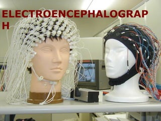
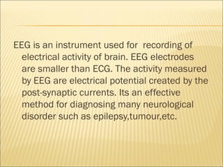
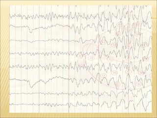
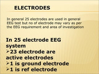
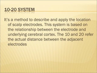
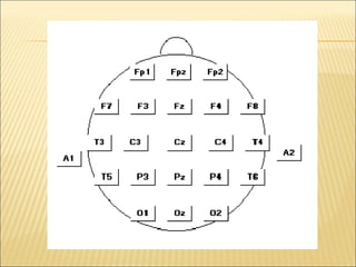
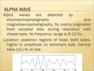
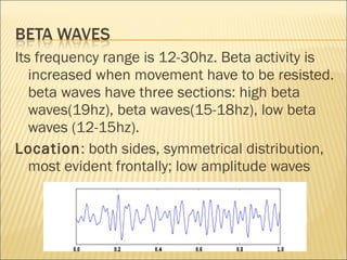
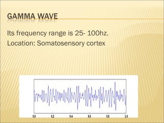
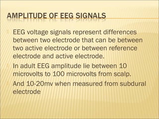
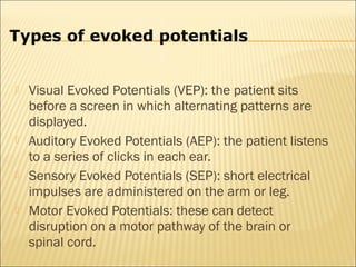
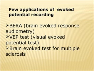
BY: LOKENDRA YADAV
ELECTROENCEPHALOGRAP
H
 ELECTROENCEPHALOGRAPH
EEG is an instrument used for recording of
  electrical activity of brain. EEG electrodes
  are smaller than ECG. The activity measured
  by EEG are electrical potential created by the
  post-synaptic currents. Its an effective
  method for diagnosing many neurological
  disorder such as epilepsy,tumour,etc.
    Biological artifact-
1.   Eye-induced artifacts
2.   Cardiac artifacts
3.   Muscle artifacts
    Environmental artifacts
ELECTRODES

In general 25 electrodes are used in general
EEG test but no of electrode may vary as per
the EEG requirement and area of investigation


In 25 electrode EEG
system
23 electrode are
active electrodes
1 is ground electrode
1 is ref electrode
It’s a method to describe and apply the location
   of scalp electrodes. This system is based on
   the relationship between the electrode and
   underlying cerebral cortex. The 10 and 20 refer
   the actual distance between the adjacent
   electrodes
BLOCK DIAGRAM
    OF EEG
    Montages
    Electrode montage selector
    Preamplifier
    Sensitivity control
    Filters
1.   Low pass filter
2.   High pass filter
3.   Notch filter
   Analog to digital convertor
   Writing unit
   Delta wave
   Theta wave
   Alpha wave
   Beta wave
   Gamma wave
Its frequency range is 1-4hz. It can be recorded
   with an electroencephalogram and usually
   associated with slow wave sleep (SWS).
Location: frontally in adults, posteriorly in
   children; high amplitude waves
Two types of theta wave described as:
Hippocampal theta wave which is observed in
  hippocampus and cortical theta wave which
  low frequency component of scalp EEG
  frequency range from 4-7Hz
Alpha waves are detected by
  electroencephalography                       and
  magnetoencephalography. Its mainly originated
  from occipital lobe during relaxation with
  closed eyes. Its frequency range is 8-12 hz.
Location: posterior regions of head, both sides,
  higher in amplitude on dominant side. Central
  sites (c3-c4) at rest .
Its frequency range is 12-30hz. Beta activity is
   increased when movement have to be resisted.
   beta waves have three sections: high beta
   waves(19hz), beta waves(15-18hz), low beta
   waves (12-15hz).
Location: both sides, symmetrical distribution,
   most evident frontally; low amplitude waves
Its frequency range is 25- 100hz.
Location: Somatosensory cortex
   EEG voltage signals represent differences
    between two electrode that can be between
    two active electrode or between reference
    electrode and active electrode.
   In adult EEG amplitude lie between 10
    microvolts to 100 microvolts from scalp.
   And 10-20mv when measured from subdural
    electrode
   Evoked potentials are used to measure the
    electrical activity in certain areas of the brain
    and spinal cord. Electrical activity is produced
    by stimulation of specific sensory nerve
    pathways.
Types of evoked potentials


   Visual Evoked Potentials (VEP): the patient sits
    before a screen in which alternating patterns are
    displayed.
   Auditory Evoked Potentials (AEP): the patient listens
    to a series of clicks in each ear.
   Sensory Evoked Potentials (SEP): short electrical
    impulses are administered on the arm or leg.
   Motor Evoked Potentials: these can detect
    disruption on a motor pathway of the brain or
    spinal cord.
Few applications of evoked
 potential recording

BERA (brain evoked response
audiometry)
VEP test (visual evoked
potential test)
Brain evoked test for multiple
sclerosis
   For rapid and accurate interpretation, it need
    constant analysis of the EEG. Therefore,
    computerized EEG signal processing to extract
    the frequency and amplitude in simple form.
   QUES…????


    QUERY..???
THANK YOU

EEG

  • 1.
  • 2.
  • 3.
    EEG is aninstrument used for recording of electrical activity of brain. EEG electrodes are smaller than ECG. The activity measured by EEG are electrical potential created by the post-synaptic currents. Its an effective method for diagnosing many neurological disorder such as epilepsy,tumour,etc.
  • 5.
    Biological artifact- 1. Eye-induced artifacts 2. Cardiac artifacts 3. Muscle artifacts  Environmental artifacts
  • 6.
    ELECTRODES In general 25electrodes are used in general EEG test but no of electrode may vary as per the EEG requirement and area of investigation In 25 electrode EEG system 23 electrode are active electrodes 1 is ground electrode 1 is ref electrode
  • 7.
    It’s a methodto describe and apply the location of scalp electrodes. This system is based on the relationship between the electrode and underlying cerebral cortex. The 10 and 20 refer the actual distance between the adjacent electrodes
  • 9.
  • 11.
    Montages  Electrode montage selector  Preamplifier  Sensitivity control  Filters 1. Low pass filter 2. High pass filter 3. Notch filter
  • 12.
    Analog to digital convertor  Writing unit
  • 13.
    Delta wave  Theta wave  Alpha wave  Beta wave  Gamma wave
  • 14.
    Its frequency rangeis 1-4hz. It can be recorded with an electroencephalogram and usually associated with slow wave sleep (SWS). Location: frontally in adults, posteriorly in children; high amplitude waves
  • 15.
    Two types oftheta wave described as: Hippocampal theta wave which is observed in hippocampus and cortical theta wave which low frequency component of scalp EEG frequency range from 4-7Hz
  • 16.
    Alpha waves aredetected by electroencephalography and magnetoencephalography. Its mainly originated from occipital lobe during relaxation with closed eyes. Its frequency range is 8-12 hz. Location: posterior regions of head, both sides, higher in amplitude on dominant side. Central sites (c3-c4) at rest .
  • 17.
    Its frequency rangeis 12-30hz. Beta activity is increased when movement have to be resisted. beta waves have three sections: high beta waves(19hz), beta waves(15-18hz), low beta waves (12-15hz). Location: both sides, symmetrical distribution, most evident frontally; low amplitude waves
  • 18.
    Its frequency rangeis 25- 100hz. Location: Somatosensory cortex
  • 19.
    EEG voltage signals represent differences between two electrode that can be between two active electrode or between reference electrode and active electrode.  In adult EEG amplitude lie between 10 microvolts to 100 microvolts from scalp.  And 10-20mv when measured from subdural electrode
  • 20.
    Evoked potentials are used to measure the electrical activity in certain areas of the brain and spinal cord. Electrical activity is produced by stimulation of specific sensory nerve pathways.
  • 21.
    Types of evokedpotentials  Visual Evoked Potentials (VEP): the patient sits before a screen in which alternating patterns are displayed.  Auditory Evoked Potentials (AEP): the patient listens to a series of clicks in each ear.  Sensory Evoked Potentials (SEP): short electrical impulses are administered on the arm or leg.  Motor Evoked Potentials: these can detect disruption on a motor pathway of the brain or spinal cord.
  • 22.
    Few applications ofevoked potential recording BERA (brain evoked response audiometry) VEP test (visual evoked potential test) Brain evoked test for multiple sclerosis
  • 23.
    For rapid and accurate interpretation, it need constant analysis of the EEG. Therefore, computerized EEG signal processing to extract the frequency and amplitude in simple form.
  • 24.
    QUES…???? QUERY..???
  • 25.