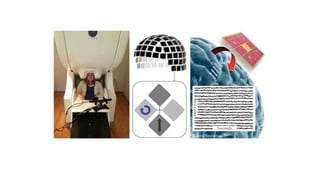
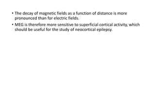
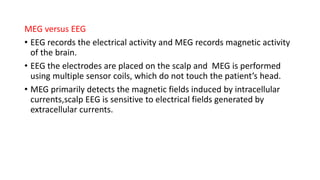
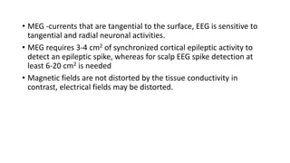
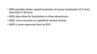
Magnetoencephalography:
Basic principles
PRESENTER- DR.PALLAV JAIN
DM RESIDENT(NEUROLOGY)
GMC,KOTA
Introduction
• Measures the magnetic fields generated by electric currents in the
brain.
• Range of femto-tesla to pico-tesla.
• Very accurate resolution of the timing of neuronal activity
• Non-invasive test.
• MEG is a direct measure of brain
function and it has a very high
temporal resolution and good
spatial resolution.
• MEG is usually combined MRI
Brain to get a good structural
perspective.
• Combination- magnetic source
imaging (MSI).
Basic Physiology
• An electric current is always associated with a magnetic field
perpendicular to its direction.
• When neurons are activated synchronously they generate electric
currents.
• The magnetic permeability of biological tissues is almost the same as
that of empty space ,Not distorted by scalp or skull.
• Magnetic fields diminish as 1/r3 with the distance of ‘r’.
• Source of the magnetic fields is dendritic current of pyramidal
neurons that fire synchronously and in parallel.
• Axonal and synaptic currents and their magnetic fields cancel out.
• The usual amplitude of magnetic fields created by the brain are
extremely small, they do not exceed a few hundred femto tesla
(10−15 T).
Recording
• One is to record these small
magnetic fields and the other is to
shield out the earth’s relatively
stronger magnetic fields.
• Technology - super-conducting
quantum interference detector
which is like a highly sensitive
magnetic field meter.
• Liquid helium which is only 3°
above absolute zero (−452°F or
−270°C).
• Attenuate the external magnetic
noise-housed inside a magnetically
shielded room.
• The actual sensors recording magnetic fields are magnetometers
and/or gradiometers.
• Magnetometers provide the best signal
• Most sensitive to deep brain sources but are also more sensitive to
competing magnetic noise.
• There are 2 types of gradiometers — axial and planar.
• Gradiometers are better at noise reduction.
MEG Basics
• Neuronal generators like equivalent current dipole (ECD)
• MEG does not detect radial dipoles but tangential dipoles are seen on
MEG
• Magnetic field of the radial dipole remains within the cranial cavity
and thus cannot be detected by sensors on the outside
• The decay of magnetic fields as a function of distance is more
pronounced than for electric fields.
• MEG is therefore more sensitive to superficial cortical activity, which
should be useful for the study of neocortical epilepsy.
MEG versus EEG
• EEG records the electrical activity and MEG records magnetic activity
of the brain.
• EEG the electrodes are placed on the scalp and MEG is performed
using multiple sensor coils, which do not touch the patient’s head.
• MEG primarily detects the magnetic fields induced by intracellular
currents,scalp EEG is sensitive to electrical fields generated by
extracellular currents.
• MEG -currents that are tangential to the surface, EEG is sensitive to
tangential and radial neuronal activities.
• MEG requires 3-4 cm2 of synchronized cortical epileptic activity to
detect an epileptic spike, whereas for scalp EEG spike detection at
least 6-20 cm2 is needed
• Magnetic fields are not distorted by the tissue conductivity in
contrast, electrical fields may be distorted.
• MEG provides better spatial resolution of source localization (2-3 mm)
than EEG (7-10 mm).
• MEG data allow for localization in three dimensions.
• MEG more sensitive to superficial cortical activity
• MEG is more expensive than an EEG.
Clinical Applications
MEG in epilepsy
• Surgical treatment -location of the seizure focus
• MEG can substantially contribute to this localization.
• MEG also helps to identify the eloquent cortex and helps determine
the area that can be safely resected during epilepsy surgery.
Source analysis
• Estimating the location of the cortical generators of neuro-magnetic
activity of interest.
• For epilepsy studies, identified IIEDs are most often used for this
purpose.
• Source analysis of slow-wave activity or fast activity is currently under
investigation
• If a seizure is recorded during a study, the onset of the seizure may
be localized using methods for spike analysis
• Source localization by the ECD modeling should be performed on all
well-defined IIEDs(spikes (20-70 ms) and sharp waves (70-200 ms)
• The clinical significance of both types of IIEDs in epileptic focus
localization is equivalent
• Several time points in the IIED waveform can be selected for source
analysis.
• Spike peak or a point on the rising phase of the spike
• Equipment and recording
• Magnetically shielded room that conforms to the current operational
and safety standards should be used.
• Reliable digitization system must be used to locate the head position.
• Determining the position of several “head position indicator” coils
while the patient is in the array.
• The ongoing waveforms of a sampling of MEG and EEG channels
should be displayed in real time to monitor the quality of the
recording.
• Displays of electrooculogram, electrocardiography (ECG) and
electromyogram may also be useful.
• Minimum duration of spontaneous MEG-EEG recording sessions
should be 30 min and preferably, this will include both wakefulness
and sleep.
• Recording during sleep should be a standard part of a spontaneous
MEG-EEG study for epilepsy because of the activating effect of sleep
on IIEDs.
• Hyperventilation may be implemented during MEG-EEG study.
Analysis of data
Waveforms of MEG and EEG for the entire recording should be visually
examined.
(1) identification of artifacts
(2) evaluation of overall data quality and integrity
(3) identification of background rhythms, asymmetries and other
background characteristics and IIEDs, including morphologic and
temporal characteristics, in both MEG and EEG.
MRI brain and co-registration
• A volumetric scan with recognized high neuroanatomic fidelity is
required.
• The field of view should be skin to skin.
• Methods of co-registration depend on MEG system and additional
software used for source localization.
• Reporting
• Clinically relevant information must be provided because source
localizations may guide intracranial electrode placement.
• Interictal discharges and when available ictal rhythms, should be
described as focal, multifocal, or generalized at a minimum.
• Source lateralization and localization, in terms of lobar or sub lobar
area should be summarized.
• Any propagation of interictal or ictal activity should also be described.
• Report should state whether the MEG-EEG source localization is
consistent with the presumed focus based on the previous EEG
findings and the patient’s seizure semiology.
• Anatomic relationship of MEG-EEG source estimates to any MRI lesion
should be described.
Pre-operative Mapping of Eloquent Cortex
• MEG provides an accurate means of functional mapping of eloquent
cortex(identification of motor, sensory, visual and auditory areas)
• Language cortex mapping can also be attempted.
• Shown functional brain tissue inside brain tumors
Other Uses
• Adult and pediatric epilepsy autism
• Schizophrenia
• Williams syndrome
• Landau-Kleffner syndrome
• Alzheimer’s disease
• Depression
• Attention deficit hyperactivity disorder
• Dyslexia
Magnetoencephalography

Magnetoencephalography

  • 1.
  • 2.
    Introduction • Measures themagnetic fields generated by electric currents in the brain. • Range of femto-tesla to pico-tesla. • Very accurate resolution of the timing of neuronal activity • Non-invasive test.
  • 3.
    • MEG isa direct measure of brain function and it has a very high temporal resolution and good spatial resolution. • MEG is usually combined MRI Brain to get a good structural perspective. • Combination- magnetic source imaging (MSI).
  • 5.
    Basic Physiology • Anelectric current is always associated with a magnetic field perpendicular to its direction. • When neurons are activated synchronously they generate electric currents. • The magnetic permeability of biological tissues is almost the same as that of empty space ,Not distorted by scalp or skull. • Magnetic fields diminish as 1/r3 with the distance of ‘r’.
  • 6.
    • Source ofthe magnetic fields is dendritic current of pyramidal neurons that fire synchronously and in parallel. • Axonal and synaptic currents and their magnetic fields cancel out. • The usual amplitude of magnetic fields created by the brain are extremely small, they do not exceed a few hundred femto tesla (10−15 T).
  • 7.
    Recording • One isto record these small magnetic fields and the other is to shield out the earth’s relatively stronger magnetic fields. • Technology - super-conducting quantum interference detector which is like a highly sensitive magnetic field meter. • Liquid helium which is only 3° above absolute zero (−452°F or −270°C). • Attenuate the external magnetic noise-housed inside a magnetically shielded room.
  • 8.
    • The actualsensors recording magnetic fields are magnetometers and/or gradiometers. • Magnetometers provide the best signal • Most sensitive to deep brain sources but are also more sensitive to competing magnetic noise. • There are 2 types of gradiometers — axial and planar. • Gradiometers are better at noise reduction.
  • 9.
    MEG Basics • Neuronalgenerators like equivalent current dipole (ECD) • MEG does not detect radial dipoles but tangential dipoles are seen on MEG • Magnetic field of the radial dipole remains within the cranial cavity and thus cannot be detected by sensors on the outside
  • 10.
    • The decayof magnetic fields as a function of distance is more pronounced than for electric fields. • MEG is therefore more sensitive to superficial cortical activity, which should be useful for the study of neocortical epilepsy.
  • 11.
    MEG versus EEG •EEG records the electrical activity and MEG records magnetic activity of the brain. • EEG the electrodes are placed on the scalp and MEG is performed using multiple sensor coils, which do not touch the patient’s head. • MEG primarily detects the magnetic fields induced by intracellular currents,scalp EEG is sensitive to electrical fields generated by extracellular currents.
  • 12.
    • MEG -currentsthat are tangential to the surface, EEG is sensitive to tangential and radial neuronal activities. • MEG requires 3-4 cm2 of synchronized cortical epileptic activity to detect an epileptic spike, whereas for scalp EEG spike detection at least 6-20 cm2 is needed • Magnetic fields are not distorted by the tissue conductivity in contrast, electrical fields may be distorted.
  • 13.
    • MEG providesbetter spatial resolution of source localization (2-3 mm) than EEG (7-10 mm). • MEG data allow for localization in three dimensions. • MEG more sensitive to superficial cortical activity • MEG is more expensive than an EEG.
  • 14.
    Clinical Applications MEG inepilepsy • Surgical treatment -location of the seizure focus • MEG can substantially contribute to this localization. • MEG also helps to identify the eloquent cortex and helps determine the area that can be safely resected during epilepsy surgery.
  • 15.
    Source analysis • Estimatingthe location of the cortical generators of neuro-magnetic activity of interest. • For epilepsy studies, identified IIEDs are most often used for this purpose. • Source analysis of slow-wave activity or fast activity is currently under investigation • If a seizure is recorded during a study, the onset of the seizure may be localized using methods for spike analysis
  • 16.
    • Source localizationby the ECD modeling should be performed on all well-defined IIEDs(spikes (20-70 ms) and sharp waves (70-200 ms) • The clinical significance of both types of IIEDs in epileptic focus localization is equivalent • Several time points in the IIED waveform can be selected for source analysis. • Spike peak or a point on the rising phase of the spike
  • 17.
    • Equipment andrecording • Magnetically shielded room that conforms to the current operational and safety standards should be used. • Reliable digitization system must be used to locate the head position. • Determining the position of several “head position indicator” coils while the patient is in the array.
  • 18.
    • The ongoingwaveforms of a sampling of MEG and EEG channels should be displayed in real time to monitor the quality of the recording. • Displays of electrooculogram, electrocardiography (ECG) and electromyogram may also be useful. • Minimum duration of spontaneous MEG-EEG recording sessions should be 30 min and preferably, this will include both wakefulness and sleep.
  • 19.
    • Recording duringsleep should be a standard part of a spontaneous MEG-EEG study for epilepsy because of the activating effect of sleep on IIEDs. • Hyperventilation may be implemented during MEG-EEG study.
  • 20.
    Analysis of data Waveformsof MEG and EEG for the entire recording should be visually examined. (1) identification of artifacts (2) evaluation of overall data quality and integrity (3) identification of background rhythms, asymmetries and other background characteristics and IIEDs, including morphologic and temporal characteristics, in both MEG and EEG.
  • 21.
    MRI brain andco-registration • A volumetric scan with recognized high neuroanatomic fidelity is required. • The field of view should be skin to skin. • Methods of co-registration depend on MEG system and additional software used for source localization.
  • 22.
    • Reporting • Clinicallyrelevant information must be provided because source localizations may guide intracranial electrode placement. • Interictal discharges and when available ictal rhythms, should be described as focal, multifocal, or generalized at a minimum. • Source lateralization and localization, in terms of lobar or sub lobar area should be summarized. • Any propagation of interictal or ictal activity should also be described.
  • 23.
    • Report shouldstate whether the MEG-EEG source localization is consistent with the presumed focus based on the previous EEG findings and the patient’s seizure semiology. • Anatomic relationship of MEG-EEG source estimates to any MRI lesion should be described.
  • 24.
    Pre-operative Mapping ofEloquent Cortex • MEG provides an accurate means of functional mapping of eloquent cortex(identification of motor, sensory, visual and auditory areas) • Language cortex mapping can also be attempted. • Shown functional brain tissue inside brain tumors
  • 25.
    Other Uses • Adultand pediatric epilepsy autism • Schizophrenia • Williams syndrome • Landau-Kleffner syndrome • Alzheimer’s disease • Depression • Attention deficit hyperactivity disorder • Dyslexia

Editor's Notes

  • #13 Magnetic fields are not distorted by the tissue conductivity of the scalp, skull, cerebrospinal fluid (CSF) and brain, in contrast, electrical fields may be distorted by the skull and CSF.
  • #14 should be useful for the study of neocortical epilepsy.
  • #18  High-pass filter of 1-4 Hz and low-pass filter of 40-70 Hz.
  • #22 The field of view should be skin to skin, that is, include the face, ears and entire scalp (sagittal orientation of slice acquisition is best) such that an accurate identification of superficial fiducial points is possible for MEG to MRI co-registration. Referring physicians should receive MEG results in the form of EEG and MEG tracings of representative spikes or sharp waves used for source analysis in addition to MSI that contain one dipole source localization and its moment per spike co registered with the patient’s brain MRI.