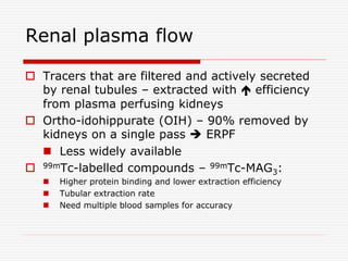
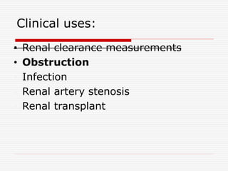
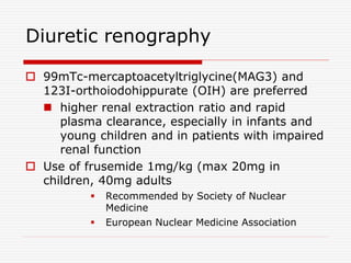
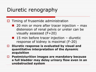
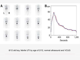
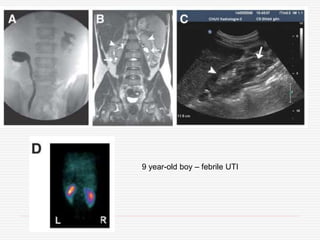
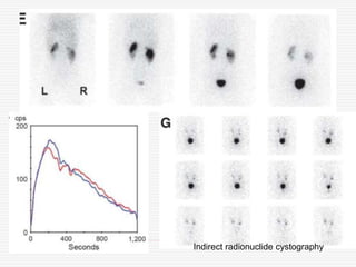
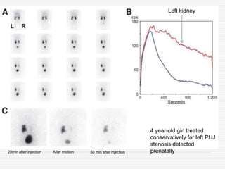
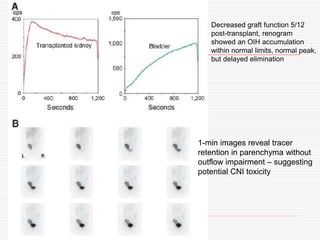
SlideShare a Scribd company logo
Nuclear Imaging in
Nephro-urology
Introduction
 Uses pharmaceuticals – labels with
radionuclides (radiopharmaceuticals) -
radiotracers
 Administers these to patients – radiation
emitted is detected
 Formation of an image using a gamma
camera or positron emission tomography
 Radionuclide imaging / nuclear
scintigraphy
 Shows the physiological function of the
system being investigated
Introduction
 Radiotracers – DTPA, MAG3, OIH etc – taken up by
kidneys, then excreted into urinary tract
 Serial frames of posterior view – acquired 20-30 min
immediately after tracer injection
 Frame rate –
 1-3s per frame for one min to assess perfusion
(perfusion phase)
 10-15s per frame for 4 min to assess function
(function phase)
 10-30s per frame to assess urinary system
(excretion phase)
Overview of renal
radiopharmaceuticals
Renal handling Radiopharmaceu
tical
Imaging Clinical use
Glomerular
filtration
51 Cr-EDTA No GFR
99mTc-DTPA Yes GFR
Tubular secretion 123I/131I-OIH Yes ERPF
99mTc-MAG3 Yes ERPF
99mTc-EC Yes ERPF
Tubular retention 99mTc-DMSA Yes Cortical imaging
99mTc-GH Yes Cortical imaging
Glomerular filtration
 Gold standard – inulin clearance
 Radionuclide of choice – 51Cr-EDTA – clearance
closest to that of inulin
 99mTc-DTPA – technetium-99m-diethylenetriamine
pentaacetic acid – correlates well with 51Cr-EDTA and
inulin
• Taken up by glomerular filtration, not
secreted/reabsorbed by tubules
 99mTc-DTPA can be used for gamma camera imaging
 Least expensive renal radiopharmaceutical
 Low radiation dose
 Small fraction may be bound to protein – not a
problem for routine measurement of GFR
 Once reaches kidney – 20% is accumulated and
remainder flows away, i.e. extraction fraction of DTPA
is 20%
Tubular secretion (1)
 p-Aminohippuric acid (PAH) – gold standard
for measurement of tubular cell function and its
clearance – effective renal plasma flow (ERPF)
 123I-OIH and 131I-OIH – cleared by tubular secretion
(small fraction by glomerular filtration)
 Clearance rate – approx 500-600ml/min
 Extraction of 131I-OIH depends on renal plasma flow
and extraction from plasma (proximal tubules)
 123I-OIH – better imaging qualities, more expensive
Tubular secretion (2)
 99mTc-MAG3
 highly protein-bound, cleared mainly by
proximal tubules
 High extraction fraction (50%) – better
scintigraphic images
 99mTc-I, I and d,d-ethylenedicysteine
(EC) – better than 99mTc-MAG3
65 year-old with contrast nephropathy on CKD – better image of TC-MAG than DTPA scan
Tubular retention
 99mTc-dimercaptosuccinic acid (DMSA)
 Excellent cortical imaging agent
 Concentrates largely in renal cortex (lesser in liver)
 Strongly bound to proteins
 At 2h post-injection : 50% retained in kidneys, no
visualization of urinary collecting system
 99mTc-glucoheptonate (GH)
 Filtered by glomerulus and bound by tubules
 Highly protein-bound – glomerular filtration partial
DMSA
 IV injection – bound to proximal tubules
 Indications:
 Assessment of kidneys in acute phase of UTI
(acute pyelonephritis)
 Assessment of kidneys in late phase of UTI –
detection of scar
 Assessment of horseshoe, solitary or ectopic
kidney
 Localisation of poor or very poorly functioning
kidney
 Assessment of renal function in the presence of
an abdominal mass
BNMS guidelines
DMSA
 Pitfalls
 Acute and chronic pyelonephritis cannot be distinguished
on the cortical scan. If a defect is present 6 months after
the last UTI then this is a scar
 A recent UTI may cause temporary reduced uptake / focal
defect and a follow-up DMSA scan should be undertaken
 The diagnosis of renal scars is difficult in the infant under
3-6 months of age because of renal immaturity. If
appropriate the scan should be delayed
 Controversies
 To obtain the highest resolution, some centres recommend
the use of a pin hole collimator, however many institutions
obtain high resolution images with clear definition between
cortex and medulla without the use of pin-hole collimation
 Currently there is no evidence to support the routine use of
SPECT in children to delineate focal defects
BNMS guidelines
Analytic methods
 Various methods
 Single-sample methods and camera-based
methods suitable for busy clinical practice
 Single-sample – single venous blood sampling
after tracer injection, and plasma radioactivity
is measured
 Plasma activity and injection dose
substituted in a predefined, empiric
equation = renal clearance
Analytic methods
 Camera-based methods – renal uptake early
after tracer injection reflects renal function
 Calculate renal function from imaging data without blood
sampling
 A region of interest (ROI) is drawn for each kidney to
estimate activity, then do a background subtraction, then
attenuation correction for depth of each kidney, then
normalized to the injection dose – finally substituted to an
empiric equation = renal clearance
 Less accurate than single-sample method
 Can assess right and left kidney function separately – split
renal function
 Can do combined single-sample and camera-based
methods
Isotopic Scan for Diagnosis of Renal Disease. Yusuke Inoue et al. Saudi Journal of Kidney Diseases and Transplantation. 15(3) 2004; 257-64
Analytic methods
 Renogram curves – overview of the time
course of renal radioactivity
 Generated by setting an ROI for each kidney
 Radioactivity in a kidney derives from renal parenchyma,
upper tract and overlapping extrarenal tissues
 Do background subtraction - renal parenchyma and upper
tract
 In normal subjects – a renogram demonstrates rapid
increase during perfusion and function phases, then rapid
decline during excretion phases
 Hypofunction flattens the slopes during the function and
excretion phases
 Obstruction causes delayed excretion
 Cannot discriminate between retention in renal
parenchyma and that in urinary tract – visual assessment
of scintigraphy images needed…
Clinical uses:
 Renal clearance measurements
 Obstruction
 Infection
 Renal artery stenosis
 Renal transplant
Renal clearance measurements
 GFR and estimation of split renal
function
 Renal Plasma Flow
Glomerular Filtration Rate
 Substances suitable for measuring
GFR
 Low protein binding
 Negligible non-renal excretion
 Freely filtered
 Chemically stable and inert
 INULIN – Gold standard
GFR
 Radioactive tracers – simplifies sample
measurements
 Single injection
 51Chromium ethylenediamine tetraacetic acid
(51Cr-EDTA) – agent of choice
 99mTc-DTPA – can be used – prepared from
kits - variability in stability and protein-binding
 Short half-life (6 hours)
 Advantage – imaging and GFR measurement
can be performed at the same time
GFR - 51Cr-EDTA
 After iv injection – EDTA rapidly diffuses
throughout bloodstream, equilibrates more
slowly with EVF
 Measure the plasma conc at 2, 3 and 4
hours after injection
 Back extrapolation to time of
injectiondistribution volume of extacellular
space, and slope of declining conc is
fractional clearance of this space  GFR
 Expressed in ml/min or normalised to body
surface area
GFR - 51Cr-EDTA
 A number of attempts to simplify GFR
measurements
 Considerable variation due to age and body
dimensions
 High errors
 All single injection methods – assume patient
in a steady state with respect to
hemodynamics and fluid exchange
 Not accurate e.g. major surgery, transfusion
or dialysis
EDTA
 GFR - plasma clearance curve - which required multiple
blood samples to be taken over a period of several hours
 Slope-intercept method – after numerous simplification
 number of sample : 1 (recommended by the
Radionuclides in Nephrology Committee on Renal
Clearance (Blaufox et al, 1996) )
 It is recommended that the plasma clearance of EDTA
from venous samples be taken as the standard measure
of GFR
 DTPA does have some technical advantages over EDTA
but normal ranges are not so well established
 Small systematic differences have been observed
between GFR measurements obtained from EDTA and
DTPA (Rehling et al, 1984, Fleming et al, 1991, Biggi et
al, 1995)
Renal plasma flow
 Tracers that are filtered and actively secreted
by renal tubules – extracted with  efficiency
from plasma perfusing kidneys
 Ortho-idohippurate (OIH) – 90% removed by
kidneys on a single pass  ERPF
 Less widely available
 99mTc-labelled compounds – 99mTc-MAG3:
 Higher protein binding and lower extraction efficiency
 Tubular extraction rate
 Need multiple blood samples for accuracy
Clinical uses:
• Renal clearance measurements
• Obstruction
• Infection
• Renal artery stenosis
• Renal transplant
Obstructive Uropathy
 Obstruction of the urinary flow
anywhere from the renal pelvis to the
urethra
 Can be acute or chronic
 In adults most commonly caused by
tumor or prostatic enlargement
(hyperplasia or malignancy)
 Need to have bilateral obstruction in
order to have renal insufficiency
Prenatal Hydronephrosis
 Prenatal hydronephrosis (collecting
system dilatation) can be identified
on prenatal ultrasound in about 1% of
patients (0.3-4.5% and is bilateral in
37-57% of cases.
 Followup is generally recommended if
the AP diameter of the collecting
system is 7-10 mm during the 3rd
trimester of pregnancy.
Prenatal Hydronephrosis
 The first follow up exam should be
performed during the 1st week after
birth- but not during the first 72
hours because of reduced urine
output after delivery.
 On post-natal follow-up, the dilatation
will resolve in 20-50% of cases.
Obstruction/Urinary tract
dilatation
 Diuretic renography – diagnostic
work-up of upper tract dilatation, and
follow-up of patients with
hydronephrosis
 Method of choice – to differentiate a
dilated unobstructed urinary system
from a true stenosis
 Can also assess urine flow and renal
function
In children:
 Sedation should be avoided – may interfere
with bladder voiding
 Increased radiation exposure to bladder
mucosa – concern
 Receiving the child with parents in a dedicated,
calm environment
 If needed – local guidelines for sedation
 Short inhalation of equimolar mixture of
nitrous oxide and oxygen
J Nucl Med 2006; 47:1819–1836
Diuretic renography
 99mTc-mercaptoacetyltriglycine(MAG3) and
123I-orthoiodohippurate (OIH) are preferred
 higher renal extraction ratio and rapid
plasma clearance, especially in infants and
young children and in patients with impaired
renal function
 Use of frusemide 1mg/kg (max 20mg in
children, 40mg adults
 Recommended by Society of Nuclear
Medicine
 European Nuclear Medicine Association
Diuretic renography
 Timing of frusemide administration
 20 min or more after tracer injection – max
distension of renal pelvis or ureter can be
visually assessed (F+20)
 15 min before tracer injection – diuretic
response of kidney is maximal (F-20)
 Diuretic response is evaluated by visual and
quantitative interpretation of the dynamic
acquisition
 Postmicturition images are mandatory because
a full bladder may delay urinary flow even in an
unobstructed system
Diuretic renography
 The role of bladder catheterization – debated -
not recommended in clinical routine practice
 Older children and adults - renography is
performed after bladder emptying
 Non–toilet-trained children, spontaneous
micturition is usually observed during the
acquisition
 Adequate functioning of the affected
kidney (GFR > 15 mL/min) and adequate
hydration are major determinants of the
response to frusemide
Limitation/pitfall of diuretic
renography
6/12 old boy, febrile UTI by age of 2/12, normal ultrasound and VCUG
9 year-old boy – febrile UTI
Indirect radionuclide cystography
4 year-old girl treated
conservatively for left PUJ
stenosis detected
prenatally
Left kidney
20min after injection After miction 50 min after injection
Clinical uses:
• Renal clearance measurements
• Obstruction
• Infection
• Renal artery stenosis
• Renal transplant
UTI
 Frequent in children
 Affect girls 2x > boys
 80% first infections diagnosed during
first 2 years of life
 5% in infants and young children –
fever of unexplained origin
 Diagnosis – urine culture
 Risk of renal damage – delay of
diagnosis/treatment, number of UTIs
UTI
 The Subcommittee on Urinary Tract
Infection of the American Academy of
Pediatric Committee on Quality
Improvement recommended imaging
(mainly sonography, VCUG, or
radionuclide cystography) of the urinary
tract in children younger than 2 y old
but considered the role of cortical renal
scintigraphy still to be unclear despite
its recognized high sensitivity
Cortical scintigraphy
 Scintigraphy with 99mTc-dimercaptosuccinic
acid (DMSA) – simple and non-invasive
 Static imaging 2-4 hours after iv injection
 Delayed or post-frusemide images in
hydronephrosis
 The sensitivity of 99mTc-DMSA for the
detection of parenchymal defects due to
infection - from 80% to 100%
 does not allow differentiation of acute
pyelonephritis from renal scars
Cortical scintigraphy
 Abnormal findings on cortical scintigraphy are found in
52%–78% of children during acute pyelonephritis, and
the risk that a renal scar will develop can reach 60%
 The role of cortical scintigraphy is still largely debated in
acute pyelonephritis but is widely accepted in the
detection of renal scars
 99mTc-DMSA scintigraphy is the reference method
for detecting renal sequelae after UTI, is more
sensitive than sonography, and should be
performed no sooner than 6 mo after the last
documented UTI
 Also used to detect scars in VUR
Radionuclide cystography
 Direct radionuclide cystograhy
 A radiologic-VCUG alternative - lower
radiation burden
 As invasive as VCUG – bladder
catheterization
 More sensitive – acquisition is continuous in
both filling and voiding phase
 Indirect radionuclide cystography
 Performed after conventional renography with 99mTc-
MAG3 or 123I-OIH – high excretion rate
 No need catheterization
 Less sensitive and specific than VCUG/direct
cystography
Clinical uses:
• Renal clearance measurements
• Obstruction
• Infection
• Renal artery stenosis
• Renal transplant
Renovascular hypertension
 3 – 5% of all hypertensive patients
 15 – 30% of referred patients for refractory
hypertension
 Renal hypoperfusion  secondary
activation of renin-angiotension system
 Stenotic or obstructive lesion within renal
artery
 Potential curable cause of hypertension
 70-90% - atherosclerosis
 10-30% - fibromuscular dysplasia
Renovascular hypertension
 RVH is a consequence of activation of RAS with
concomitant release of angiotensin II (a
vasoconstrictor) and aldosterone (leading to
plasma volume expansion) to maintain
physiologic renal perfusion by increasing blood
pressure
 Clinical features:
 abdominal bruits, rapid onset of hypertension,
refractory hypertension, unilateral renal atrophy,
azotemia (esp when worsened by ACEi or ARB),
unexplained azotemia/hypokemia
 Episodes of flash pulmonary edema in patients with
relatively well-preserved systolic function
 Gold standard – Renal angiography
Captopril-enhanced renography
 To determine which patients can expect normalization of
BP or improvement of BP control after revascularization
 ACEi reduce the conversion of angiotensin I 
angiotensin II - diminishing the vasoconstriction of the
postglomerular efferent arteriole and decreasing the
GFR, which can be detected by scintigraphy
 Both glomerulus-filtered (99mTc-DTPA) and tubule-
secreted (99mTc-MAG3 or 123I-OIH) - currently used
 ACEi – captopril (25-50mg) orally 60min before
renography; or iv enalaprilat (40mg/kg) over
325min >15min before renography
Captopril-enhanced renography
 Known pitfalls  erroneous results are
 food ingestion within 4 h before receiving
captopril,
 dehydration, hypotension,
 or a full bladder impairing drainage
 ACEi enhanced renography now is rarely used
as the primary imaging tool
 Most reliable in predicting recovery in patients
with FMD
35 year-old : Doppler ultrasound
showing parvus-tardus pattern with
collapsed resistance index
Normal right kidney
Left kidney
Right kidney
75 year-old lady
– OIH study
Typical diagnostic workup for
RVH
Doppler sonography, CT
angiography or MRA
ACE inhibitor
renography
Renal
angiography
and treatment
Clinical uses:
• Renal clearance measurements
• Obstruction
• Infection
• Renal artery stenosis
• Renal transplant
Renal transplantation
 Comprehensive evaluation of renal transplants – in
differential diagnosis of medical and surgical
complications in early post-op and in long-term follow
up
 Selection of patients for biopsy and for various drug
regimens
 Anuric ATN – improving indices of renal function (ERPF,
uptake of tubular tracers) – indicates resolution of tubular
injury
 The protocol: a flow study, scintigram of kidneys, prevoid
and postvoid bladder image, injection site image,
time/acitivity curves of graft and bladder, and quantitative
data of perfusion, function and tracer transit
Report of the Radionuclides in Nehrourology Committee for
evaluationof transplanted kidney (review of techniques).
Dubovsky et al. Semin Nucl Med. 1999 Apr;29(2):175-88
Renal transplantation
 Flow study – 99mmercaptoacetyltriglycine or DTPA
 Quantitative analysis and function phase should
include images and time/activity curves
 Serial studies: decline in function and poor
perfusion indicative of acute rejection
 A normally appearing scintigram without cortical
retention, low function – chronic rejection
 Diuretic renogram - to exclude obstruction
Report of the Radionuclides in Nehrourology Committee for
evaluationof transplanted kidney (review of techniques).
Dubovsky et al. Semin Nucl Med. 1999 Apr;29(2):175-88
Renal transplantation
 A baseline study should be performed
within 1 – 2 days of operation – to
allow comparison of serial studies
because of deteriorating renal
function
 DTPA – tracer of choice in early
stages
 DTPA/MAG3 can be used later stages
Indications for renography in
transplantation
Renography in transplantation
 Ultrasound – 1st line evaluation in
renal graft dysfunction
 Renography – must be available on
emergency basis
 Non-visualization – irremediable loss of function
 May not differentiate rejection from ischemia
(RAS)
 ACEi renography – help determine whether
arterial hypertension is dependent on RAS
 Help in diagnosis of urinary complications –
obstruction or leak
Graft dysfunction
1d post-
transplantation in
a 41 y-old
woman
5d post-transplantation
6d post-transplantation – sudden anuria and abdominal pain
Decreased graft function 5/12
post-transplant, renogram
showed an OIH accumulation
within normal limits, normal peak,
but delayed elimination
1-min images reveal tracer
retention in parenchyma without
outflow impairment – suggesting
potential CNI toxicity
Conclusion
 Role of nuclear medicine – in investigation of
renal parenchymal function and upper urinary
tract abnormalities
 Radiation burden low
 Do not require sedation or specific patient
preparation
 Easy to perform
 Knowledge of renal patho-physiology and
recognition of limitation and technical pitfalls
essential

More Related Content

What's hot

HIDA Scan
HIDA ScanHIDA Scan
MRI IN UROLOGY
MRI IN UROLOGYMRI IN UROLOGY
MRI IN UROLOGY
GovtRoyapettahHospit
 
Mri in urology
Mri in urologyMri in urology
Mri in urology
Prateek Laddha
 
Diagnotic Imaging of Nephrocalcinosis
Diagnotic Imaging of NephrocalcinosisDiagnotic Imaging of Nephrocalcinosis
Diagnotic Imaging of Nephrocalcinosis
Mohamed M.A. Zaitoun
 
Scans.. Dr.Padmesh
Scans.. Dr.PadmeshScans.. Dr.Padmesh
Scans.. Dr.Padmesh
Dr Padmesh Vadakepat
 
CT Urography
CT UrographyCT Urography
Ct mri urography
Ct mri urographyCt mri urography
Ct mri urography
Dev Lakhera
 
Dtpa in pujo
Dtpa in pujoDtpa in pujo
Dtpa in pujo
Praveen Ganji
 
Renal ultrasound nephrology day almansoura aldawly dr aboelfetoh
Renal ultrasound    nephrology  day almansoura aldawly  dr aboelfetohRenal ultrasound    nephrology  day almansoura aldawly  dr aboelfetoh
Renal ultrasound nephrology day almansoura aldawly dr aboelfetoh
FarragBahbah
 
CT urography
CT urography CT urography
CT urography
SabitaMandal1
 
Isotope
IsotopeIsotope
Isotope
FarragBahbah
 
Mrcp Radiology
Mrcp RadiologyMrcp Radiology
Mrcp Radiology
kunalj000
 
Intravenous urography
Intravenous urographyIntravenous urography
Intravenous urography
RamanGhimire3
 
MCU- Micturating cysto-urethrogram
MCU- Micturating cysto-urethrogramMCU- Micturating cysto-urethrogram
MCU- Micturating cysto-urethrogram
Dr. Mohit Goel
 
Radionuclides in urology
Radionuclides in urologyRadionuclides in urology
Radionuclides in urology
Roshan Shetty
 
Magnetic resonance cholangiopancreatography ppt
Magnetic resonance cholangiopancreatography pptMagnetic resonance cholangiopancreatography ppt
Magnetic resonance cholangiopancreatography ppt
Anjan Dangal
 
Mri nephrology 2017
Mri nephrology 2017Mri nephrology 2017
Mri nephrology 2017
FarragBahbah
 
Ultrasound
UltrasoundUltrasound
UltrasoundRad Tech
 
Nuclear endocrinology
Nuclear endocrinologyNuclear endocrinology
Nuclear endocrinologyLudwig Rivero
 

What's hot (20)

HIDA Scan
HIDA ScanHIDA Scan
HIDA Scan
 
MRI IN UROLOGY
MRI IN UROLOGYMRI IN UROLOGY
MRI IN UROLOGY
 
Mri in urology
Mri in urologyMri in urology
Mri in urology
 
Diagnotic Imaging of Nephrocalcinosis
Diagnotic Imaging of NephrocalcinosisDiagnotic Imaging of Nephrocalcinosis
Diagnotic Imaging of Nephrocalcinosis
 
Scans.. Dr.Padmesh
Scans.. Dr.PadmeshScans.. Dr.Padmesh
Scans.. Dr.Padmesh
 
CT Urography
CT UrographyCT Urography
CT Urography
 
Ct mri urography
Ct mri urographyCt mri urography
Ct mri urography
 
Dtpa in pujo
Dtpa in pujoDtpa in pujo
Dtpa in pujo
 
Renal ultrasound nephrology day almansoura aldawly dr aboelfetoh
Renal ultrasound    nephrology  day almansoura aldawly  dr aboelfetohRenal ultrasound    nephrology  day almansoura aldawly  dr aboelfetoh
Renal ultrasound nephrology day almansoura aldawly dr aboelfetoh
 
CT urography
CT urography CT urography
CT urography
 
Isotope
IsotopeIsotope
Isotope
 
Mrcp Radiology
Mrcp RadiologyMrcp Radiology
Mrcp Radiology
 
Intravenous urography
Intravenous urographyIntravenous urography
Intravenous urography
 
MCU- Micturating cysto-urethrogram
MCU- Micturating cysto-urethrogramMCU- Micturating cysto-urethrogram
MCU- Micturating cysto-urethrogram
 
Radionuclides in urology
Radionuclides in urologyRadionuclides in urology
Radionuclides in urology
 
Diuretic renography
Diuretic renographyDiuretic renography
Diuretic renography
 
Magnetic resonance cholangiopancreatography ppt
Magnetic resonance cholangiopancreatography pptMagnetic resonance cholangiopancreatography ppt
Magnetic resonance cholangiopancreatography ppt
 
Mri nephrology 2017
Mri nephrology 2017Mri nephrology 2017
Mri nephrology 2017
 
Ultrasound
UltrasoundUltrasound
Ultrasound
 
Nuclear endocrinology
Nuclear endocrinologyNuclear endocrinology
Nuclear endocrinology
 

Similar to Nuclear medicine in nerphology

Renal Dynamic Scan - Isotope Scan - DTPA
Renal Dynamic Scan - Isotope Scan - DTPARenal Dynamic Scan - Isotope Scan - DTPA
Renal Dynamic Scan - Isotope Scan - DTPA
BhanumurthyKaushikMa
 
Neuclar medicine lecture -.pptx
Neuclar medicine lecture -.pptxNeuclar medicine lecture -.pptx
Neuclar medicine lecture -.pptx
Aya Faroug
 
Renal Scan
Renal ScanRenal Scan
Renal Scans, Scintigraphy.pptx
Renal Scans, Scintigraphy.pptxRenal Scans, Scintigraphy.pptx
Renal Scans, Scintigraphy.pptx
Muhammad Jalal Khan
 
RENAL SCINTIGRAPHY.pptx
RENAL SCINTIGRAPHY.pptxRENAL SCINTIGRAPHY.pptx
RENAL SCINTIGRAPHY.pptx
sahalzain1
 
Renal biopsy seminar
Renal biopsy seminarRenal biopsy seminar
Renal biopsy seminar
Vishal Golay
 
ganesh babu ct uro presentation.pptx
ganesh babu     ct uro presentation.pptxganesh babu     ct uro presentation.pptx
ganesh babu ct uro presentation.pptx
shasshankk12345
 
Investigation of Biliary Tract
Investigation of Biliary Tract Investigation of Biliary Tract
Investigation of Biliary Tract
Scholars of Medical Imaging BPKIHS Dharan
 
Investigation of Biliary Tract
Investigation of Biliary Tract Investigation of Biliary Tract
Investigation of Biliary Tract
Sujan Poudel
 
nuclear urology.pptx
nuclear urology.pptxnuclear urology.pptx
nuclear urology.pptx
Islah Raoof
 
Nuclear imaging in dentistry
Nuclear imaging in dentistryNuclear imaging in dentistry
Nuclear imaging in dentistry
Mammootty Ik
 
Standard versus tubeless mini percutaneous nephrolithotomy
Standard versus tubeless mini percutaneous nephrolithotomyStandard versus tubeless mini percutaneous nephrolithotomy
Standard versus tubeless mini percutaneous nephrolithotomy
Youttam Laudari
 
Skeleton systems examination in medical
Skeleton systems examination in medical Skeleton systems examination in medical
Skeleton systems examination in medical
C L GUPTA EYE INSTITUTE MORADABAD UTTER PRADESH
 
Molecular imaging and therapy in prostate cancer
Molecular imaging and therapy in prostate cancerMolecular imaging and therapy in prostate cancer
Molecular imaging and therapy in prostate cancer
Molecular Imaging Society of India (MISI)
 
medicine.Kidney 2.(dr.ala)
medicine.Kidney 2.(dr.ala)medicine.Kidney 2.(dr.ala)
medicine.Kidney 2.(dr.ala)student
 
GIT and RENAL SYSTEM NUCLEAR MEDICINE PROCEDURES
GIT and RENAL SYSTEM  NUCLEAR MEDICINE PROCEDURESGIT and RENAL SYSTEM  NUCLEAR MEDICINE PROCEDURES
GIT and RENAL SYSTEM NUCLEAR MEDICINE PROCEDURESVipin Kumar
 
Nuclear Medicine Procedures-3
Nuclear Medicine Procedures-3Nuclear Medicine Procedures-3
Nuclear Medicine Procedures-3
Ashraf Zytoon
 
IVP by Dr.Anil.ppt
IVP by Dr.Anil.pptIVP by Dr.Anil.ppt
IVP by Dr.Anil.ppt
anilrawat684816
 
Renal trauma kidney injury
Renal trauma kidney injuryRenal trauma kidney injury
Renal trauma kidney injury
Rojan Adhikari
 
Magnetic Resonance Cholangiopancreatography- MRCP
Magnetic Resonance Cholangiopancreatography- MRCPMagnetic Resonance Cholangiopancreatography- MRCP
Magnetic Resonance Cholangiopancreatography- MRCP
Nitish Virmani
 

Similar to Nuclear medicine in nerphology (20)

Renal Dynamic Scan - Isotope Scan - DTPA
Renal Dynamic Scan - Isotope Scan - DTPARenal Dynamic Scan - Isotope Scan - DTPA
Renal Dynamic Scan - Isotope Scan - DTPA
 
Neuclar medicine lecture -.pptx
Neuclar medicine lecture -.pptxNeuclar medicine lecture -.pptx
Neuclar medicine lecture -.pptx
 
Renal Scan
Renal ScanRenal Scan
Renal Scan
 
Renal Scans, Scintigraphy.pptx
Renal Scans, Scintigraphy.pptxRenal Scans, Scintigraphy.pptx
Renal Scans, Scintigraphy.pptx
 
RENAL SCINTIGRAPHY.pptx
RENAL SCINTIGRAPHY.pptxRENAL SCINTIGRAPHY.pptx
RENAL SCINTIGRAPHY.pptx
 
Renal biopsy seminar
Renal biopsy seminarRenal biopsy seminar
Renal biopsy seminar
 
ganesh babu ct uro presentation.pptx
ganesh babu     ct uro presentation.pptxganesh babu     ct uro presentation.pptx
ganesh babu ct uro presentation.pptx
 
Investigation of Biliary Tract
Investigation of Biliary Tract Investigation of Biliary Tract
Investigation of Biliary Tract
 
Investigation of Biliary Tract
Investigation of Biliary Tract Investigation of Biliary Tract
Investigation of Biliary Tract
 
nuclear urology.pptx
nuclear urology.pptxnuclear urology.pptx
nuclear urology.pptx
 
Nuclear imaging in dentistry
Nuclear imaging in dentistryNuclear imaging in dentistry
Nuclear imaging in dentistry
 
Standard versus tubeless mini percutaneous nephrolithotomy
Standard versus tubeless mini percutaneous nephrolithotomyStandard versus tubeless mini percutaneous nephrolithotomy
Standard versus tubeless mini percutaneous nephrolithotomy
 
Skeleton systems examination in medical
Skeleton systems examination in medical Skeleton systems examination in medical
Skeleton systems examination in medical
 
Molecular imaging and therapy in prostate cancer
Molecular imaging and therapy in prostate cancerMolecular imaging and therapy in prostate cancer
Molecular imaging and therapy in prostate cancer
 
medicine.Kidney 2.(dr.ala)
medicine.Kidney 2.(dr.ala)medicine.Kidney 2.(dr.ala)
medicine.Kidney 2.(dr.ala)
 
GIT and RENAL SYSTEM NUCLEAR MEDICINE PROCEDURES
GIT and RENAL SYSTEM  NUCLEAR MEDICINE PROCEDURESGIT and RENAL SYSTEM  NUCLEAR MEDICINE PROCEDURES
GIT and RENAL SYSTEM NUCLEAR MEDICINE PROCEDURES
 
Nuclear Medicine Procedures-3
Nuclear Medicine Procedures-3Nuclear Medicine Procedures-3
Nuclear Medicine Procedures-3
 
IVP by Dr.Anil.ppt
IVP by Dr.Anil.pptIVP by Dr.Anil.ppt
IVP by Dr.Anil.ppt
 
Renal trauma kidney injury
Renal trauma kidney injuryRenal trauma kidney injury
Renal trauma kidney injury
 
Magnetic Resonance Cholangiopancreatography- MRCP
Magnetic Resonance Cholangiopancreatography- MRCPMagnetic Resonance Cholangiopancreatography- MRCP
Magnetic Resonance Cholangiopancreatography- MRCP
 

More from Lokender Yadav

Nuclear imaging and PET physics
Nuclear imaging and PET physicsNuclear imaging and PET physics
Nuclear imaging and PET physics
Lokender Yadav
 
Clinical applications of CBCT
Clinical applications of CBCTClinical applications of CBCT
Clinical applications of CBCT
Lokender Yadav
 
Dental lab basics & CAD CAM
Dental lab basics & CAD CAMDental lab basics & CAD CAM
Dental lab basics & CAD CAM
Lokender Yadav
 
Training development
Training developmentTraining development
Training development
Lokender Yadav
 
Satellite
SatelliteSatellite
Satellite
Lokender Yadav
 
Remote sensing
Remote sensingRemote sensing
Remote sensing
Lokender Yadav
 
Dicom
DicomDicom
Chromotherapy
ChromotherapyChromotherapy
Chromotherapy
Lokender Yadav
 
Dental Light Cure
Dental Light CureDental Light Cure
Dental Light Cure
Lokender Yadav
 
Basics of Lasers
Basics of Lasers Basics of Lasers
Basics of Lasers
Lokender Yadav
 
Soft Skills
Soft Skills Soft Skills
Soft Skills
Lokender Yadav
 
Ethical Hacking & Network Security
Ethical Hacking & Network Security Ethical Hacking & Network Security
Ethical Hacking & Network Security
Lokender Yadav
 
How to handle sales objections
How to handle sales objections  How to handle sales objections
How to handle sales objections
Lokender Yadav
 
Solar energy business opportunity
Solar energy business opportunitySolar energy business opportunity
Solar energy business opportunity
Lokender Yadav
 
Nuclear medicine in gastroenterology
Nuclear medicine in gastroenterologyNuclear medicine in gastroenterology
Nuclear medicine in gastroenterologyLokender Yadav
 
Nuclear Medicine in Thyroidology
Nuclear Medicine in ThyroidologyNuclear Medicine in Thyroidology
Nuclear Medicine in ThyroidologyLokender Yadav
 
Role of nuclear medicine
Role of nuclear medicineRole of nuclear medicine
Role of nuclear medicine
Lokender Yadav
 
secrets of presentation skill
secrets of presentation skillsecrets of presentation skill
secrets of presentation skillLokender Yadav
 

More from Lokender Yadav (20)

Nuclear imaging and PET physics
Nuclear imaging and PET physicsNuclear imaging and PET physics
Nuclear imaging and PET physics
 
Clinical applications of CBCT
Clinical applications of CBCTClinical applications of CBCT
Clinical applications of CBCT
 
Dental lab basics & CAD CAM
Dental lab basics & CAD CAMDental lab basics & CAD CAM
Dental lab basics & CAD CAM
 
Training development
Training developmentTraining development
Training development
 
Satellite
SatelliteSatellite
Satellite
 
Remote sensing
Remote sensingRemote sensing
Remote sensing
 
Dicom
DicomDicom
Dicom
 
Chromotherapy
ChromotherapyChromotherapy
Chromotherapy
 
Dental Light Cure
Dental Light CureDental Light Cure
Dental Light Cure
 
Basics of Lasers
Basics of Lasers Basics of Lasers
Basics of Lasers
 
Soft Skills
Soft Skills Soft Skills
Soft Skills
 
Ethical Hacking & Network Security
Ethical Hacking & Network Security Ethical Hacking & Network Security
Ethical Hacking & Network Security
 
How to handle sales objections
How to handle sales objections  How to handle sales objections
How to handle sales objections
 
Solar energy business opportunity
Solar energy business opportunitySolar energy business opportunity
Solar energy business opportunity
 
Nuclear medicine in gastroenterology
Nuclear medicine in gastroenterologyNuclear medicine in gastroenterology
Nuclear medicine in gastroenterology
 
Nuclear Medicine in Thyroidology
Nuclear Medicine in ThyroidologyNuclear Medicine in Thyroidology
Nuclear Medicine in Thyroidology
 
Role of nuclear medicine
Role of nuclear medicineRole of nuclear medicine
Role of nuclear medicine
 
Physics of ct mri
Physics of ct mriPhysics of ct mri
Physics of ct mri
 
secrets of presentation skill
secrets of presentation skillsecrets of presentation skill
secrets of presentation skill
 
Patient Safety
Patient SafetyPatient Safety
Patient Safety
 

Recently uploaded

ARTHROLOGY PPT NCISM SYLLABUS AYURVEDA STUDENTS
ARTHROLOGY PPT NCISM SYLLABUS AYURVEDA STUDENTSARTHROLOGY PPT NCISM SYLLABUS AYURVEDA STUDENTS
ARTHROLOGY PPT NCISM SYLLABUS AYURVEDA STUDENTS
Dr. Vinay Pareek
 
planning for change nursing Management ppt
planning for change nursing Management pptplanning for change nursing Management ppt
planning for change nursing Management ppt
Thangamjayarani
 
HOT NEW PRODUCT! BIG SALES FAST SHIPPING NOW FROM CHINA!! EU KU DB BK substit...
HOT NEW PRODUCT! BIG SALES FAST SHIPPING NOW FROM CHINA!! EU KU DB BK substit...HOT NEW PRODUCT! BIG SALES FAST SHIPPING NOW FROM CHINA!! EU KU DB BK substit...
HOT NEW PRODUCT! BIG SALES FAST SHIPPING NOW FROM CHINA!! EU KU DB BK substit...
GL Anaacs
 
How STIs Influence the Development of Pelvic Inflammatory Disease.pptx
How STIs Influence the Development of Pelvic Inflammatory Disease.pptxHow STIs Influence the Development of Pelvic Inflammatory Disease.pptx
How STIs Influence the Development of Pelvic Inflammatory Disease.pptx
FFragrant
 
ANATOMY AND PHYSIOLOGY OF URINARY SYSTEM.pptx
ANATOMY AND PHYSIOLOGY OF URINARY SYSTEM.pptxANATOMY AND PHYSIOLOGY OF URINARY SYSTEM.pptx
ANATOMY AND PHYSIOLOGY OF URINARY SYSTEM.pptx
Swetaba Besh
 
Physiology of Chemical Sensation of smell.pdf
Physiology of Chemical Sensation of smell.pdfPhysiology of Chemical Sensation of smell.pdf
Physiology of Chemical Sensation of smell.pdf
MedicoseAcademics
 
NVBDCP.pptx Nation vector borne disease control program
NVBDCP.pptx Nation vector borne disease control programNVBDCP.pptx Nation vector borne disease control program
NVBDCP.pptx Nation vector borne disease control program
Sapna Thakur
 
basicmodesofventilation2022-220313203758.pdf
basicmodesofventilation2022-220313203758.pdfbasicmodesofventilation2022-220313203758.pdf
basicmodesofventilation2022-220313203758.pdf
aljamhori teaching hospital
 
Pharma Pcd Franchise in Jharkhand - Yodley Lifesciences
Pharma Pcd Franchise in Jharkhand - Yodley LifesciencesPharma Pcd Franchise in Jharkhand - Yodley Lifesciences
Pharma Pcd Franchise in Jharkhand - Yodley Lifesciences
Yodley Lifesciences
 
Phone Us ❤85270-49040❤ #ℂall #gIRLS In Surat By Surat @ℂall @Girls Hotel With...
Phone Us ❤85270-49040❤ #ℂall #gIRLS In Surat By Surat @ℂall @Girls Hotel With...Phone Us ❤85270-49040❤ #ℂall #gIRLS In Surat By Surat @ℂall @Girls Hotel With...
Phone Us ❤85270-49040❤ #ℂall #gIRLS In Surat By Surat @ℂall @Girls Hotel With...
Savita Shen $i11
 
Tom Selleck Health: A Comprehensive Look at the Iconic Actor’s Wellness Journey
Tom Selleck Health: A Comprehensive Look at the Iconic Actor’s Wellness JourneyTom Selleck Health: A Comprehensive Look at the Iconic Actor’s Wellness Journey
Tom Selleck Health: A Comprehensive Look at the Iconic Actor’s Wellness Journey
greendigital
 
Ozempic: Preoperative Management of Patients on GLP-1 Receptor Agonists
Ozempic: Preoperative Management of Patients on GLP-1 Receptor Agonists  Ozempic: Preoperative Management of Patients on GLP-1 Receptor Agonists
Ozempic: Preoperative Management of Patients on GLP-1 Receptor Agonists
Saeid Safari
 
How to Give Better Lectures: Some Tips for Doctors
How to Give Better Lectures: Some Tips for DoctorsHow to Give Better Lectures: Some Tips for Doctors
How to Give Better Lectures: Some Tips for Doctors
LanceCatedral
 
heat stroke and heat exhaustion in children
heat stroke and heat exhaustion in childrenheat stroke and heat exhaustion in children
heat stroke and heat exhaustion in children
SumeraAhmad5
 
For Better Surat #ℂall #Girl Service ❤85270-49040❤ Surat #ℂall #Girls
For Better Surat #ℂall #Girl Service ❤85270-49040❤ Surat #ℂall #GirlsFor Better Surat #ℂall #Girl Service ❤85270-49040❤ Surat #ℂall #Girls
For Better Surat #ℂall #Girl Service ❤85270-49040❤ Surat #ℂall #Girls
Savita Shen $i11
 
New Drug Discovery and Development .....
New Drug Discovery and Development .....New Drug Discovery and Development .....
New Drug Discovery and Development .....
NEHA GUPTA
 
Pharynx and Clinical Correlations BY Dr.Rabia Inam Gandapore.pptx
Pharynx and Clinical Correlations BY Dr.Rabia Inam Gandapore.pptxPharynx and Clinical Correlations BY Dr.Rabia Inam Gandapore.pptx
Pharynx and Clinical Correlations BY Dr.Rabia Inam Gandapore.pptx
Dr. Rabia Inam Gandapore
 
Maxilla, Mandible & Hyoid Bone & Clinical Correlations by Dr. RIG.pptx
Maxilla, Mandible & Hyoid Bone & Clinical Correlations by Dr. RIG.pptxMaxilla, Mandible & Hyoid Bone & Clinical Correlations by Dr. RIG.pptx
Maxilla, Mandible & Hyoid Bone & Clinical Correlations by Dr. RIG.pptx
Dr. Rabia Inam Gandapore
 
Light House Retreats: Plant Medicine Retreat Europe
Light House Retreats: Plant Medicine Retreat EuropeLight House Retreats: Plant Medicine Retreat Europe
Light House Retreats: Plant Medicine Retreat Europe
Lighthouse Retreat
 
Dehradun #ℂall #gIRLS Oyo Hotel 9719300533 #ℂall #gIRL in Dehradun
Dehradun #ℂall #gIRLS Oyo Hotel 9719300533 #ℂall #gIRL in DehradunDehradun #ℂall #gIRLS Oyo Hotel 9719300533 #ℂall #gIRL in Dehradun
Dehradun #ℂall #gIRLS Oyo Hotel 9719300533 #ℂall #gIRL in Dehradun
chandankumarsmartiso
 

Recently uploaded (20)

ARTHROLOGY PPT NCISM SYLLABUS AYURVEDA STUDENTS
ARTHROLOGY PPT NCISM SYLLABUS AYURVEDA STUDENTSARTHROLOGY PPT NCISM SYLLABUS AYURVEDA STUDENTS
ARTHROLOGY PPT NCISM SYLLABUS AYURVEDA STUDENTS
 
planning for change nursing Management ppt
planning for change nursing Management pptplanning for change nursing Management ppt
planning for change nursing Management ppt
 
HOT NEW PRODUCT! BIG SALES FAST SHIPPING NOW FROM CHINA!! EU KU DB BK substit...
HOT NEW PRODUCT! BIG SALES FAST SHIPPING NOW FROM CHINA!! EU KU DB BK substit...HOT NEW PRODUCT! BIG SALES FAST SHIPPING NOW FROM CHINA!! EU KU DB BK substit...
HOT NEW PRODUCT! BIG SALES FAST SHIPPING NOW FROM CHINA!! EU KU DB BK substit...
 
How STIs Influence the Development of Pelvic Inflammatory Disease.pptx
How STIs Influence the Development of Pelvic Inflammatory Disease.pptxHow STIs Influence the Development of Pelvic Inflammatory Disease.pptx
How STIs Influence the Development of Pelvic Inflammatory Disease.pptx
 
ANATOMY AND PHYSIOLOGY OF URINARY SYSTEM.pptx
ANATOMY AND PHYSIOLOGY OF URINARY SYSTEM.pptxANATOMY AND PHYSIOLOGY OF URINARY SYSTEM.pptx
ANATOMY AND PHYSIOLOGY OF URINARY SYSTEM.pptx
 
Physiology of Chemical Sensation of smell.pdf
Physiology of Chemical Sensation of smell.pdfPhysiology of Chemical Sensation of smell.pdf
Physiology of Chemical Sensation of smell.pdf
 
NVBDCP.pptx Nation vector borne disease control program
NVBDCP.pptx Nation vector borne disease control programNVBDCP.pptx Nation vector borne disease control program
NVBDCP.pptx Nation vector borne disease control program
 
basicmodesofventilation2022-220313203758.pdf
basicmodesofventilation2022-220313203758.pdfbasicmodesofventilation2022-220313203758.pdf
basicmodesofventilation2022-220313203758.pdf
 
Pharma Pcd Franchise in Jharkhand - Yodley Lifesciences
Pharma Pcd Franchise in Jharkhand - Yodley LifesciencesPharma Pcd Franchise in Jharkhand - Yodley Lifesciences
Pharma Pcd Franchise in Jharkhand - Yodley Lifesciences
 
Phone Us ❤85270-49040❤ #ℂall #gIRLS In Surat By Surat @ℂall @Girls Hotel With...
Phone Us ❤85270-49040❤ #ℂall #gIRLS In Surat By Surat @ℂall @Girls Hotel With...Phone Us ❤85270-49040❤ #ℂall #gIRLS In Surat By Surat @ℂall @Girls Hotel With...
Phone Us ❤85270-49040❤ #ℂall #gIRLS In Surat By Surat @ℂall @Girls Hotel With...
 
Tom Selleck Health: A Comprehensive Look at the Iconic Actor’s Wellness Journey
Tom Selleck Health: A Comprehensive Look at the Iconic Actor’s Wellness JourneyTom Selleck Health: A Comprehensive Look at the Iconic Actor’s Wellness Journey
Tom Selleck Health: A Comprehensive Look at the Iconic Actor’s Wellness Journey
 
Ozempic: Preoperative Management of Patients on GLP-1 Receptor Agonists
Ozempic: Preoperative Management of Patients on GLP-1 Receptor Agonists  Ozempic: Preoperative Management of Patients on GLP-1 Receptor Agonists
Ozempic: Preoperative Management of Patients on GLP-1 Receptor Agonists
 
How to Give Better Lectures: Some Tips for Doctors
How to Give Better Lectures: Some Tips for DoctorsHow to Give Better Lectures: Some Tips for Doctors
How to Give Better Lectures: Some Tips for Doctors
 
heat stroke and heat exhaustion in children
heat stroke and heat exhaustion in childrenheat stroke and heat exhaustion in children
heat stroke and heat exhaustion in children
 
For Better Surat #ℂall #Girl Service ❤85270-49040❤ Surat #ℂall #Girls
For Better Surat #ℂall #Girl Service ❤85270-49040❤ Surat #ℂall #GirlsFor Better Surat #ℂall #Girl Service ❤85270-49040❤ Surat #ℂall #Girls
For Better Surat #ℂall #Girl Service ❤85270-49040❤ Surat #ℂall #Girls
 
New Drug Discovery and Development .....
New Drug Discovery and Development .....New Drug Discovery and Development .....
New Drug Discovery and Development .....
 
Pharynx and Clinical Correlations BY Dr.Rabia Inam Gandapore.pptx
Pharynx and Clinical Correlations BY Dr.Rabia Inam Gandapore.pptxPharynx and Clinical Correlations BY Dr.Rabia Inam Gandapore.pptx
Pharynx and Clinical Correlations BY Dr.Rabia Inam Gandapore.pptx
 
Maxilla, Mandible & Hyoid Bone & Clinical Correlations by Dr. RIG.pptx
Maxilla, Mandible & Hyoid Bone & Clinical Correlations by Dr. RIG.pptxMaxilla, Mandible & Hyoid Bone & Clinical Correlations by Dr. RIG.pptx
Maxilla, Mandible & Hyoid Bone & Clinical Correlations by Dr. RIG.pptx
 
Light House Retreats: Plant Medicine Retreat Europe
Light House Retreats: Plant Medicine Retreat EuropeLight House Retreats: Plant Medicine Retreat Europe
Light House Retreats: Plant Medicine Retreat Europe
 
Dehradun #ℂall #gIRLS Oyo Hotel 9719300533 #ℂall #gIRL in Dehradun
Dehradun #ℂall #gIRLS Oyo Hotel 9719300533 #ℂall #gIRL in DehradunDehradun #ℂall #gIRLS Oyo Hotel 9719300533 #ℂall #gIRL in Dehradun
Dehradun #ℂall #gIRLS Oyo Hotel 9719300533 #ℂall #gIRL in Dehradun
 

Nuclear medicine in nerphology

  • 2. Introduction  Uses pharmaceuticals – labels with radionuclides (radiopharmaceuticals) - radiotracers  Administers these to patients – radiation emitted is detected  Formation of an image using a gamma camera or positron emission tomography  Radionuclide imaging / nuclear scintigraphy  Shows the physiological function of the system being investigated
  • 3. Introduction  Radiotracers – DTPA, MAG3, OIH etc – taken up by kidneys, then excreted into urinary tract  Serial frames of posterior view – acquired 20-30 min immediately after tracer injection  Frame rate –  1-3s per frame for one min to assess perfusion (perfusion phase)  10-15s per frame for 4 min to assess function (function phase)  10-30s per frame to assess urinary system (excretion phase)
  • 4. Overview of renal radiopharmaceuticals Renal handling Radiopharmaceu tical Imaging Clinical use Glomerular filtration 51 Cr-EDTA No GFR 99mTc-DTPA Yes GFR Tubular secretion 123I/131I-OIH Yes ERPF 99mTc-MAG3 Yes ERPF 99mTc-EC Yes ERPF Tubular retention 99mTc-DMSA Yes Cortical imaging 99mTc-GH Yes Cortical imaging
  • 5. Glomerular filtration  Gold standard – inulin clearance  Radionuclide of choice – 51Cr-EDTA – clearance closest to that of inulin  99mTc-DTPA – technetium-99m-diethylenetriamine pentaacetic acid – correlates well with 51Cr-EDTA and inulin • Taken up by glomerular filtration, not secreted/reabsorbed by tubules  99mTc-DTPA can be used for gamma camera imaging  Least expensive renal radiopharmaceutical  Low radiation dose  Small fraction may be bound to protein – not a problem for routine measurement of GFR  Once reaches kidney – 20% is accumulated and remainder flows away, i.e. extraction fraction of DTPA is 20%
  • 6. Tubular secretion (1)  p-Aminohippuric acid (PAH) – gold standard for measurement of tubular cell function and its clearance – effective renal plasma flow (ERPF)  123I-OIH and 131I-OIH – cleared by tubular secretion (small fraction by glomerular filtration)  Clearance rate – approx 500-600ml/min  Extraction of 131I-OIH depends on renal plasma flow and extraction from plasma (proximal tubules)  123I-OIH – better imaging qualities, more expensive
  • 7. Tubular secretion (2)  99mTc-MAG3  highly protein-bound, cleared mainly by proximal tubules  High extraction fraction (50%) – better scintigraphic images  99mTc-I, I and d,d-ethylenedicysteine (EC) – better than 99mTc-MAG3
  • 8. 65 year-old with contrast nephropathy on CKD – better image of TC-MAG than DTPA scan
  • 9. Tubular retention  99mTc-dimercaptosuccinic acid (DMSA)  Excellent cortical imaging agent  Concentrates largely in renal cortex (lesser in liver)  Strongly bound to proteins  At 2h post-injection : 50% retained in kidneys, no visualization of urinary collecting system  99mTc-glucoheptonate (GH)  Filtered by glomerulus and bound by tubules  Highly protein-bound – glomerular filtration partial
  • 10. DMSA  IV injection – bound to proximal tubules  Indications:  Assessment of kidneys in acute phase of UTI (acute pyelonephritis)  Assessment of kidneys in late phase of UTI – detection of scar  Assessment of horseshoe, solitary or ectopic kidney  Localisation of poor or very poorly functioning kidney  Assessment of renal function in the presence of an abdominal mass BNMS guidelines
  • 11. DMSA  Pitfalls  Acute and chronic pyelonephritis cannot be distinguished on the cortical scan. If a defect is present 6 months after the last UTI then this is a scar  A recent UTI may cause temporary reduced uptake / focal defect and a follow-up DMSA scan should be undertaken  The diagnosis of renal scars is difficult in the infant under 3-6 months of age because of renal immaturity. If appropriate the scan should be delayed  Controversies  To obtain the highest resolution, some centres recommend the use of a pin hole collimator, however many institutions obtain high resolution images with clear definition between cortex and medulla without the use of pin-hole collimation  Currently there is no evidence to support the routine use of SPECT in children to delineate focal defects BNMS guidelines
  • 12. Analytic methods  Various methods  Single-sample methods and camera-based methods suitable for busy clinical practice  Single-sample – single venous blood sampling after tracer injection, and plasma radioactivity is measured  Plasma activity and injection dose substituted in a predefined, empiric equation = renal clearance
  • 13. Analytic methods  Camera-based methods – renal uptake early after tracer injection reflects renal function  Calculate renal function from imaging data without blood sampling  A region of interest (ROI) is drawn for each kidney to estimate activity, then do a background subtraction, then attenuation correction for depth of each kidney, then normalized to the injection dose – finally substituted to an empiric equation = renal clearance  Less accurate than single-sample method  Can assess right and left kidney function separately – split renal function  Can do combined single-sample and camera-based methods Isotopic Scan for Diagnosis of Renal Disease. Yusuke Inoue et al. Saudi Journal of Kidney Diseases and Transplantation. 15(3) 2004; 257-64
  • 14. Analytic methods  Renogram curves – overview of the time course of renal radioactivity  Generated by setting an ROI for each kidney  Radioactivity in a kidney derives from renal parenchyma, upper tract and overlapping extrarenal tissues  Do background subtraction - renal parenchyma and upper tract  In normal subjects – a renogram demonstrates rapid increase during perfusion and function phases, then rapid decline during excretion phases  Hypofunction flattens the slopes during the function and excretion phases  Obstruction causes delayed excretion  Cannot discriminate between retention in renal parenchyma and that in urinary tract – visual assessment of scintigraphy images needed…
  • 15.
  • 16.
  • 17. Clinical uses:  Renal clearance measurements  Obstruction  Infection  Renal artery stenosis  Renal transplant
  • 18. Renal clearance measurements  GFR and estimation of split renal function  Renal Plasma Flow
  • 19. Glomerular Filtration Rate  Substances suitable for measuring GFR  Low protein binding  Negligible non-renal excretion  Freely filtered  Chemically stable and inert  INULIN – Gold standard
  • 20. GFR  Radioactive tracers – simplifies sample measurements  Single injection  51Chromium ethylenediamine tetraacetic acid (51Cr-EDTA) – agent of choice  99mTc-DTPA – can be used – prepared from kits - variability in stability and protein-binding  Short half-life (6 hours)  Advantage – imaging and GFR measurement can be performed at the same time
  • 21. GFR - 51Cr-EDTA  After iv injection – EDTA rapidly diffuses throughout bloodstream, equilibrates more slowly with EVF  Measure the plasma conc at 2, 3 and 4 hours after injection  Back extrapolation to time of injectiondistribution volume of extacellular space, and slope of declining conc is fractional clearance of this space  GFR  Expressed in ml/min or normalised to body surface area
  • 22. GFR - 51Cr-EDTA  A number of attempts to simplify GFR measurements  Considerable variation due to age and body dimensions  High errors  All single injection methods – assume patient in a steady state with respect to hemodynamics and fluid exchange  Not accurate e.g. major surgery, transfusion or dialysis
  • 23. EDTA  GFR - plasma clearance curve - which required multiple blood samples to be taken over a period of several hours  Slope-intercept method – after numerous simplification  number of sample : 1 (recommended by the Radionuclides in Nephrology Committee on Renal Clearance (Blaufox et al, 1996) )  It is recommended that the plasma clearance of EDTA from venous samples be taken as the standard measure of GFR  DTPA does have some technical advantages over EDTA but normal ranges are not so well established  Small systematic differences have been observed between GFR measurements obtained from EDTA and DTPA (Rehling et al, 1984, Fleming et al, 1991, Biggi et al, 1995)
  • 24. Renal plasma flow  Tracers that are filtered and actively secreted by renal tubules – extracted with  efficiency from plasma perfusing kidneys  Ortho-idohippurate (OIH) – 90% removed by kidneys on a single pass  ERPF  Less widely available  99mTc-labelled compounds – 99mTc-MAG3:  Higher protein binding and lower extraction efficiency  Tubular extraction rate  Need multiple blood samples for accuracy
  • 25. Clinical uses: • Renal clearance measurements • Obstruction • Infection • Renal artery stenosis • Renal transplant
  • 26. Obstructive Uropathy  Obstruction of the urinary flow anywhere from the renal pelvis to the urethra  Can be acute or chronic  In adults most commonly caused by tumor or prostatic enlargement (hyperplasia or malignancy)  Need to have bilateral obstruction in order to have renal insufficiency
  • 27. Prenatal Hydronephrosis  Prenatal hydronephrosis (collecting system dilatation) can be identified on prenatal ultrasound in about 1% of patients (0.3-4.5% and is bilateral in 37-57% of cases.  Followup is generally recommended if the AP diameter of the collecting system is 7-10 mm during the 3rd trimester of pregnancy.
  • 28. Prenatal Hydronephrosis  The first follow up exam should be performed during the 1st week after birth- but not during the first 72 hours because of reduced urine output after delivery.  On post-natal follow-up, the dilatation will resolve in 20-50% of cases.
  • 29. Obstruction/Urinary tract dilatation  Diuretic renography – diagnostic work-up of upper tract dilatation, and follow-up of patients with hydronephrosis  Method of choice – to differentiate a dilated unobstructed urinary system from a true stenosis  Can also assess urine flow and renal function
  • 30. In children:  Sedation should be avoided – may interfere with bladder voiding  Increased radiation exposure to bladder mucosa – concern  Receiving the child with parents in a dedicated, calm environment  If needed – local guidelines for sedation  Short inhalation of equimolar mixture of nitrous oxide and oxygen J Nucl Med 2006; 47:1819–1836
  • 31. Diuretic renography  99mTc-mercaptoacetyltriglycine(MAG3) and 123I-orthoiodohippurate (OIH) are preferred  higher renal extraction ratio and rapid plasma clearance, especially in infants and young children and in patients with impaired renal function  Use of frusemide 1mg/kg (max 20mg in children, 40mg adults  Recommended by Society of Nuclear Medicine  European Nuclear Medicine Association
  • 32. Diuretic renography  Timing of frusemide administration  20 min or more after tracer injection – max distension of renal pelvis or ureter can be visually assessed (F+20)  15 min before tracer injection – diuretic response of kidney is maximal (F-20)  Diuretic response is evaluated by visual and quantitative interpretation of the dynamic acquisition  Postmicturition images are mandatory because a full bladder may delay urinary flow even in an unobstructed system
  • 33. Diuretic renography  The role of bladder catheterization – debated - not recommended in clinical routine practice  Older children and adults - renography is performed after bladder emptying  Non–toilet-trained children, spontaneous micturition is usually observed during the acquisition  Adequate functioning of the affected kidney (GFR > 15 mL/min) and adequate hydration are major determinants of the response to frusemide
  • 35. 6/12 old boy, febrile UTI by age of 2/12, normal ultrasound and VCUG
  • 36.
  • 37. 9 year-old boy – febrile UTI
  • 39. 4 year-old girl treated conservatively for left PUJ stenosis detected prenatally Left kidney 20min after injection After miction 50 min after injection
  • 40.
  • 41. Clinical uses: • Renal clearance measurements • Obstruction • Infection • Renal artery stenosis • Renal transplant
  • 42. UTI  Frequent in children  Affect girls 2x > boys  80% first infections diagnosed during first 2 years of life  5% in infants and young children – fever of unexplained origin  Diagnosis – urine culture  Risk of renal damage – delay of diagnosis/treatment, number of UTIs
  • 43. UTI  The Subcommittee on Urinary Tract Infection of the American Academy of Pediatric Committee on Quality Improvement recommended imaging (mainly sonography, VCUG, or radionuclide cystography) of the urinary tract in children younger than 2 y old but considered the role of cortical renal scintigraphy still to be unclear despite its recognized high sensitivity
  • 44. Cortical scintigraphy  Scintigraphy with 99mTc-dimercaptosuccinic acid (DMSA) – simple and non-invasive  Static imaging 2-4 hours after iv injection  Delayed or post-frusemide images in hydronephrosis  The sensitivity of 99mTc-DMSA for the detection of parenchymal defects due to infection - from 80% to 100%  does not allow differentiation of acute pyelonephritis from renal scars
  • 45. Cortical scintigraphy  Abnormal findings on cortical scintigraphy are found in 52%–78% of children during acute pyelonephritis, and the risk that a renal scar will develop can reach 60%  The role of cortical scintigraphy is still largely debated in acute pyelonephritis but is widely accepted in the detection of renal scars  99mTc-DMSA scintigraphy is the reference method for detecting renal sequelae after UTI, is more sensitive than sonography, and should be performed no sooner than 6 mo after the last documented UTI  Also used to detect scars in VUR
  • 46.
  • 47.
  • 48.
  • 49. Radionuclide cystography  Direct radionuclide cystograhy  A radiologic-VCUG alternative - lower radiation burden  As invasive as VCUG – bladder catheterization  More sensitive – acquisition is continuous in both filling and voiding phase  Indirect radionuclide cystography  Performed after conventional renography with 99mTc- MAG3 or 123I-OIH – high excretion rate  No need catheterization  Less sensitive and specific than VCUG/direct cystography
  • 50.
  • 51.
  • 52.
  • 53.
  • 54.
  • 55. Clinical uses: • Renal clearance measurements • Obstruction • Infection • Renal artery stenosis • Renal transplant
  • 56. Renovascular hypertension  3 – 5% of all hypertensive patients  15 – 30% of referred patients for refractory hypertension  Renal hypoperfusion  secondary activation of renin-angiotension system  Stenotic or obstructive lesion within renal artery  Potential curable cause of hypertension  70-90% - atherosclerosis  10-30% - fibromuscular dysplasia
  • 57. Renovascular hypertension  RVH is a consequence of activation of RAS with concomitant release of angiotensin II (a vasoconstrictor) and aldosterone (leading to plasma volume expansion) to maintain physiologic renal perfusion by increasing blood pressure  Clinical features:  abdominal bruits, rapid onset of hypertension, refractory hypertension, unilateral renal atrophy, azotemia (esp when worsened by ACEi or ARB), unexplained azotemia/hypokemia  Episodes of flash pulmonary edema in patients with relatively well-preserved systolic function  Gold standard – Renal angiography
  • 58.
  • 59. Captopril-enhanced renography  To determine which patients can expect normalization of BP or improvement of BP control after revascularization  ACEi reduce the conversion of angiotensin I  angiotensin II - diminishing the vasoconstriction of the postglomerular efferent arteriole and decreasing the GFR, which can be detected by scintigraphy  Both glomerulus-filtered (99mTc-DTPA) and tubule- secreted (99mTc-MAG3 or 123I-OIH) - currently used  ACEi – captopril (25-50mg) orally 60min before renography; or iv enalaprilat (40mg/kg) over 325min >15min before renography
  • 60. Captopril-enhanced renography  Known pitfalls  erroneous results are  food ingestion within 4 h before receiving captopril,  dehydration, hypotension,  or a full bladder impairing drainage  ACEi enhanced renography now is rarely used as the primary imaging tool  Most reliable in predicting recovery in patients with FMD
  • 61. 35 year-old : Doppler ultrasound showing parvus-tardus pattern with collapsed resistance index Normal right kidney
  • 63.
  • 64. 75 year-old lady – OIH study
  • 65.
  • 66.
  • 67. Typical diagnostic workup for RVH Doppler sonography, CT angiography or MRA ACE inhibitor renography Renal angiography and treatment
  • 68. Clinical uses: • Renal clearance measurements • Obstruction • Infection • Renal artery stenosis • Renal transplant
  • 69. Renal transplantation  Comprehensive evaluation of renal transplants – in differential diagnosis of medical and surgical complications in early post-op and in long-term follow up  Selection of patients for biopsy and for various drug regimens  Anuric ATN – improving indices of renal function (ERPF, uptake of tubular tracers) – indicates resolution of tubular injury  The protocol: a flow study, scintigram of kidneys, prevoid and postvoid bladder image, injection site image, time/acitivity curves of graft and bladder, and quantitative data of perfusion, function and tracer transit Report of the Radionuclides in Nehrourology Committee for evaluationof transplanted kidney (review of techniques). Dubovsky et al. Semin Nucl Med. 1999 Apr;29(2):175-88
  • 70. Renal transplantation  Flow study – 99mmercaptoacetyltriglycine or DTPA  Quantitative analysis and function phase should include images and time/activity curves  Serial studies: decline in function and poor perfusion indicative of acute rejection  A normally appearing scintigram without cortical retention, low function – chronic rejection  Diuretic renogram - to exclude obstruction Report of the Radionuclides in Nehrourology Committee for evaluationof transplanted kidney (review of techniques). Dubovsky et al. Semin Nucl Med. 1999 Apr;29(2):175-88
  • 71. Renal transplantation  A baseline study should be performed within 1 – 2 days of operation – to allow comparison of serial studies because of deteriorating renal function  DTPA – tracer of choice in early stages  DTPA/MAG3 can be used later stages
  • 72. Indications for renography in transplantation
  • 73. Renography in transplantation  Ultrasound – 1st line evaluation in renal graft dysfunction  Renography – must be available on emergency basis  Non-visualization – irremediable loss of function  May not differentiate rejection from ischemia (RAS)  ACEi renography – help determine whether arterial hypertension is dependent on RAS  Help in diagnosis of urinary complications – obstruction or leak
  • 75.
  • 76.
  • 78. 6d post-transplantation – sudden anuria and abdominal pain
  • 79. Decreased graft function 5/12 post-transplant, renogram showed an OIH accumulation within normal limits, normal peak, but delayed elimination 1-min images reveal tracer retention in parenchyma without outflow impairment – suggesting potential CNI toxicity
  • 80. Conclusion  Role of nuclear medicine – in investigation of renal parenchymal function and upper urinary tract abnormalities  Radiation burden low  Do not require sedation or specific patient preparation  Easy to perform  Knowledge of renal patho-physiology and recognition of limitation and technical pitfalls essential