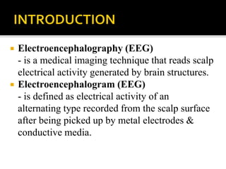
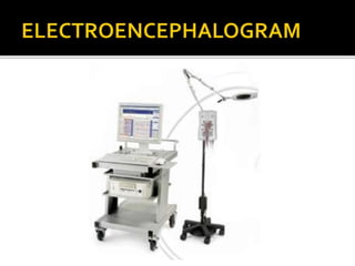
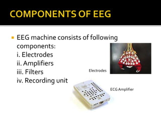
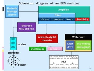
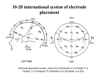
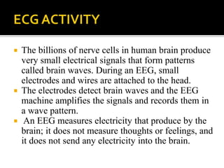
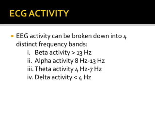
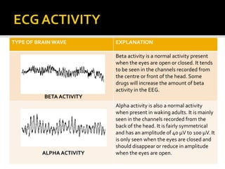
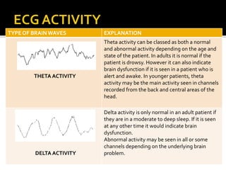
PREPARED BY:
THARSHINI ELANGOVAN 08DEU13F2031
SHALENI KAVIRAJAN 08DEU13F2013
LECTURER:
PN IDA MARIA BT MOHDYUSOFF
 Electroencephalography (EEG)
- is a medical imaging technique that reads scalp
electrical activity generated by brain structures.
 Electroencephalogram (EEG)
- is defined as electrical activity of an
alternating type recorded from the scalp surface
after being picked up by metal electrodes &
conductive media.
 Diagnosis of epilepsy
 Monitor alertness, coma and brain death.
 Locate areas of damage in the brain following
head injuries, stroke, tumor and etc.
 Control anesthesia depth.
 Investigate sleep disorder and physiology.
 EEG machine consists of following
components:
i. Electrodes
ii. Amplifiers
iii. Filters
iv. Recording unit
Electrodes
ECG Amplifier
 EEG electrodes
-> Small metal discs usually
made of stainless, tin, god or
silver covered with a silver
chloride coating.
-> Placed on the scalp in special
positions.These positions are
specified using the
International 10/20 system.
-> Each electrode site is labeled
with a letter and a number.The
letters refers to the area of
brain underlying the electrode
(e.g; F-Frontal lobe andT-
Temporal lobe). Even numbers
denote the right side of the
head and odd numbers denote
the left side of the head.
 Electrode gel
-> acts as a malleable extension
of the electrode, so that the
movement of the electrodes
cables are less likely to produce
artifacts.
->The gel maximizes skin
contact and allows for a low-
resistance recording through
the skin.
->The electrolyte gel is injected
into each cavity until a small
amount out the hole in the
mount.With a moderate
amount of downward pressure,
the syringe with the blunt
needle is rapidly rocked back
and forth.
Types of EEG
machine
Analog
Digital
 Patient will be asked to lie on their back on a bed or
table or relax in a chair with eyes closed.
 The EEG technologist will attach several flat metal
discs (electrodes) to different places on patients’ head,
using a sticky paste to hold the electrodes in place.
 A cap with fixed electrodes may be placed on the head
instead of individual electrodes. In rare cases, these
electrodes may be attached to the scalp with tiny
needles.
 During the recording, patients have to lie with eyes
closed and they are not allowed to talk to the
technologist unless they need to do so.
 The recording may be stopped from time to time to
allow patient to stretch and reposition themselves.
 EEG system covers all parts of brain by
placing electrodes on all parts of brain.
Electrode
Placement
System
10-20
International
System (21
electrodes)
10-10
International
System (64
electrodes)
 The billions of nerve cells in human brain produce
very small electrical signals that form patterns
called brain waves. During an EEG, small
electrodes and wires are attached to the head.
 The electrodes detect brain waves and the EEG
machine amplifies the signals and records them in
a wave pattern.
 An EEG measures electricity that produce by the
brain; it does not measure thoughts or feelings, and
it does not send any electricity into the brain.
 EEG activity can be broken down into 4
distinct frequency bands:
i. Beta activity > 13 Hz
ii. Alpha activity 8 Hz-13 Hz
iii.Theta activity 4 Hz-7 Hz
iv. Delta activity < 4 Hz
TYPE OF BRAIN WAVE EXPLANATION
BETA ACTIVITY
Beta activity is a normal activity present
when the eyes are open or closed. It tends
to be seen in the channels recorded from
the centre or front of the head. Some
drugs will increase the amount of beta
activity in the EEG.
ALPHA ACTIVITY
Alpha activity is also a normal activity
when present in waking adults. It is mainly
seen in the channels recorded from the
back of the head. It is fairly symmetrical
and has an amplitude of 40 µV to 100 µV. It
is only seen when the eyes are closed and
should disappear or reduce in amplitude
when the eyes are open.
TYPE OF BRAIN WAVES EXPLANATION
THETA ACTIVITY
Theta activity can be classed as both a normal
and abnormal activity depending on the age and
state of the patient. In adults it is normal if the
patient is drowsy. However it can also indicate
brain dysfunction if it is seen in a patient who is
alert and awake. In younger patients, theta
activity may be the main activity seen in channels
recorded from the back and central areas of the
head.
DELTA ACTIVITY
Delta activity is only normal in an adult patient if
they are in a moderate to deep sleep. If it is seen
at any other time it would indicate brain
dysfunction.
Abnormal activity may be seen in all or some
channels depending on the underlying brain
problem.

Electroencephalogram

  • 1.
    PREPARED BY: THARSHINI ELANGOVAN08DEU13F2031 SHALENI KAVIRAJAN 08DEU13F2013 LECTURER: PN IDA MARIA BT MOHDYUSOFF
  • 2.
     Electroencephalography (EEG) -is a medical imaging technique that reads scalp electrical activity generated by brain structures.  Electroencephalogram (EEG) - is defined as electrical activity of an alternating type recorded from the scalp surface after being picked up by metal electrodes & conductive media.
  • 4.
     Diagnosis ofepilepsy  Monitor alertness, coma and brain death.  Locate areas of damage in the brain following head injuries, stroke, tumor and etc.  Control anesthesia depth.  Investigate sleep disorder and physiology.
  • 5.
     EEG machineconsists of following components: i. Electrodes ii. Amplifiers iii. Filters iv. Recording unit Electrodes ECG Amplifier
  • 7.
     EEG electrodes ->Small metal discs usually made of stainless, tin, god or silver covered with a silver chloride coating. -> Placed on the scalp in special positions.These positions are specified using the International 10/20 system. -> Each electrode site is labeled with a letter and a number.The letters refers to the area of brain underlying the electrode (e.g; F-Frontal lobe andT- Temporal lobe). Even numbers denote the right side of the head and odd numbers denote the left side of the head.
  • 8.
     Electrode gel ->acts as a malleable extension of the electrode, so that the movement of the electrodes cables are less likely to produce artifacts. ->The gel maximizes skin contact and allows for a low- resistance recording through the skin. ->The electrolyte gel is injected into each cavity until a small amount out the hole in the mount.With a moderate amount of downward pressure, the syringe with the blunt needle is rapidly rocked back and forth.
  • 9.
  • 10.
     Patient willbe asked to lie on their back on a bed or table or relax in a chair with eyes closed.  The EEG technologist will attach several flat metal discs (electrodes) to different places on patients’ head, using a sticky paste to hold the electrodes in place.  A cap with fixed electrodes may be placed on the head instead of individual electrodes. In rare cases, these electrodes may be attached to the scalp with tiny needles.  During the recording, patients have to lie with eyes closed and they are not allowed to talk to the technologist unless they need to do so.  The recording may be stopped from time to time to allow patient to stretch and reposition themselves.
  • 11.
     EEG systemcovers all parts of brain by placing electrodes on all parts of brain. Electrode Placement System 10-20 International System (21 electrodes) 10-10 International System (64 electrodes)
  • 14.
     The billionsof nerve cells in human brain produce very small electrical signals that form patterns called brain waves. During an EEG, small electrodes and wires are attached to the head.  The electrodes detect brain waves and the EEG machine amplifies the signals and records them in a wave pattern.  An EEG measures electricity that produce by the brain; it does not measure thoughts or feelings, and it does not send any electricity into the brain.
  • 15.
     EEG activitycan be broken down into 4 distinct frequency bands: i. Beta activity > 13 Hz ii. Alpha activity 8 Hz-13 Hz iii.Theta activity 4 Hz-7 Hz iv. Delta activity < 4 Hz
  • 16.
    TYPE OF BRAINWAVE EXPLANATION BETA ACTIVITY Beta activity is a normal activity present when the eyes are open or closed. It tends to be seen in the channels recorded from the centre or front of the head. Some drugs will increase the amount of beta activity in the EEG. ALPHA ACTIVITY Alpha activity is also a normal activity when present in waking adults. It is mainly seen in the channels recorded from the back of the head. It is fairly symmetrical and has an amplitude of 40 µV to 100 µV. It is only seen when the eyes are closed and should disappear or reduce in amplitude when the eyes are open.
  • 17.
    TYPE OF BRAINWAVES EXPLANATION THETA ACTIVITY Theta activity can be classed as both a normal and abnormal activity depending on the age and state of the patient. In adults it is normal if the patient is drowsy. However it can also indicate brain dysfunction if it is seen in a patient who is alert and awake. In younger patients, theta activity may be the main activity seen in channels recorded from the back and central areas of the head. DELTA ACTIVITY Delta activity is only normal in an adult patient if they are in a moderate to deep sleep. If it is seen at any other time it would indicate brain dysfunction. Abnormal activity may be seen in all or some channels depending on the underlying brain problem.