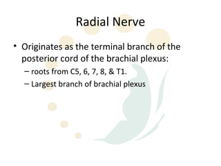
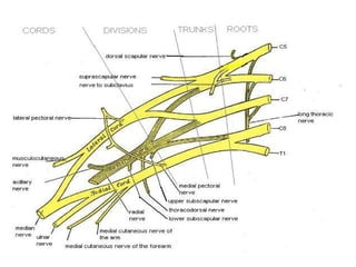
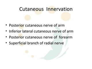
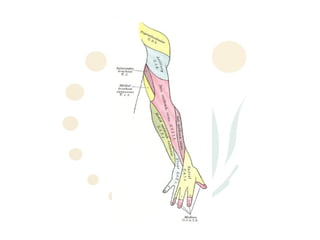
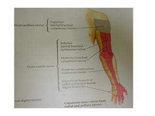
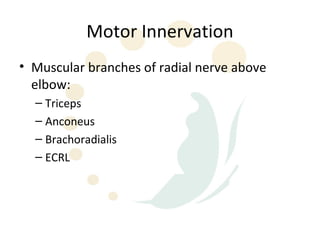
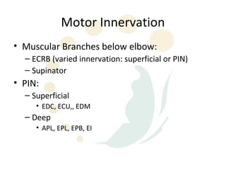
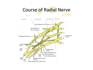
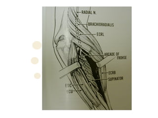
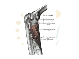
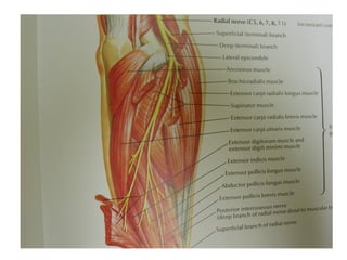
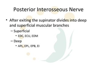
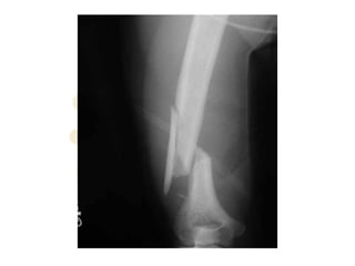
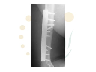
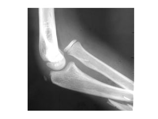
Radial Nerve Anatomy
     Episode 1
Radial Nerve
• Originates as the terminal branch of the
  posterior cord of the brachial plexus:
  – roots from C5, 6, 7, 8, & T1.
  – Largest branch of brachial plexus
Cutaneous Innervation

•   Posterior cutaneous nerve of arm
•   Inferior lateral cutaneous nerve of arm
•   Posterior cutaneous nerve of forearm
•   Superficial branch of radial nerve
Motor Innervation
• Muscular branches of radial nerve above
  elbow:
  – Triceps
  – Anconeus
  – Brachoradialis
  – ECRL
Motor Innervation
• Muscular Branches below elbow:
  – ECRB (varied innervation: superficial or PIN)
  – Supinator
• PIN:
  – Superficial
     • EDC, ECU,, EDM
  – Deep
     • APL, EPL, EPB, EI
Course of Radial Nerve
• Largest terminal branch of posterior cord
• Enters posterior aspect of humerus through lower
  triangular interval
   – Teres major (superior)
   – Long head triceps (medial)
   – Humerus (lateral)
• Gives posterior cutaneous nerve of arm in axilla
Course of Radial Nerve
• Comes to lie in distal part of spiral groove with
  profundi brachii artery
   – Beneath lateral head of triceps and proximal to origin of
     medial head
• Gives branches to triceps, anconeus and inferior
  lateral cutaneous nerve of arm
• Through lateral intermuscular septum 10-12cm
  above lateral epicondyle
Course of Radial Nerve
• In anterior compartment of arm lies between
  brachialis and brachioradialis
  – 1-3 accessory branches to brachialis
  – Large branch to BR (sometimes this branch given
    by superficial radial below elbow)
• ECRL generally innervated proximal to elbow
  joint
Course of Radial Nerve
• Enters the forearm anterior to lateral epicondyle
   – More specifically over articulation between capitulum and
     radial head
• ECRB innervated distal to elbow joint either PIN or
  superficial branch

• At some point 3cm above or below divides into:
   – Superficial radial
   – PIN
Course of Superficial Radial Nerve
• Runs over supinator,PT and FDS
• Lies under BR with radial artery on its ulnar side from
  1/3 of the way down forearm
• Passes posteriorly through tendon of BR proximal to
  radial styloid
• Passes over tendons of snuffbox
• Terminates as cutaneous branches to dorsum of
  hand and lateral 3.5 digits short of nailbeds
Posterior Interosseous Nerve
• Gains access to posterior forearm by diving
  posterior and laterally through ligament of
  Frohse and the interval between the two
  heads of supinator
• Arcade of Frohse is a fibrous arch originating
  lateral epicondyle superior part of superficial
  layer of supinator
  – German Anatomist, Fritz Frohse (1871-1916)
Posterior Interosseous Nerve
• After exiting the supinator divides into deep
  and superficial muscular branches
  – Superficial
     • EDC, ECU, EDM
  – Deep
     • APL, EPL, EPB, EI
Radial Nerve Compression Sites
• PIN
  – As it traverses the radial tunnel it encounters 4
    sites of compression
  – “Radial tunnel syndrome”
     • entrapment neuropathy
Radial Tunnel Syndrome
1. Fibrous bands
  •   Tight fibrous bands anterior to radial head at entrance of
      tunnel
1. Arcade of Frohse
  •   Most common
1. Leash of Henry
  •   Radial recurrent vx that fan out over radial nn
1. Medial border of ECRB
Cheiralgia paresthetica
• Described by Robert Wartenberg 1932
• Superficial radial nerve neuritis
  – Wartenberg Syndrome
• Causes:
  – Trauma, iatrogenic, compression (muscle anomalies,
    fascial bands, thrombosis radial recurrent artery, tumour,
    haemorrhage)
• Symptoms:
  – Dysaesthesia over distribution
Radial Nerve Palsy
• Causes:
  – Fractures esp humerus, Monteggia
  – Penetrating injury/trauma
  – “Saturday Night Palsy” “Crutches”
  – Iatrogenic
  – tumour
Episode II
Radial Nerve Tendon Transfers

Radial nerve-anatomy

  • 2.
  • 3.
    Radial Nerve • Originatesas the terminal branch of the posterior cord of the brachial plexus: – roots from C5, 6, 7, 8, & T1. – Largest branch of brachial plexus
  • 5.
    Cutaneous Innervation • Posterior cutaneous nerve of arm • Inferior lateral cutaneous nerve of arm • Posterior cutaneous nerve of forearm • Superficial branch of radial nerve
  • 8.
    Motor Innervation • Muscularbranches of radial nerve above elbow: – Triceps – Anconeus – Brachoradialis – ECRL
  • 9.
    Motor Innervation • MuscularBranches below elbow: – ECRB (varied innervation: superficial or PIN) – Supinator • PIN: – Superficial • EDC, ECU,, EDM – Deep • APL, EPL, EPB, EI
  • 10.
  • 11.
    Course of RadialNerve • Largest terminal branch of posterior cord • Enters posterior aspect of humerus through lower triangular interval – Teres major (superior) – Long head triceps (medial) – Humerus (lateral) • Gives posterior cutaneous nerve of arm in axilla
  • 13.
    Course of RadialNerve • Comes to lie in distal part of spiral groove with profundi brachii artery – Beneath lateral head of triceps and proximal to origin of medial head • Gives branches to triceps, anconeus and inferior lateral cutaneous nerve of arm • Through lateral intermuscular septum 10-12cm above lateral epicondyle
  • 15.
    Course of RadialNerve • In anterior compartment of arm lies between brachialis and brachioradialis – 1-3 accessory branches to brachialis – Large branch to BR (sometimes this branch given by superficial radial below elbow) • ECRL generally innervated proximal to elbow joint
  • 17.
    Course of RadialNerve • Enters the forearm anterior to lateral epicondyle – More specifically over articulation between capitulum and radial head • ECRB innervated distal to elbow joint either PIN or superficial branch • At some point 3cm above or below divides into: – Superficial radial – PIN
  • 19.
    Course of SuperficialRadial Nerve • Runs over supinator,PT and FDS • Lies under BR with radial artery on its ulnar side from 1/3 of the way down forearm • Passes posteriorly through tendon of BR proximal to radial styloid • Passes over tendons of snuffbox • Terminates as cutaneous branches to dorsum of hand and lateral 3.5 digits short of nailbeds
  • 22.
    Posterior Interosseous Nerve •Gains access to posterior forearm by diving posterior and laterally through ligament of Frohse and the interval between the two heads of supinator • Arcade of Frohse is a fibrous arch originating lateral epicondyle superior part of superficial layer of supinator – German Anatomist, Fritz Frohse (1871-1916)
  • 26.
    Posterior Interosseous Nerve •After exiting the supinator divides into deep and superficial muscular branches – Superficial • EDC, ECU, EDM – Deep • APL, EPL, EPB, EI
  • 27.
    Radial Nerve CompressionSites • PIN – As it traverses the radial tunnel it encounters 4 sites of compression – “Radial tunnel syndrome” • entrapment neuropathy
  • 28.
    Radial Tunnel Syndrome 1.Fibrous bands • Tight fibrous bands anterior to radial head at entrance of tunnel 1. Arcade of Frohse • Most common 1. Leash of Henry • Radial recurrent vx that fan out over radial nn 1. Medial border of ECRB
  • 29.
    Cheiralgia paresthetica • Describedby Robert Wartenberg 1932 • Superficial radial nerve neuritis – Wartenberg Syndrome • Causes: – Trauma, iatrogenic, compression (muscle anomalies, fascial bands, thrombosis radial recurrent artery, tumour, haemorrhage) • Symptoms: – Dysaesthesia over distribution
  • 30.
    Radial Nerve Palsy •Causes: – Fractures esp humerus, Monteggia – Penetrating injury/trauma – “Saturday Night Palsy” “Crutches” – Iatrogenic – tumour
  • 36.
    Episode II Radial NerveTendon Transfers