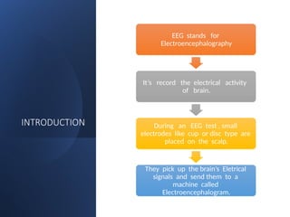
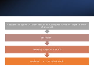
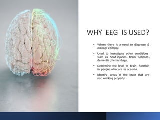
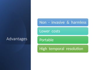
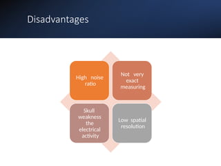
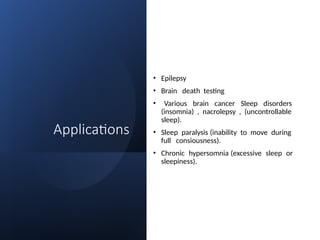
ELECTROENCEPHALOGRAPH
Y (EEG)
MUKESH SUNDARARAJAN
Components
INTRODUCTION REQUIREMENTS HISTORY WORKING
ADVANTAGES DISADVANTAGES APPLICATIONS USES
INTRODUCTION
EEG stands for
Electroencephalography
It’s record the electrical activity
of brain.
During an EEG test , small
electrodes like cup or disc type are
placed on the scalp.
They pick up the brain’s Eletrical
signals and send them to a
machine called
Electroencephalogram.
amplitude = 2 to 200 micro volt.
frequency range = 0.1 to 100
EEG waves
It records the signals as wavy lines on to a computer screen or paper in order
of microvolt.
REQUIREMENTS
EEG (8/16
channels).
Silver cup
electrodes /
metallic bridge
electrodes.
Electrode jelly.
Rubber cap.
Quiet dark
comfortable
room.
Skin pencil &
measuring tape.
HISTORY OF EEG
In 1890 - Adolf Beck (polish physiologist) of Poland, investigations of
spontaneous electrical activity of the brain of Rabbits & Dogs.
Discovered electrical activity of brain by probing the surface of
exposed brains (cerebral hemispheres) of animals (Rabbits & Monkeys).
In 1875 - Richard caton (physician) from Germany,
• In 1920 - Hans Berger (Austrian psychiatrist) was the first to record EEG
tracings from human beings.He Discovered waves at 10HZ.(Alpha
waves because they were the first he isolated in the human EEG).
• In 1935 - Forester & Alteberger they first use of intra operative EEG.
• In 1950 - Herbert jasper & Wilder penfield developed this technique,
using ECoG for localization &surgical treatment of epilepsy
Older Generation Current Generation
PROCEDURE
• A standard non – invasive EEG takes about 1 hour.The patient will be
positioned on a padded bed or table , or in comfortable chair.
• To measure the electrical activity in various part of the brain , a nurse or
EEG technician will attach 16 to 20 electrodes to the scalp.
• The brain generates electrical impulses that these electrodes will pick
up. Then a temporary glue will be used to attach them to the skin , no
gain will be involved.
TYPES OF LOBES
• Frontal lobes:
Emotional & motor control.
• Parietal lobes:
Sensory purpose (pain , touch, pressure).
• Temporal lobes:
Auditory system (hear & sound).
• Occipital lobes:
Visual purpose (see & eye).
WHY EEG IS USED?
• Where there is a need to diagnose &
manage epilepsy.
• Used to investigate other conditions
such as head injuries , brain tumours ,
dementia , hemorrhage.
• Determine the level of brain function
in people who are in a coma.
• Identify areas of the brain that are
not working properly.
TYPES OF
WAVES
• Alpha waves
• Beta waves
• Delta waves
• Theta waves
Alpha waves
• Alpha waves are found in normal persons (resting state), when they
are awake .
• They occur in occipital region .
• Alpha waves are 8 - 13 HZ.
• Alpha waves are present, they indicate a calm & releaxed state.
Beta waves
• Beta waves are recorded from parietal & frontal region of scalp.
• Divided into 2 types.
Beta - 1 which is inhibited by cerebral activity.
Beta - 2 excited by mental activity
like tension.
• Beta waves are 13 - 30 HZ.
• Beta waves indicate can alert such as when you focus on solving
a problem.
Theta waves
• Theta waves are recorded from temporal region of scalp from children.
• They occur stress & frustration.
• Theta waves are 4 - 8 HZ.
• Theta waves observed when you are day dreaming & drowsiness.
• Brain disorders (Adult , children , premature , serious brain disorders).
• Emotional questions are asked get in the theta waves.
Delta waves
• Delta waves are recorded from cortex region.
• They occur deep sleep in premature babies & incases of brain
diseases.
• Delta waves are 0.5 - 4 HZ.
• Delta waves observed when you are day dreaming & drowsiness.
• 2 - 3 seconds gets in the waves.
Advantages
Non - invasive & harmless
Lower costs
Portable
High temporal resolution
Disadvantages
High noise
ratio
Not very
exact
measuring
Skull
weakness
the
electrical
activity
Low spatial
resolution
Applications
• Epilepsy
• Brain death testing
• Various brain cancer Sleep disorders
(insomnia) , nacrolepsy , (uncontrollable
sleep).
• Sleep paralysis (inability to move during
full consiousness).
• Chronic hypersomnia (excessive sleep or
sleepiness).
THANK YOU

Brief introduction on Electro encephalography-SSM.pptx

  • 1.
  • 2.
    Components INTRODUCTION REQUIREMENTS HISTORYWORKING ADVANTAGES DISADVANTAGES APPLICATIONS USES
  • 3.
    INTRODUCTION EEG stands for Electroencephalography It’srecord the electrical activity of brain. During an EEG test , small electrodes like cup or disc type are placed on the scalp. They pick up the brain’s Eletrical signals and send them to a machine called Electroencephalogram.
  • 4.
    amplitude = 2to 200 micro volt. frequency range = 0.1 to 100 EEG waves It records the signals as wavy lines on to a computer screen or paper in order of microvolt.
  • 5.
    REQUIREMENTS EEG (8/16 channels). Silver cup electrodes/ metallic bridge electrodes. Electrode jelly. Rubber cap. Quiet dark comfortable room. Skin pencil & measuring tape.
  • 6.
    HISTORY OF EEG In1890 - Adolf Beck (polish physiologist) of Poland, investigations of spontaneous electrical activity of the brain of Rabbits & Dogs. Discovered electrical activity of brain by probing the surface of exposed brains (cerebral hemispheres) of animals (Rabbits & Monkeys). In 1875 - Richard caton (physician) from Germany,
  • 7.
    • In 1920- Hans Berger (Austrian psychiatrist) was the first to record EEG tracings from human beings.He Discovered waves at 10HZ.(Alpha waves because they were the first he isolated in the human EEG). • In 1935 - Forester & Alteberger they first use of intra operative EEG. • In 1950 - Herbert jasper & Wilder penfield developed this technique, using ECoG for localization &surgical treatment of epilepsy
  • 8.
  • 10.
    PROCEDURE • A standardnon – invasive EEG takes about 1 hour.The patient will be positioned on a padded bed or table , or in comfortable chair. • To measure the electrical activity in various part of the brain , a nurse or EEG technician will attach 16 to 20 electrodes to the scalp. • The brain generates electrical impulses that these electrodes will pick up. Then a temporary glue will be used to attach them to the skin , no gain will be involved.
  • 11.
    TYPES OF LOBES •Frontal lobes: Emotional & motor control. • Parietal lobes: Sensory purpose (pain , touch, pressure). • Temporal lobes: Auditory system (hear & sound). • Occipital lobes: Visual purpose (see & eye).
  • 13.
    WHY EEG ISUSED? • Where there is a need to diagnose & manage epilepsy. • Used to investigate other conditions such as head injuries , brain tumours , dementia , hemorrhage. • Determine the level of brain function in people who are in a coma. • Identify areas of the brain that are not working properly.
  • 14.
    TYPES OF WAVES • Alphawaves • Beta waves • Delta waves • Theta waves
  • 15.
    Alpha waves • Alphawaves are found in normal persons (resting state), when they are awake . • They occur in occipital region . • Alpha waves are 8 - 13 HZ. • Alpha waves are present, they indicate a calm & releaxed state.
  • 16.
    Beta waves • Betawaves are recorded from parietal & frontal region of scalp. • Divided into 2 types. Beta - 1 which is inhibited by cerebral activity. Beta - 2 excited by mental activity like tension. • Beta waves are 13 - 30 HZ. • Beta waves indicate can alert such as when you focus on solving a problem.
  • 17.
    Theta waves • Thetawaves are recorded from temporal region of scalp from children. • They occur stress & frustration. • Theta waves are 4 - 8 HZ. • Theta waves observed when you are day dreaming & drowsiness. • Brain disorders (Adult , children , premature , serious brain disorders). • Emotional questions are asked get in the theta waves.
  • 18.
    Delta waves • Deltawaves are recorded from cortex region. • They occur deep sleep in premature babies & incases of brain diseases. • Delta waves are 0.5 - 4 HZ. • Delta waves observed when you are day dreaming & drowsiness. • 2 - 3 seconds gets in the waves.
  • 20.
    Advantages Non - invasive& harmless Lower costs Portable High temporal resolution
  • 21.
  • 22.
    Applications • Epilepsy • Braindeath testing • Various brain cancer Sleep disorders (insomnia) , nacrolepsy , (uncontrollable sleep). • Sleep paralysis (inability to move during full consiousness). • Chronic hypersomnia (excessive sleep or sleepiness).
  • 23.