Downloaded 463 times
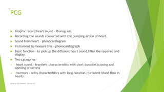
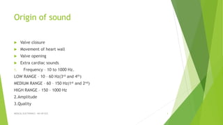


Phonocardiography (PCG) is a method for recording heart sounds, capturing the sounds associated with heart pumping actions via a phonocardiograph. It distinguishes between heart sounds and murmurs, measuring frequency, amplitude, and quality across different sound categories. The document details instrumentation, recording techniques, types of microphones, and their specific applications in medical electronics, including fetal and esophageal PCG.