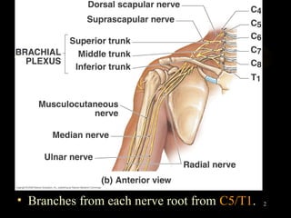
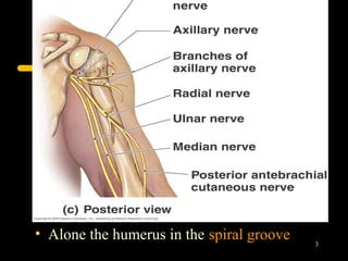
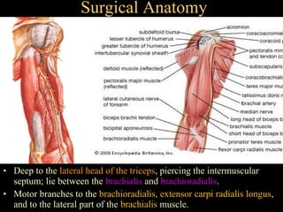
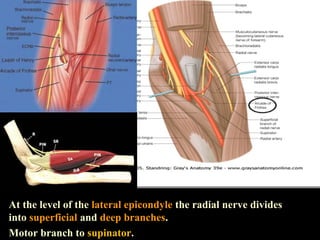
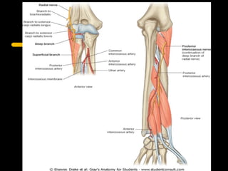
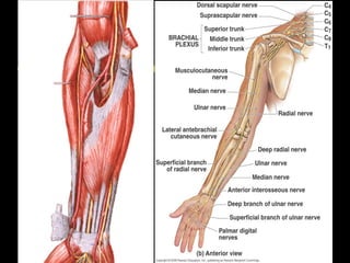
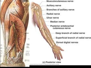
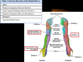
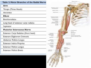
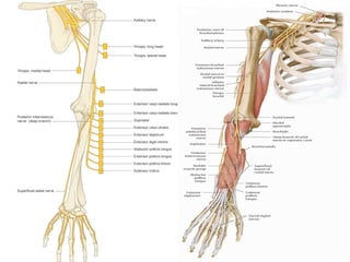
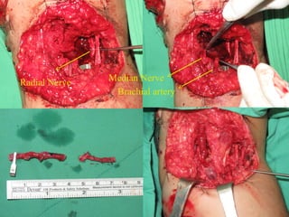
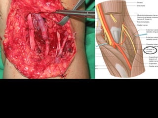
Radial nerve
• Surgical anatomy
• Motor and sensory innervation
• Clinical symptoms and sign
• Clinical case illustration
• Branches from each nerve root from C5/T1.   2
• Alone the humerus in the spiral groove
                                           3
Surgical Anatomy


• Deep to the lateral head of the triceps, piercing the intermuscular
  septum; lie between the brachialis and brachioradialis.
• Motor branches to the brachioradialis, extensor carpi radialis longus,
  and to the lateral part of the brachialis muscle.
At the level of the lateral epicondyle the radial nerve divides
into superficial and deep branches.
Motor branch to supinator.
8
Anatomy
Summary
• PIN arises from radial nerve. Radial nerve pulak
  arises from posterior cord of the brachial plexus
• It passes posterior to the axillary artery between
  long and media heads of triceps muscle, to lie in
  the spiral groove between medial & lateral heads
  of triceps muscle.
• Here it is accompanied by the profunda brachii
  artery before it pierces it lateral intermuscular
  septum of the lower third humerus to run between
  brachialis & brachioradialis
• At the lateral of epicondyle humerus, it gives rises
  to PIN & superficial radial nerve
• Radial nerve supply all the extensor muscle of forearm &
   arm. However it also supply brachioradialis, which is
   flexor of elbow when forearm pronated
• Damage to the nerve in the spiral groove causes
  wrist drop but no loss of elbow extension, as fibres of
  triceps remain intact proximal to this site
• Only damage in the axilla will causes loss of elbow
   extension & wrist drop
• Damage to posterior interosseus nerve (PIN) does not
   cause wrist drop because extensor carpi radialis
   longus receives its innervation from the main radial
   nerve
• Pin only cause unable to extend metacarpophalangeal
   joint  finger drop.
Clinical case presentation
M/34 左手遭玻璃切割傷
Radial Nerve   Median Nerve
                Brachial artery
Radial nerve injury

Radial nerve injury

  • 1.
    Radial nerve • Surgicalanatomy • Motor and sensory innervation • Clinical symptoms and sign • Clinical case illustration
  • 2.
    • Branches fromeach nerve root from C5/T1. 2
  • 3.
    • Alone thehumerus in the spiral groove 3
  • 4.
    Surgical Anatomy • Deepto the lateral head of the triceps, piercing the intermuscular septum; lie between the brachialis and brachioradialis. • Motor branches to the brachioradialis, extensor carpi radialis longus, and to the lateral part of the brachialis muscle.
  • 5.
    At the levelof the lateral epicondyle the radial nerve divides into superficial and deep branches. Motor branch to supinator.
  • 8.
  • 11.
  • 12.
    Summary • PIN arisesfrom radial nerve. Radial nerve pulak arises from posterior cord of the brachial plexus • It passes posterior to the axillary artery between long and media heads of triceps muscle, to lie in the spiral groove between medial & lateral heads of triceps muscle. • Here it is accompanied by the profunda brachii artery before it pierces it lateral intermuscular septum of the lower third humerus to run between brachialis & brachioradialis • At the lateral of epicondyle humerus, it gives rises to PIN & superficial radial nerve
  • 13.
    • Radial nervesupply all the extensor muscle of forearm & arm. However it also supply brachioradialis, which is flexor of elbow when forearm pronated • Damage to the nerve in the spiral groove causes wrist drop but no loss of elbow extension, as fibres of triceps remain intact proximal to this site • Only damage in the axilla will causes loss of elbow extension & wrist drop • Damage to posterior interosseus nerve (PIN) does not cause wrist drop because extensor carpi radialis longus receives its innervation from the main radial nerve • Pin only cause unable to extend metacarpophalangeal joint  finger drop.
  • 14.
    Clinical case presentation M/34左手遭玻璃切割傷
  • 15.
    Radial Nerve Median Nerve Brachial artery