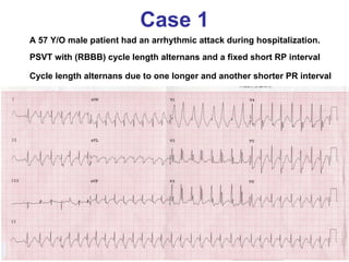
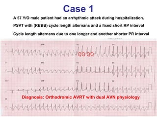
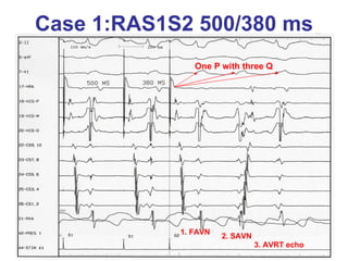
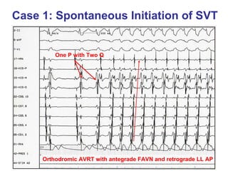
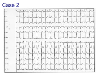
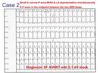
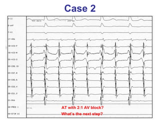
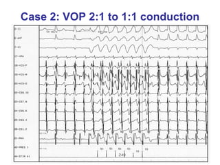
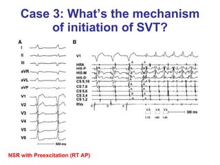
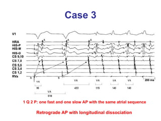
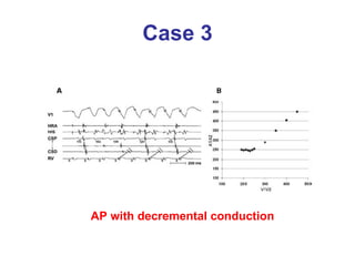
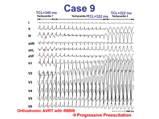
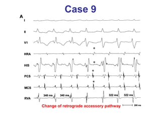
ECG Tracing
Case 1 A 57 Y/O male patient had an arrhythmic attack during hospitalization.  PSVT with (RBBB) cycle length alternans and a fixed short RP interval Cycle length alternans due to one longer and another shorter PR interval
Case 1 A 57 Y/O male patient had an arrhythmic attack during hospitalization.  PSVT with (RBBB) cycle length alternans and a fixed short RP interval Cycle length alternans due to one longer and another shorter PR interval Diagnosis: Orthodromic AVRT with dual AVN physiology
Case 1:RAS1S2 500/380 ms One P with three Q 1. FAVN 2. SAVN 3. AVRT echo
Case 1: Spontaneous Initiation of SVT Orthodromic AVRT with antegrade FAVN and retrograde LL AP One P with Two Q
Case 2
Case 2 Small & narrow P wave  RA & LA depolarization simultaneously  Diagnosis: SF AVNRT with 2:1 AV block A P wave in the midpoint between the two QRS beats
Case 2 AT with 2:1 AV block? What’s the next step?
Case 2: VOP 2:1 to 1:1 conduction
Case 3: What’s the mechanism of initiation of SVT? NSR with Preexcitation (RT AP)
Case 3 1 Q 2 P: one fast and one slow AP with the same atrial sequence Retrograde AP with longitudinal dissociation
Case 3 AP with decremental conduction
Case 4: W  N QRS tachycardia
Case 4: W  N QRS tachycardia VA dissociation VT H PSVT No H Wide to Narrow
Case 4 PPI-TCL=130 ms (>115 ms) V  A  V Diagnosis: FS AVNRT
Atypical AVNRT vs Septal AVRT (Michaud GF et al. JACC 2001)
Case 5 Two different retrograde P waves: two different pathways Pseudo R’ Pseudo S
Case 5 VA=102 ms VA=72 ms HV=178 ms HV=190 ms AVRT AVNRT alternatively
Case 6 RAS1S2 induced SF AVNRT
Case 6: After successful modification of SAVN RA burst induce narrow SVT with VA dissociation
Case 6:VOP terminate tachycardia Diagnosis: Junctional Tachycardia after ablation of SAVN
Case 7 PSVT with cycle length alternans and electrical alternans A fixed RP interval suggesting orthodromic AVRT Cycle length alternans due to dual AVN physiology Diagnosis: Orthodromic AVRT with dual AVN physiology long short
Case 7 NSR with intermittent preexcitation (RT AP)
Case 7 Diagnosis: Orthodromic AVRT using RT AP and Dual AVN physiology A fixed VA interval using RT AP Two different AH interval through fast and slow AVN
Case 8 Wide QRS complex tachycardia? (VA 1:1 conduction) PSVT with RBBB? (Atypical RBBB) Idiopathic LV-VT? (RBBB+LAD) Preexcitated tachycardia? (no delta wave)
Case 8: 60 seconds after adenosine Wide QRS=  Narrow QRS
Case 8: 65 seconds after adenosine H WQRS NQRS SF AVNRT No H V earlier VT
Case 9  Progressive Preexcitation Orthodromic AVRT with RBBB TCL=340 ms TCL=322 ms TCL=322 ms
Case 9 Change of retrograde accessory pathway
Case 10 Narrow QRS complex tachycardia with VA dissociation (V>A)
Case 10 Diagnosis: One VPC induced SF AVNRT with VA 2:1 conduction
謝謝聆聽 敬請指教

Ecg tracings teaching

  • 1.
  • 2.
    Case 1 A57 Y/O male patient had an arrhythmic attack during hospitalization. PSVT with (RBBB) cycle length alternans and a fixed short RP interval Cycle length alternans due to one longer and another shorter PR interval
  • 3.
    Case 1 A57 Y/O male patient had an arrhythmic attack during hospitalization. PSVT with (RBBB) cycle length alternans and a fixed short RP interval Cycle length alternans due to one longer and another shorter PR interval Diagnosis: Orthodromic AVRT with dual AVN physiology
  • 4.
    Case 1:RAS1S2 500/380ms One P with three Q 1. FAVN 2. SAVN 3. AVRT echo
  • 5.
    Case 1: SpontaneousInitiation of SVT Orthodromic AVRT with antegrade FAVN and retrograde LL AP One P with Two Q
  • 6.
  • 7.
    Case 2 Small& narrow P wave  RA & LA depolarization simultaneously Diagnosis: SF AVNRT with 2:1 AV block A P wave in the midpoint between the two QRS beats
  • 8.
    Case 2 ATwith 2:1 AV block? What’s the next step?
  • 9.
    Case 2: VOP2:1 to 1:1 conduction
  • 10.
    Case 3: What’sthe mechanism of initiation of SVT? NSR with Preexcitation (RT AP)
  • 11.
    Case 3 1Q 2 P: one fast and one slow AP with the same atrial sequence Retrograde AP with longitudinal dissociation
  • 12.
    Case 3 APwith decremental conduction
  • 13.
    Case 4: W N QRS tachycardia
  • 14.
    Case 4: W N QRS tachycardia VA dissociation VT H PSVT No H Wide to Narrow
  • 15.
    Case 4 PPI-TCL=130ms (>115 ms) V  A  V Diagnosis: FS AVNRT
  • 16.
    Atypical AVNRT vsSeptal AVRT (Michaud GF et al. JACC 2001)
  • 17.
    Case 5 Twodifferent retrograde P waves: two different pathways Pseudo R’ Pseudo S
  • 18.
    Case 5 VA=102ms VA=72 ms HV=178 ms HV=190 ms AVRT AVNRT alternatively
  • 19.
    Case 6 RAS1S2induced SF AVNRT
  • 20.
    Case 6: Aftersuccessful modification of SAVN RA burst induce narrow SVT with VA dissociation
  • 21.
    Case 6:VOP terminatetachycardia Diagnosis: Junctional Tachycardia after ablation of SAVN
  • 22.
    Case 7 PSVTwith cycle length alternans and electrical alternans A fixed RP interval suggesting orthodromic AVRT Cycle length alternans due to dual AVN physiology Diagnosis: Orthodromic AVRT with dual AVN physiology long short
  • 23.
    Case 7 NSRwith intermittent preexcitation (RT AP)
  • 24.
    Case 7 Diagnosis:Orthodromic AVRT using RT AP and Dual AVN physiology A fixed VA interval using RT AP Two different AH interval through fast and slow AVN
  • 25.
    Case 8 WideQRS complex tachycardia? (VA 1:1 conduction) PSVT with RBBB? (Atypical RBBB) Idiopathic LV-VT? (RBBB+LAD) Preexcitated tachycardia? (no delta wave)
  • 26.
    Case 8: 60seconds after adenosine Wide QRS=  Narrow QRS
  • 27.
    Case 8: 65seconds after adenosine H WQRS NQRS SF AVNRT No H V earlier VT
  • 28.
    Case 9 Progressive Preexcitation Orthodromic AVRT with RBBB TCL=340 ms TCL=322 ms TCL=322 ms
  • 29.
    Case 9 Changeof retrograde accessory pathway
  • 30.
    Case 10 NarrowQRS complex tachycardia with VA dissociation (V>A)
  • 31.
    Case 10 Diagnosis:One VPC induced SF AVNRT with VA 2:1 conduction
  • 32.