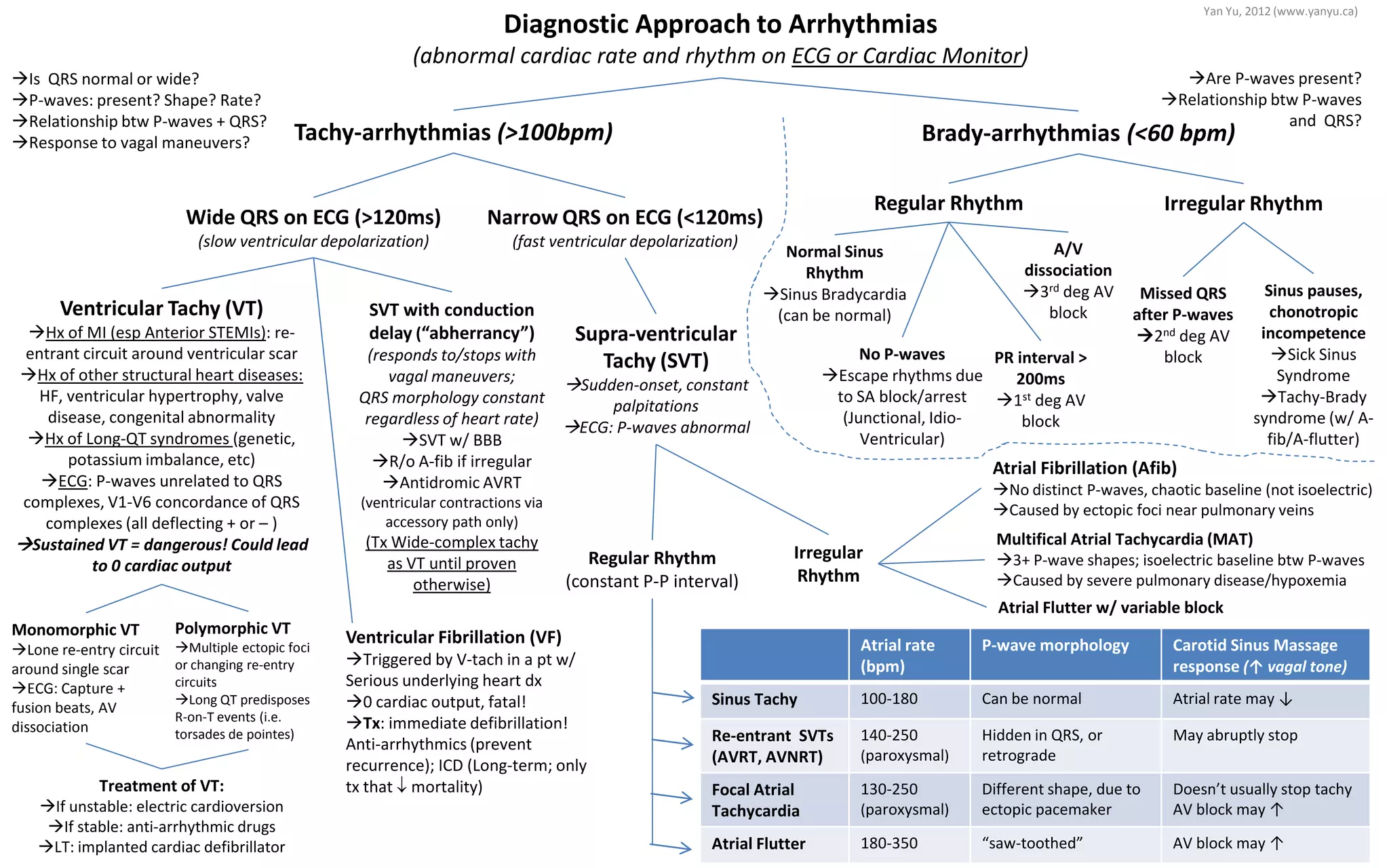
SVT with conduction
delay (“abherrancy”)
(responds to/stops with
vagal maneuvers;
QRS morphology constant
regardless of heart rate)
SVT w/ BBB
R/o A-fib if irregular
Antidromic AVRT
(ventricular contractions via
accessory path only)
(Tx Wide-complex tachy
as VT until proven
otherwise)
Brady-arrhythmias (<60 bpm)Tachy-arrhythmias (>100bpm)
Diagnostic Approach to Arrhythmias
(abnormal cardiac rate and rhythm on ECG or Cardiac Monitor)
Wide QRS on ECG (>120ms)
(slow ventricular depolarization)
Regular Rhythm Irregular Rhythm
Ventricular Tachy (VT)
Hx of MI (esp Anterior STEMIs): re-
entrant circuit around ventricular scar
Hx of other structural heart diseases:
HF, ventricular hypertrophy, valve
disease, congenital abnormality
Hx of Long-QT syndromes (genetic,
potassium imbalance, etc)
ECG: P-waves unrelated to QRS
complexes, V1-V6 concordance of QRS
complexes (all deflecting + or – )
Sustained VT = dangerous! Could lead
to 0 cardiac output
Narrow QRS on ECG (<120ms)
(fast ventricular depolarization)
Polymorphic VT
Multiple ectopic foci
or changing re-entry
circuits
Long QT predisposes
R-on-T events (i.e.
torsades de pointes)
Monomorphic VT
Lone re-entry circuit
around single scar
ECG: Capture +
fusion beats, AV
dissociation
Supra-ventricular
Tachy (SVT)
Sudden-onset, constant
palpitations
ECG: P-waves abnormal
Regular Rhythm
(constant P-P interval)
Irregular
Rhythm
Multifical Atrial Tachycardia (MAT)
3+ P-wave shapes; isoelectric baseline btw P-waves
Caused by severe pulmonary disease/hypoxemia
Atrial Flutter w/ variable block
Atrial rate
(bpm)
P-wave morphology Carotid Sinus Massage
response (↑ vagal tone)
Sinus Tachy 100-180 Can be normal Atrial rate may ↓
Re-entrant SVTs
(AVRT, AVNRT)
140-250
(paroxysmal)
Hidden in QRS, or
retrograde
May abruptly stop
Focal Atrial
Tachycardia
130-250
(paroxysmal)
Different shape, due to
ectopic pacemaker
Doesn’t usually stop tachy
AV block may ↑
Atrial Flutter 180-350 “saw-toothed” AV block may ↑
Atrial Fibrillation (Afib)
No distinct P-waves, chaotic baseline (not isoelectric)
Caused by ectopic foci near pulmonary veins
Sinus pauses,
chonotropic
incompetence
Sick Sinus
Syndrome
Tachy-Brady
syndrome (w/ A-
fib/A-flutter)
Missed QRS
after P-waves
2nd deg AV
blockNo P-waves
Escape rhythms due
to SA block/arrest
(Junctional, Idio-
Ventricular)
Normal Sinus
Rhythm
Sinus Bradycardia
(can be normal)
PR interval >
200ms
1st deg AV
block
A/V
dissociation
3rd deg AV
block
Are P-waves present?
Relationship btw P-waves
and QRS?
Is QRS normal or wide?
P-waves: present? Shape? Rate?
Relationship btw P-waves + QRS?
Response to vagal maneuvers?
Ventricular Fibrillation (VF)
Triggered by V-tach in a pt w/
Serious underlying heart dx
0 cardiac output, fatal!
Tx: immediate defibrillation!
Anti-arrhythmics (prevent
recurrence); ICD (Long-term; only
tx that ↓ mortality)Treatment of VT:
If unstable: electric cardioversion
If stable: anti-arrhythmic drugs
LT: implanted cardiac defibrillator
Yan Yu, 2012 (www.yanyu.ca)

Approach toarrhythmias

  • 1.
    SVT with conduction delay(“abherrancy”) (responds to/stops with vagal maneuvers; QRS morphology constant regardless of heart rate) SVT w/ BBB R/o A-fib if irregular Antidromic AVRT (ventricular contractions via accessory path only) (Tx Wide-complex tachy as VT until proven otherwise) Brady-arrhythmias (<60 bpm)Tachy-arrhythmias (>100bpm) Diagnostic Approach to Arrhythmias (abnormal cardiac rate and rhythm on ECG or Cardiac Monitor) Wide QRS on ECG (>120ms) (slow ventricular depolarization) Regular Rhythm Irregular Rhythm Ventricular Tachy (VT) Hx of MI (esp Anterior STEMIs): re- entrant circuit around ventricular scar Hx of other structural heart diseases: HF, ventricular hypertrophy, valve disease, congenital abnormality Hx of Long-QT syndromes (genetic, potassium imbalance, etc) ECG: P-waves unrelated to QRS complexes, V1-V6 concordance of QRS complexes (all deflecting + or – ) Sustained VT = dangerous! Could lead to 0 cardiac output Narrow QRS on ECG (<120ms) (fast ventricular depolarization) Polymorphic VT Multiple ectopic foci or changing re-entry circuits Long QT predisposes R-on-T events (i.e. torsades de pointes) Monomorphic VT Lone re-entry circuit around single scar ECG: Capture + fusion beats, AV dissociation Supra-ventricular Tachy (SVT) Sudden-onset, constant palpitations ECG: P-waves abnormal Regular Rhythm (constant P-P interval) Irregular Rhythm Multifical Atrial Tachycardia (MAT) 3+ P-wave shapes; isoelectric baseline btw P-waves Caused by severe pulmonary disease/hypoxemia Atrial Flutter w/ variable block Atrial rate (bpm) P-wave morphology Carotid Sinus Massage response (↑ vagal tone) Sinus Tachy 100-180 Can be normal Atrial rate may ↓ Re-entrant SVTs (AVRT, AVNRT) 140-250 (paroxysmal) Hidden in QRS, or retrograde May abruptly stop Focal Atrial Tachycardia 130-250 (paroxysmal) Different shape, due to ectopic pacemaker Doesn’t usually stop tachy AV block may ↑ Atrial Flutter 180-350 “saw-toothed” AV block may ↑ Atrial Fibrillation (Afib) No distinct P-waves, chaotic baseline (not isoelectric) Caused by ectopic foci near pulmonary veins Sinus pauses, chonotropic incompetence Sick Sinus Syndrome Tachy-Brady syndrome (w/ A- fib/A-flutter) Missed QRS after P-waves 2nd deg AV blockNo P-waves Escape rhythms due to SA block/arrest (Junctional, Idio- Ventricular) Normal Sinus Rhythm Sinus Bradycardia (can be normal) PR interval > 200ms 1st deg AV block A/V dissociation 3rd deg AV block Are P-waves present? Relationship btw P-waves and QRS? Is QRS normal or wide? P-waves: present? Shape? Rate? Relationship btw P-waves + QRS? Response to vagal maneuvers? Ventricular Fibrillation (VF) Triggered by V-tach in a pt w/ Serious underlying heart dx 0 cardiac output, fatal! Tx: immediate defibrillation! Anti-arrhythmics (prevent recurrence); ICD (Long-term; only tx that ↓ mortality)Treatment of VT: If unstable: electric cardioversion If stable: anti-arrhythmic drugs LT: implanted cardiac defibrillator Yan Yu, 2012 (www.yanyu.ca)