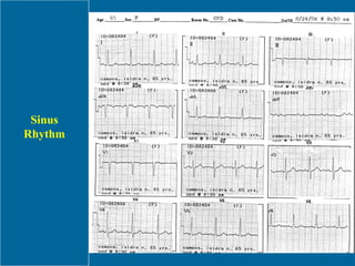
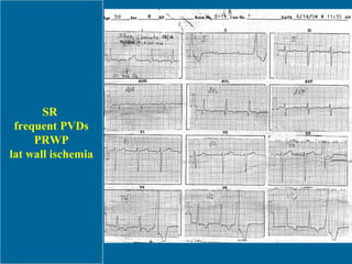
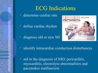
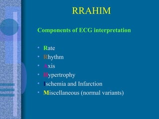
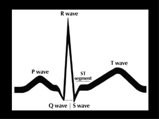
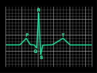
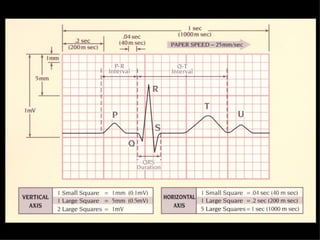
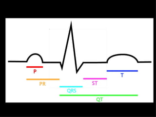
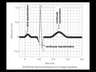
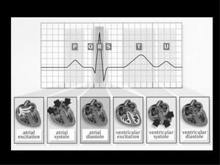
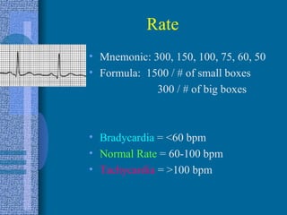
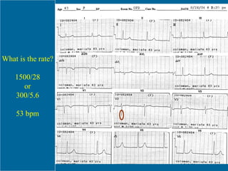
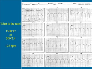
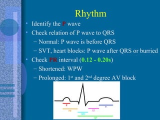
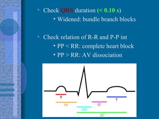
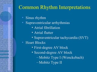
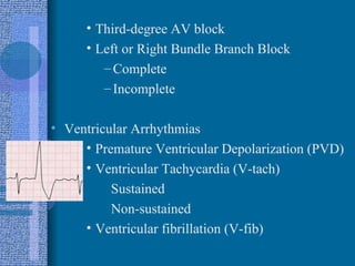
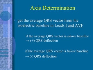
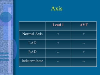
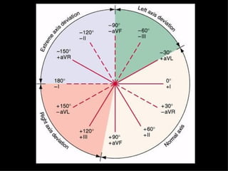
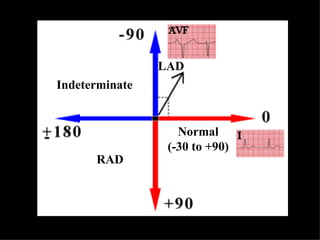
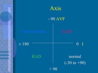
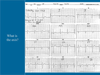
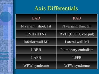
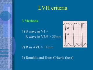
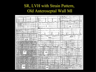
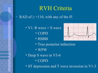
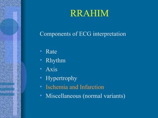
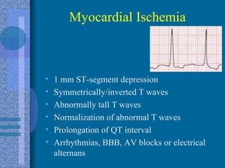
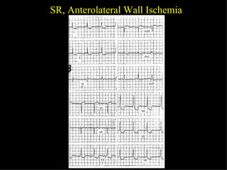
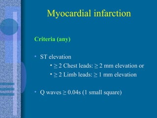
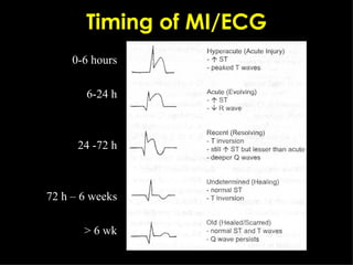
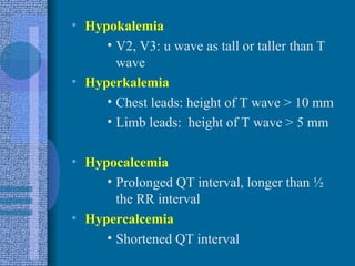
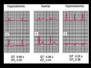
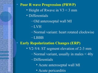
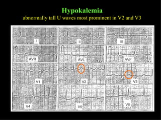
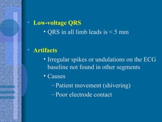
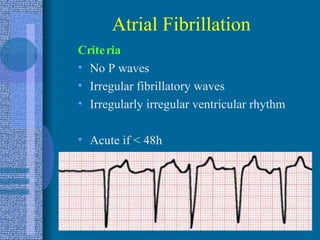
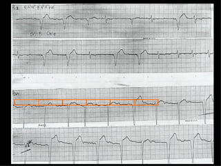
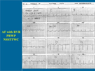
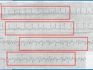
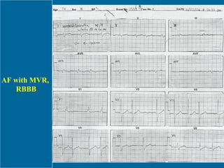
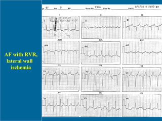
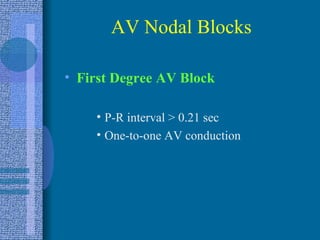
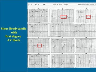
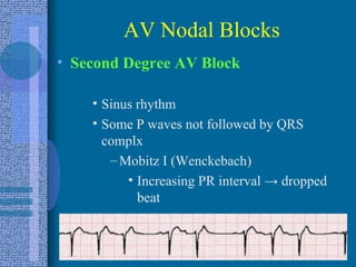
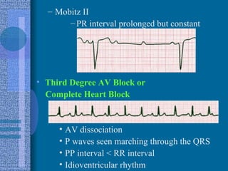
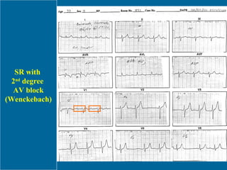
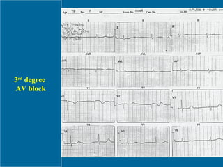
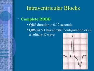
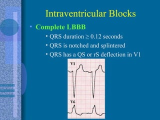
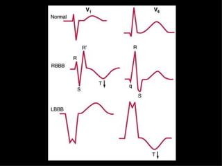
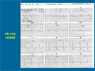
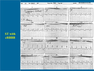
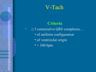
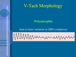
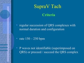
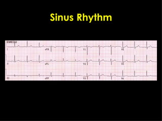
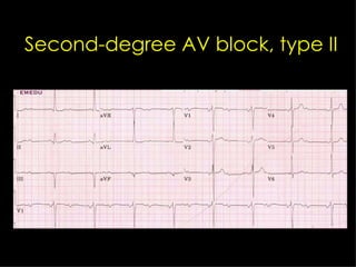
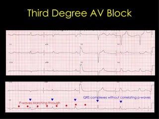
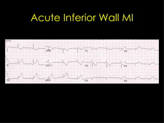
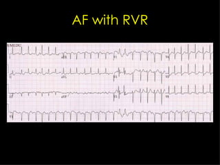
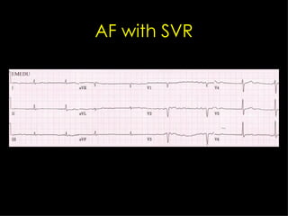
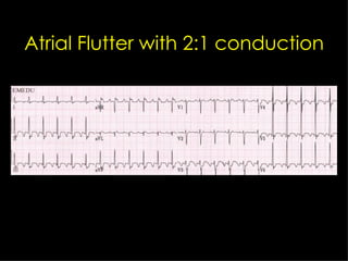
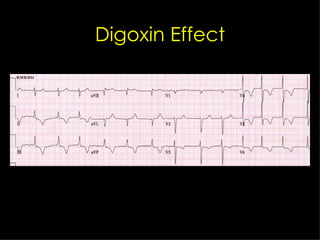
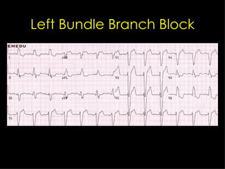
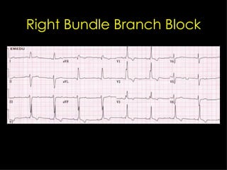
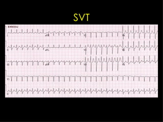
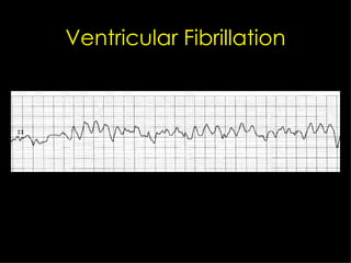
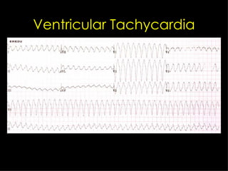
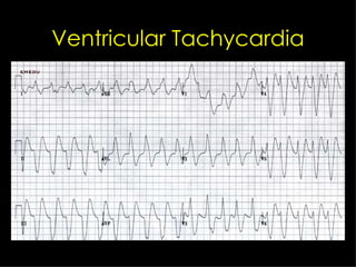
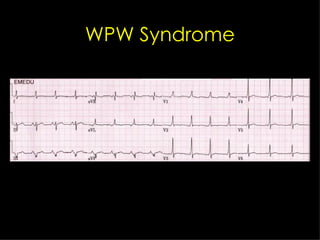
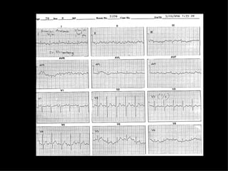
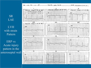
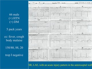
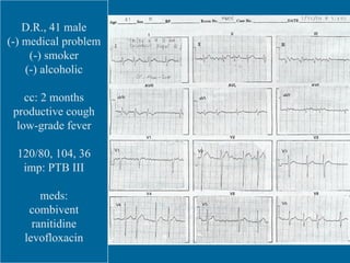
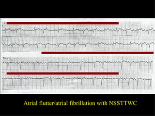
Electrocardiographic Cases Crisbert I. Cualteros, M.D.
Sinus Rhythm
SR  frequent PVDs PRWP lat wall ischemia
ECG Indications determine cardiac rate define cardiac rhythm diagnose old or new MI identify intracardiac conduction disturbances aid in the diagnosis of IHD, pericarditis, myocarditis, electrolyte abnormalities and pacemaker malfunction
V1 = 4th ICS, R sternal border V2 = 4th ICS, L sternal border V3 = halfway between V2 and V4 V4 = 5th ICS, L MCL V5 = 5th ICS, anterior axillary line V6 = 5th ICS, L mid-midaxillary line V3R = halfway between V1 and V4R V4R =  5th ICS, R MCL  Lead Locations
Correspondence Anteroseptal wall V1-V3 Lateral wall V5, V6 Anterior wall V3, V4 Septal wall V1, V2 High lateral wall I, aVL Inferior wall II, III, aVF Area Leads
Area Leads RV wall V3R and V4R Posterior LV wall Mirror image of V 1/2 Diffuse/global/massive Almost all leads Inferolateral wall V5, V6, II, III, aVF Anterolateral wall V3-V6, I, aVL
 
RRAHIM Components of ECG interpretation R ate R hythm A xis H ypertrophy I schemia and Infarction M iscellaneous (normal variants)


Rate Mnemonic: 300, 150, 100, 75, 60, 50 Formula:  1500 / # of small boxes   300 / # of big boxes Bradycardia  = <60 bpm Normal Rate  = 60-100 bpm Tachycardia  = >100 bpm
What is the rate? 1500/28 or 300/5.6 53 bpm
What is the rate? 1500/12 or 300/2.4 125 bpm
Rhythm Identify the  P  wave Check relation of P wave to QRS Normal: P wave is before QRS SVT, heart blocks: P wave after QRS or burried Check   PR  interval ( 0.12 - 0.20s ) Shortened: WPW Prolonged: 1 st  and 2 nd  degree AV block
Check  QRS  duration  (< 0.10 s ) Widened: bundle branch blocks Check relation of R-R and P-P int PP < RR: complete heart block PP > RR: AV dissociation
Common Rhythm Interpretations Sinus rhythm Supraventricular arrhythmias Atrial fibrillation Atrial flutter Supraventricular tachycardia (SVT) Heart Blocks First-degree AV block Second-degree AV block Mobitz Type I (Wenckebach) Mobitz Type II
Third-degree AV block Left or Right Bundle Branch Block Complete Incomplete Ventricular Arrhythmias Premature Ventricular Depolarization (PVD) Ventricular Tachycardia (V-tach) Sustained Non-sustained Ventricular fibrillation (V-fib)
Axis Determination get the average QRS vector from the isoelectric baseline in Leads  I and AVF if the average QRS vector is  above  baseline  ->  (+) QRS deflection if the average QRS vector is  below  baseline ->   (-) QRS deflection
Axis -- -- indeterminate + -- RAD -- + LAD + + Normal Axis AVF Lead 1
 
Normal (-30 to +90) RAD Indeterminate LAD
Axis   - 90  AVF indeterminate   LAD ± 180  0  I   RAD   normal      (-30 to +90)   + 90
What is  the axis?
Axis Differentials WPW syndrome WPW syndrome LPFB LAFB Pulmonary embolism LBBB Lateral wall MI Inferior wall MI RVH (COPD, cor pul) LVH (HTN) N variant: thin, tall N variant: short, fat RAD LAD
Hypertrophy Six Possibilities No hypertrophy LVH RVH LAE RAE combination
LVH criteria 3 Methods 1) S wave in V1 +  R wave in V5/6 > 35mm  2) R in AVL > 11mm 3) Romhilt and Estes Criteria (best)
SR, LVH with Strain Pattern,  Old Anteroseptal Wall MI
RVH Criteria RAD of  ≥  +110, with any of the ff: V1: R wave > S wave COPD RBBB True posterior infarction WPW Deep S wave in V5-6 COPD ST depression and T wave inversion in V1-3
LAE (p mitrale) 2 Methods V1: wide  terminal  component of P  wave  ≥  1 mm wide (0.04 s) and  ≥  1 mm deep Any lead: P wave wider than 0.12s  or with a  ≥  1 mm notch in the middle
ST, RAD, LAE, RVH
RAE (p pulmonale) 2 Methods V1: tall  initial  component of P wave  ≥  2mm wide and  ≥  2 mm tall Any Lead: P wave  ≥  2.5 mm tall
RRAHIM Components of ECG interpretation Rate Rhythm Axis Hypertrophy Ischemia and Infarction Miscellaneous (normal variants)
Myocardial Ischemia 1 mm ST-segment depression Symmetrically/inverted T waves Abnormally tall T waves Normalization of abnormal T waves Prolongation of QT interval Arrhythmias, BBB, AV blocks or electrical alternans
SR, Anterolateral Wall Ischemia
Myocardial infarction Criteria (any) ST elevation  ≥   2 Chest leads:  ≥  2 mm elevation or ≥   2 Limb leads:  ≥  1 mm elevation Q waves  ≥  0.04s (1 small square)
Timing of MI/ECG 0-6 hours 6-24 h 24 -72 h 72 h – 6 weeks > 6 wk
Differentials for ST elevation Acute pericarditis Ventricular aneurysm Severe LV wall hypokinesia Early repolarization changes Variant (prinzmetal) angina
Q waves never significant in aVR not significant in V1 unless with abnormalities in other precordial leads not significant in III unless with abnormalities in II, aVF more reliable if associated with ST changes Not significant in V1-V3 if (+)  LBBB , but  significant  if (+)  RBBB
Criteria for Pathologic Q waves ≥   0.04 sec in duration ≥   25% of the R wave amplitude
Recent Anteroseptal Wall MI
SR, Acute Inferior Wall MI
RRAHIM Components of ECG interpretation R ate R hythm A xis H ypertrophy I schemia and Infarction M iscellaneous (normal variants)
Hypokalemia V2, V3: u wave as tall or taller than T wave Hyperkalemia Chest leads: height of T wave > 10 mm Limb leads:  height of T wave > 5 mm Hypocalcemia Prolonged QT interval, longer than ½ the RR interval Hypercalcemia Shortened QT interval
 
Poor R wave Progression (PRWP) Height of Rwave in V3 < 3 mm Differentials Old anteroseptal wall MI LVH Normal variant: heart rotated clockwise LBBB Early Repolarization Changes (ERP) V2-V4: ST segment elevation of 2-3 mm Normal variant, usually in males < 40y Differentials Acute anteroseptal wall MI Acute pericarditis
SR, ERP
SR, ERP
SR, PRWP
Hyperkalemia tall, peaked T waves usually >10 mm in the chest leads
Hypokalemia abnormally tall U waves most prominent in V2 and V3
Low-voltage QRS QRS in all limb leads is < 5 mm Artifacts Irregular spikes or undulations on the ECG baseline not found in other segments Causes Patient movement (shivering) Poor electrode contact
Atrial Fibrillation Criteria No P waves Irregular fibrillatory waves Irregularly irregular ventricular rhythm Acute if < 48h
Top 5 causes of AF  (EVICT) E thanol (Holiday Heart Syndrome) V alvular heart disease (MS) I HD C ardiomyopathy T hyrotoxicosis
 
AF with RVR PRWP NSSTTWC
 
AF with MVR,  RBBB
AF with RVR, lateral wall  ischemia
AV Nodal Blocks First Degree AV Block P-R interval > 0.21 sec One-to-one AV conduction
Sinus Bradycardia with first degree  AV block
AV Nodal Blocks Second Degree AV Block Sinus rhythm Some P waves not followed by QRS complx Mobitz I (Wenckebach) Increasing PR interval  ->  dropped beat
Mobitz II PR interval prolonged but constant Third Degree AV Block or  Complete Heart Block AV dissociation P waves seen marching through the QRS PP interval < RR interval Idioventricular rhythm
SR with  2 nd  degree  AV block (Wenckebach)
3 rd  degree  AV block
Intraventricular Blocks Complete RBBB QRS duration  ≥  0.12 seconds QRS in V1 has an rsR’ configuration or is a solitary R wave
Intraventricular Blocks Complete LBBB QRS duration  ≥  0.12 seconds QRS is notched and splintered QRS has a QS or rS deflection in V1
.
SR with cRBBB
ST with cRBBB
V-Tach Criteria ≥   3 consecutive QRS complexes…  of uniform configuration of ventricular origin > 100 bpm
Monomorphic Sustained VT: > 30 s Hemodynamic compromise Requires intervention for termination  Non-sustained V-Tach Morphology
Polymorphic beat to beat variation in QRS complexes V-Tach Morphology
SupraV Tach Criteria regular succession of QRS complexes with normal duration and configuration rate 150 – 250 bpm P waves not identifiable (superimposed on QRS) or preceed / succeed the QRS complex
 
 
Sinus Rhythm
Second-degree AV block, type II
Third Degree AV Block
Acute Inferior Wall MI
AF with RVR
AF with SVR
Anteroseptal Wall MI
Atrial Flutter with 2:1 conduction
Digoxin Effect
ERP
First-Degree AV block, SB
Frequent PVCs in Bigeminy
Hyperkalemia
Left Bundle Branch Block
Right Bundle Branch Block
SVT
Ventricular Fibrillation
Ventricular Tachycardia
WPW Syndrome
 
SR LAE LVH with strain  Pattern ERP vs Acute injury pattern in the anteroseptal wall
SR, LAH, LAE, IVCD, LVH, lateral wall ischemia and/or strain. NSSTTWC, inferior wall. ERP vs.  acute injury, anteroseptal wall. 66 male (+) HTN (+) DM 5 pack years cc: fever, cough body malaise 150/80, 88, 20 trop I negative SR, LAE, with an acute injury pattern in the anteroseptal wall
D.R., 41 male (-) medical problem (-) smoker (-) alcoholic cc: 2 months productive cough low-grade fever 120/80, 104, 36 imp: PTB III meds: combivent ranitidine levofloxacin
Atrial flutter/atrial fibrillation with NSSTTWC
Thank You! http://crisbertcualteros.page.tl

ELECTROCARDIOGRAM

  • 1.
  • 2.
  • 3.
    SR frequentPVDs PRWP lat wall ischemia
  • 4.
    ECG Indications determinecardiac rate define cardiac rhythm diagnose old or new MI identify intracardiac conduction disturbances aid in the diagnosis of IHD, pericarditis, myocarditis, electrolyte abnormalities and pacemaker malfunction
  • 5.
    V1 = 4thICS, R sternal border V2 = 4th ICS, L sternal border V3 = halfway between V2 and V4 V4 = 5th ICS, L MCL V5 = 5th ICS, anterior axillary line V6 = 5th ICS, L mid-midaxillary line V3R = halfway between V1 and V4R V4R = 5th ICS, R MCL Lead Locations
  • 6.
    Correspondence Anteroseptal wallV1-V3 Lateral wall V5, V6 Anterior wall V3, V4 Septal wall V1, V2 High lateral wall I, aVL Inferior wall II, III, aVF Area Leads
  • 7.
    Area Leads RVwall V3R and V4R Posterior LV wall Mirror image of V 1/2 Diffuse/global/massive Almost all leads Inferolateral wall V5, V6, II, III, aVF Anterolateral wall V3-V6, I, aVL
  • 8.
  • 9.
    RRAHIM Components ofECG interpretation R ate R hythm A xis H ypertrophy I schemia and Infarction M iscellaneous (normal variants)
  • 10.
  • 11.
  • 12.
  • 13.
  • 14.
  • 15.
  • 16.
    Rate Mnemonic: 300,150, 100, 75, 60, 50 Formula: 1500 / # of small boxes 300 / # of big boxes Bradycardia = <60 bpm Normal Rate = 60-100 bpm Tachycardia = >100 bpm
  • 17.
    What is therate? 1500/28 or 300/5.6 53 bpm
  • 18.
    What is therate? 1500/12 or 300/2.4 125 bpm
  • 19.
    Rhythm Identify the P wave Check relation of P wave to QRS Normal: P wave is before QRS SVT, heart blocks: P wave after QRS or burried Check PR interval ( 0.12 - 0.20s ) Shortened: WPW Prolonged: 1 st and 2 nd degree AV block
  • 20.
    Check QRS duration (< 0.10 s ) Widened: bundle branch blocks Check relation of R-R and P-P int PP < RR: complete heart block PP > RR: AV dissociation
  • 21.
    Common Rhythm InterpretationsSinus rhythm Supraventricular arrhythmias Atrial fibrillation Atrial flutter Supraventricular tachycardia (SVT) Heart Blocks First-degree AV block Second-degree AV block Mobitz Type I (Wenckebach) Mobitz Type II
  • 22.
    Third-degree AV blockLeft or Right Bundle Branch Block Complete Incomplete Ventricular Arrhythmias Premature Ventricular Depolarization (PVD) Ventricular Tachycardia (V-tach) Sustained Non-sustained Ventricular fibrillation (V-fib)
  • 23.
    Axis Determination getthe average QRS vector from the isoelectric baseline in Leads I and AVF if the average QRS vector is above baseline -> (+) QRS deflection if the average QRS vector is below baseline -> (-) QRS deflection
  • 24.
    Axis -- --indeterminate + -- RAD -- + LAD + + Normal Axis AVF Lead 1
  • 25.
  • 26.
    Normal (-30 to+90) RAD Indeterminate LAD
  • 27.
    Axis - 90 AVF indeterminate LAD ± 180 0 I RAD normal (-30 to +90) + 90
  • 28.
    What is the axis?
  • 29.
    Axis Differentials WPWsyndrome WPW syndrome LPFB LAFB Pulmonary embolism LBBB Lateral wall MI Inferior wall MI RVH (COPD, cor pul) LVH (HTN) N variant: thin, tall N variant: short, fat RAD LAD
  • 30.
    Hypertrophy Six PossibilitiesNo hypertrophy LVH RVH LAE RAE combination
  • 31.
    LVH criteria 3Methods 1) S wave in V1 + R wave in V5/6 > 35mm 2) R in AVL > 11mm 3) Romhilt and Estes Criteria (best)
  • 32.
    SR, LVH withStrain Pattern, Old Anteroseptal Wall MI
  • 33.
    RVH Criteria RADof ≥ +110, with any of the ff: V1: R wave > S wave COPD RBBB True posterior infarction WPW Deep S wave in V5-6 COPD ST depression and T wave inversion in V1-3
  • 34.
    LAE (p mitrale)2 Methods V1: wide terminal component of P wave ≥ 1 mm wide (0.04 s) and ≥ 1 mm deep Any lead: P wave wider than 0.12s or with a ≥ 1 mm notch in the middle
  • 35.
  • 36.
    RAE (p pulmonale)2 Methods V1: tall initial component of P wave ≥ 2mm wide and ≥ 2 mm tall Any Lead: P wave ≥ 2.5 mm tall
  • 37.
    RRAHIM Components ofECG interpretation Rate Rhythm Axis Hypertrophy Ischemia and Infarction Miscellaneous (normal variants)
  • 38.
    Myocardial Ischemia 1mm ST-segment depression Symmetrically/inverted T waves Abnormally tall T waves Normalization of abnormal T waves Prolongation of QT interval Arrhythmias, BBB, AV blocks or electrical alternans
  • 39.
  • 40.
    Myocardial infarction Criteria(any) ST elevation ≥ 2 Chest leads: ≥ 2 mm elevation or ≥ 2 Limb leads: ≥ 1 mm elevation Q waves ≥ 0.04s (1 small square)
  • 41.
    Timing of MI/ECG0-6 hours 6-24 h 24 -72 h 72 h – 6 weeks > 6 wk
  • 42.
    Differentials for STelevation Acute pericarditis Ventricular aneurysm Severe LV wall hypokinesia Early repolarization changes Variant (prinzmetal) angina
  • 43.
    Q waves neversignificant in aVR not significant in V1 unless with abnormalities in other precordial leads not significant in III unless with abnormalities in II, aVF more reliable if associated with ST changes Not significant in V1-V3 if (+) LBBB , but significant if (+) RBBB
  • 44.
    Criteria for PathologicQ waves ≥ 0.04 sec in duration ≥ 25% of the R wave amplitude
  • 45.
  • 46.
  • 47.
    RRAHIM Components ofECG interpretation R ate R hythm A xis H ypertrophy I schemia and Infarction M iscellaneous (normal variants)
  • 48.
    Hypokalemia V2, V3:u wave as tall or taller than T wave Hyperkalemia Chest leads: height of T wave > 10 mm Limb leads: height of T wave > 5 mm Hypocalcemia Prolonged QT interval, longer than ½ the RR interval Hypercalcemia Shortened QT interval
  • 49.
  • 50.
    Poor R waveProgression (PRWP) Height of Rwave in V3 < 3 mm Differentials Old anteroseptal wall MI LVH Normal variant: heart rotated clockwise LBBB Early Repolarization Changes (ERP) V2-V4: ST segment elevation of 2-3 mm Normal variant, usually in males < 40y Differentials Acute anteroseptal wall MI Acute pericarditis
  • 51.
  • 52.
  • 53.
  • 54.
    Hyperkalemia tall, peakedT waves usually >10 mm in the chest leads
  • 55.
    Hypokalemia abnormally tallU waves most prominent in V2 and V3
  • 56.
    Low-voltage QRS QRSin all limb leads is < 5 mm Artifacts Irregular spikes or undulations on the ECG baseline not found in other segments Causes Patient movement (shivering) Poor electrode contact
  • 57.
    Atrial Fibrillation CriteriaNo P waves Irregular fibrillatory waves Irregularly irregular ventricular rhythm Acute if < 48h
  • 58.
    Top 5 causesof AF (EVICT) E thanol (Holiday Heart Syndrome) V alvular heart disease (MS) I HD C ardiomyopathy T hyrotoxicosis
  • 59.
  • 60.
    AF with RVRPRWP NSSTTWC
  • 61.
  • 62.
  • 63.
    AF with RVR,lateral wall ischemia
  • 64.
    AV Nodal BlocksFirst Degree AV Block P-R interval > 0.21 sec One-to-one AV conduction
  • 65.
    Sinus Bradycardia withfirst degree AV block
  • 66.
    AV Nodal BlocksSecond Degree AV Block Sinus rhythm Some P waves not followed by QRS complx Mobitz I (Wenckebach) Increasing PR interval -> dropped beat
  • 67.
    Mobitz II PRinterval prolonged but constant Third Degree AV Block or Complete Heart Block AV dissociation P waves seen marching through the QRS PP interval < RR interval Idioventricular rhythm
  • 68.
    SR with 2 nd degree AV block (Wenckebach)
  • 69.
    3 rd degree AV block
  • 70.
    Intraventricular Blocks CompleteRBBB QRS duration ≥ 0.12 seconds QRS in V1 has an rsR’ configuration or is a solitary R wave
  • 71.
    Intraventricular Blocks CompleteLBBB QRS duration ≥ 0.12 seconds QRS is notched and splintered QRS has a QS or rS deflection in V1
  • 72.
  • 73.
  • 74.
  • 75.
    V-Tach Criteria ≥ 3 consecutive QRS complexes… of uniform configuration of ventricular origin > 100 bpm
  • 76.
    Monomorphic Sustained VT:> 30 s Hemodynamic compromise Requires intervention for termination Non-sustained V-Tach Morphology
  • 77.
    Polymorphic beat tobeat variation in QRS complexes V-Tach Morphology
  • 78.
    SupraV Tach Criteriaregular succession of QRS complexes with normal duration and configuration rate 150 – 250 bpm P waves not identifiable (superimposed on QRS) or preceed / succeed the QRS complex
  • 79.
  • 80.
  • 81.
  • 82.
  • 83.
  • 84.
  • 85.
  • 86.
  • 87.
  • 88.
    Atrial Flutter with2:1 conduction
  • 89.
  • 90.
  • 91.
  • 92.
  • 93.
  • 94.
  • 95.
  • 96.
  • 97.
  • 98.
  • 99.
  • 100.
  • 101.
  • 102.
    SR LAE LVHwith strain Pattern ERP vs Acute injury pattern in the anteroseptal wall
  • 103.
    SR, LAH, LAE,IVCD, LVH, lateral wall ischemia and/or strain. NSSTTWC, inferior wall. ERP vs. acute injury, anteroseptal wall. 66 male (+) HTN (+) DM 5 pack years cc: fever, cough body malaise 150/80, 88, 20 trop I negative SR, LAE, with an acute injury pattern in the anteroseptal wall
  • 104.
    D.R., 41 male(-) medical problem (-) smoker (-) alcoholic cc: 2 months productive cough low-grade fever 120/80, 104, 36 imp: PTB III meds: combivent ranitidine levofloxacin
  • 105.
  • 106.