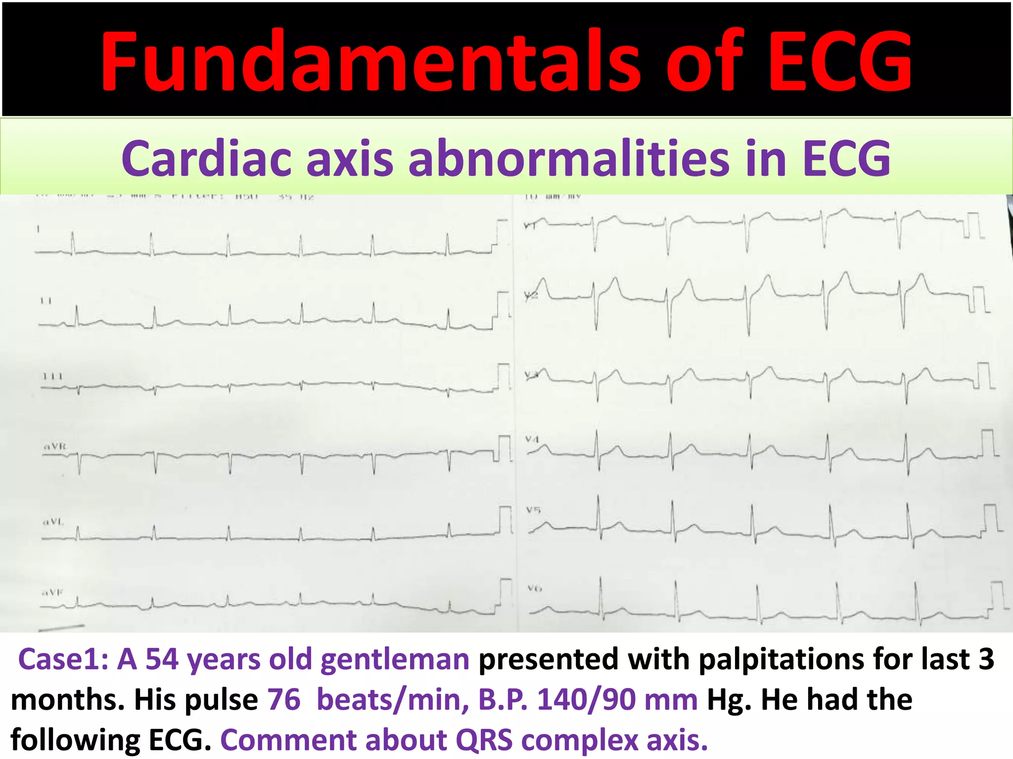
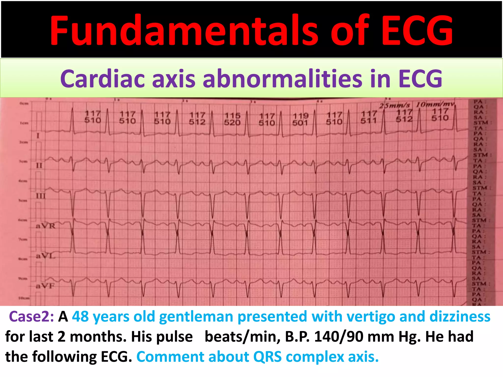
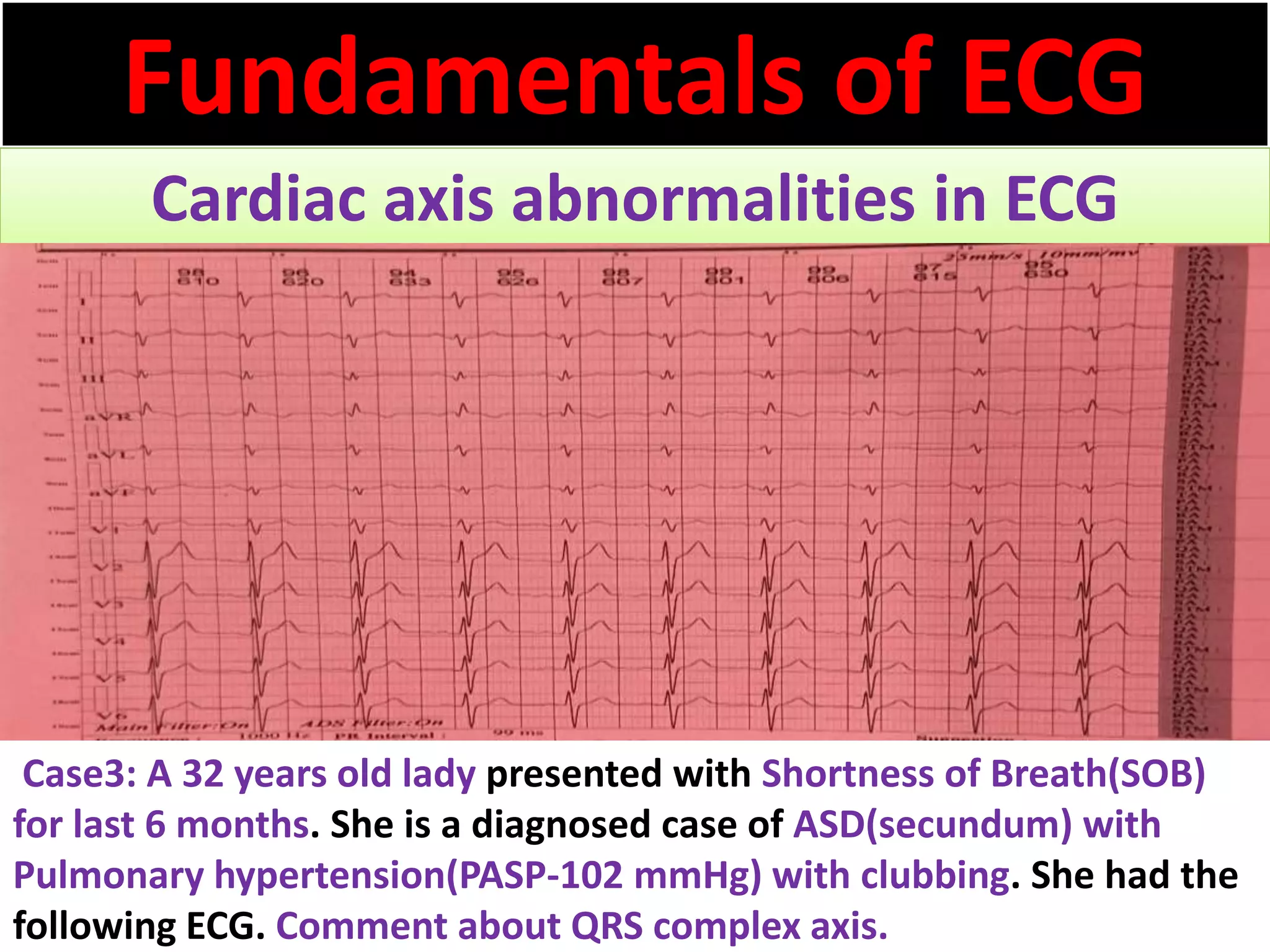
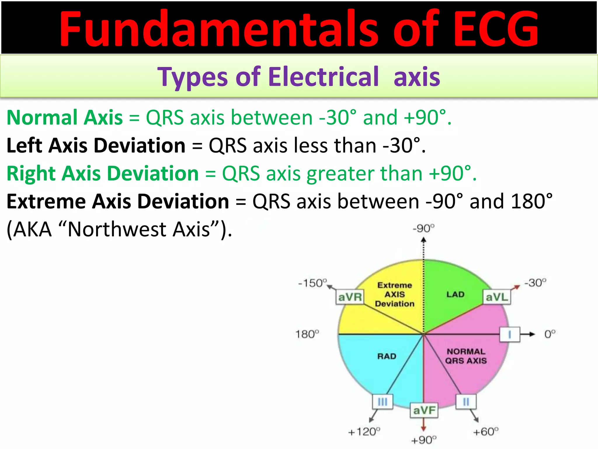
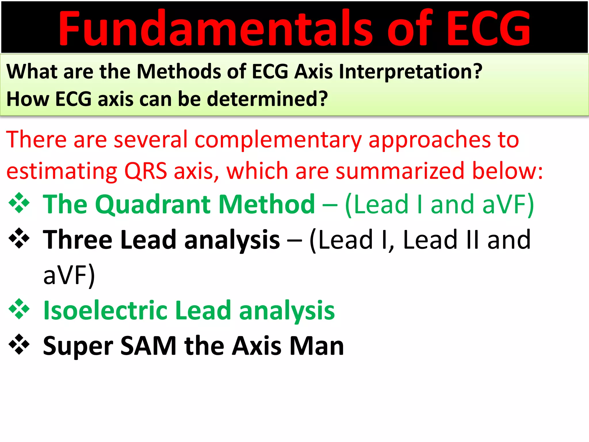
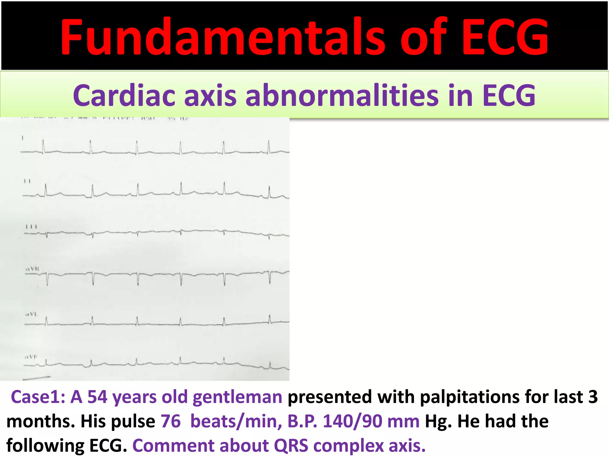
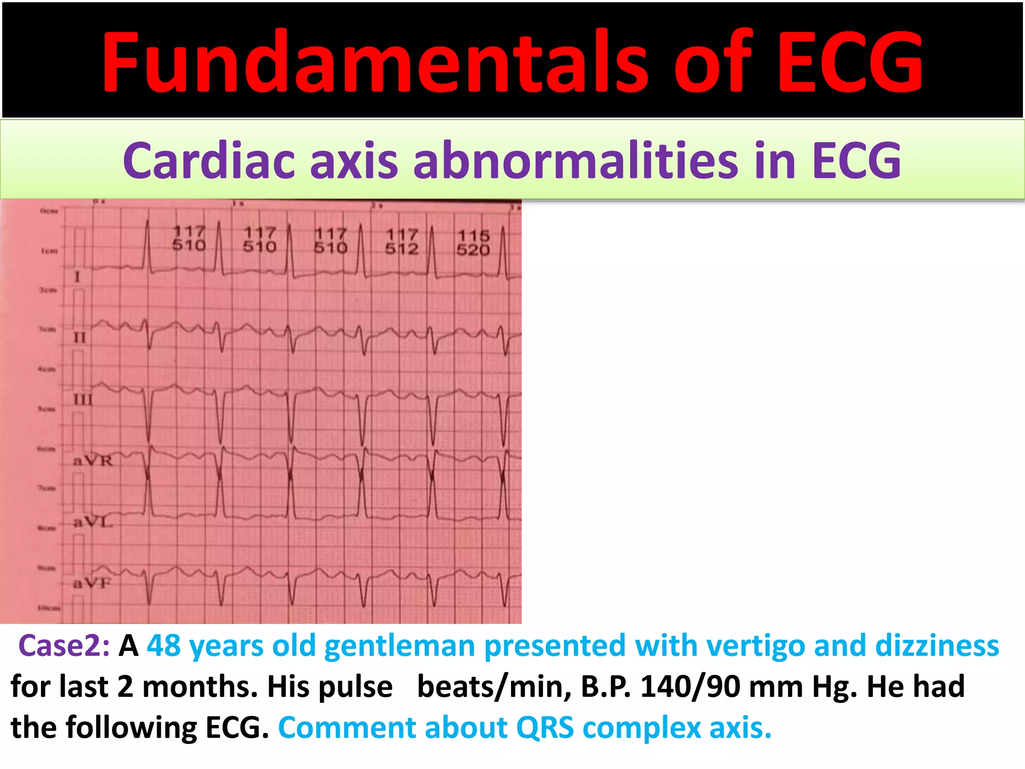
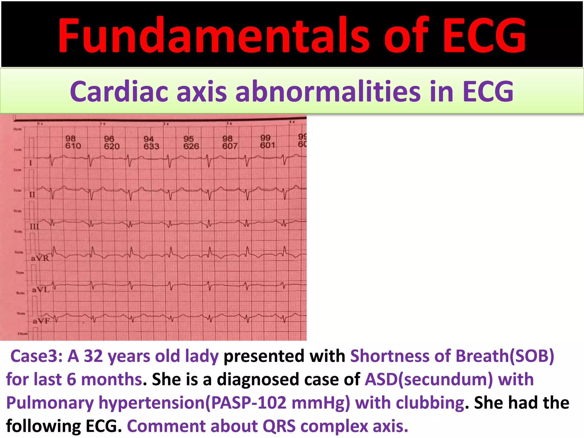
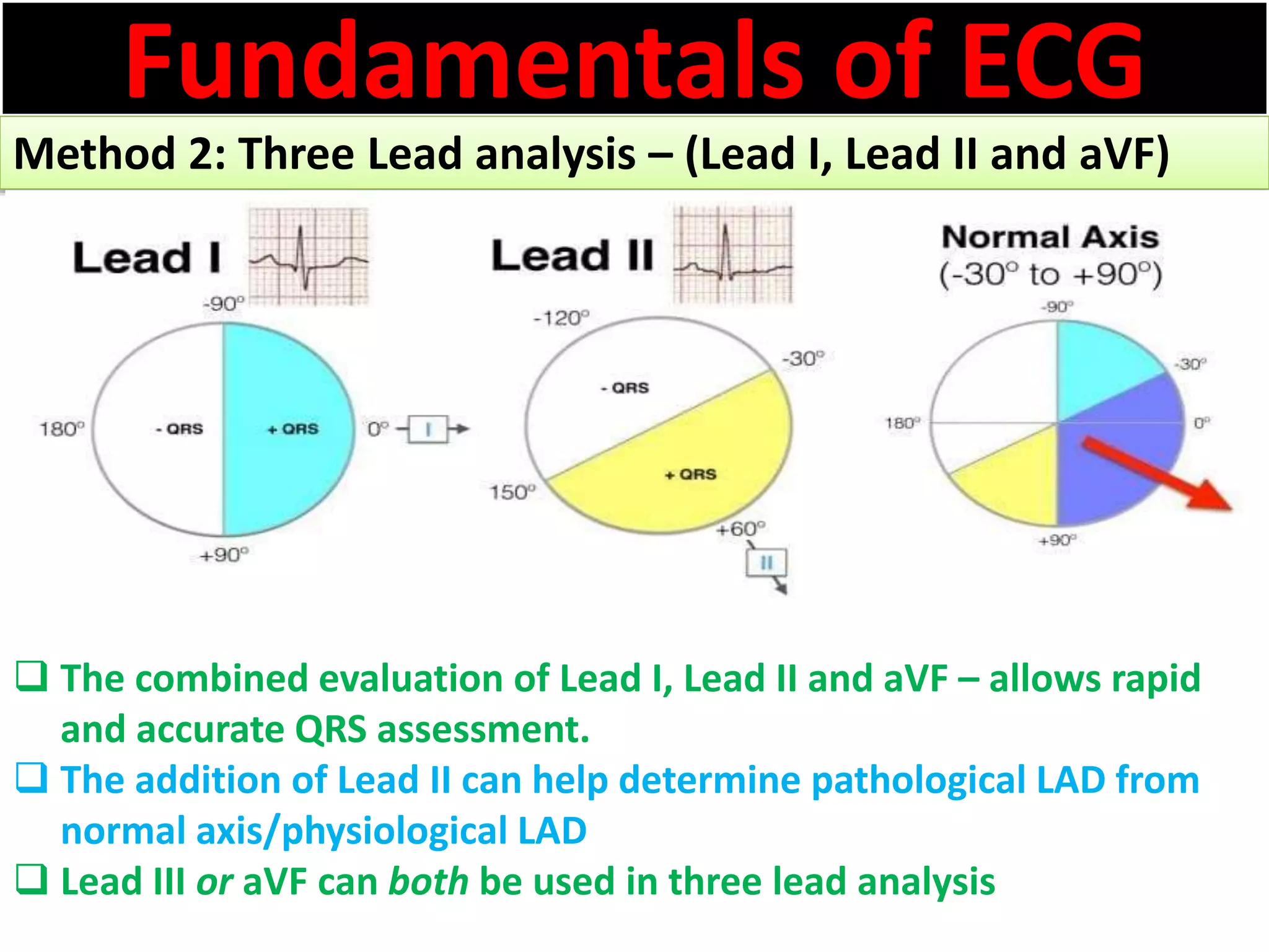
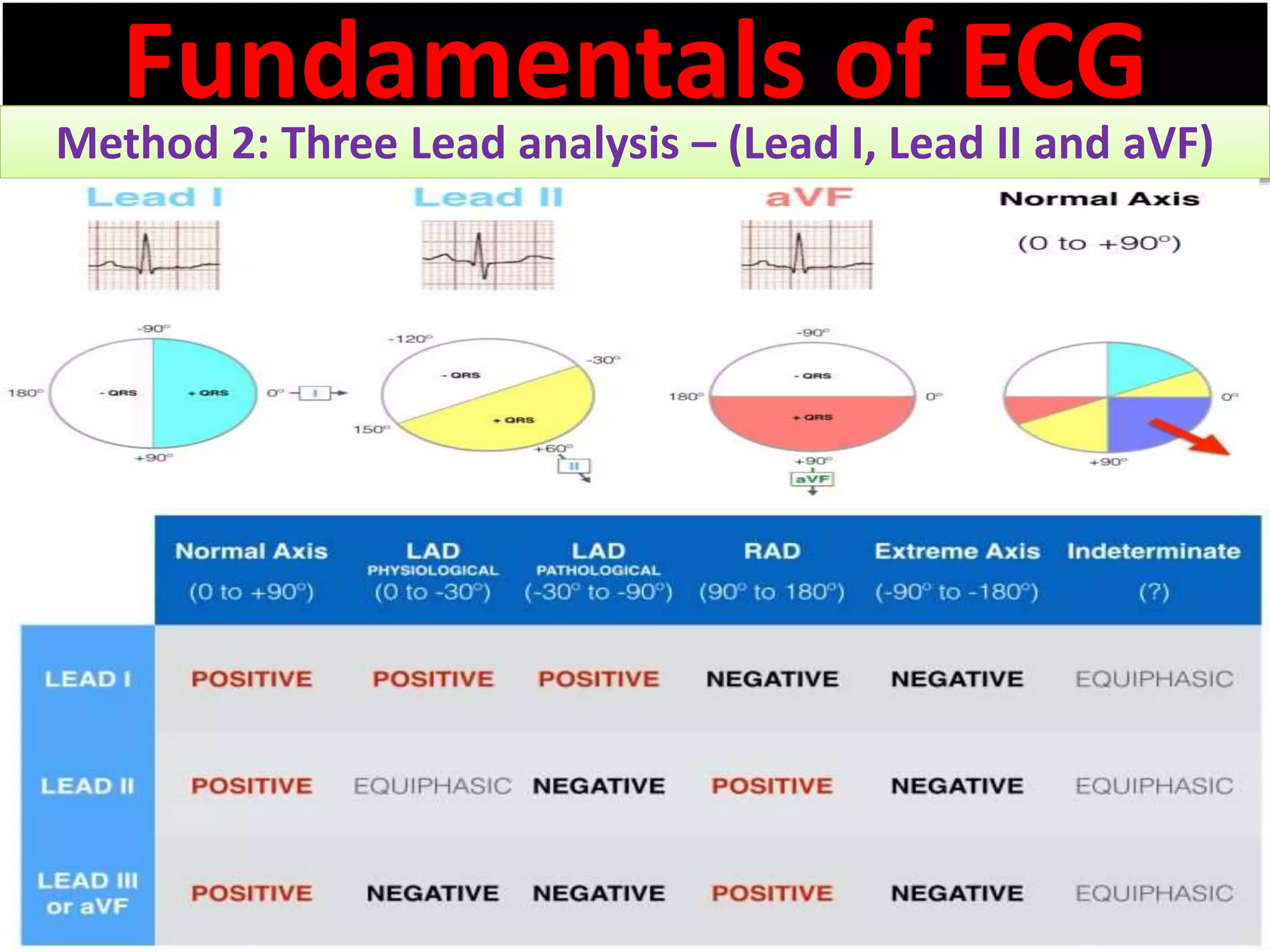
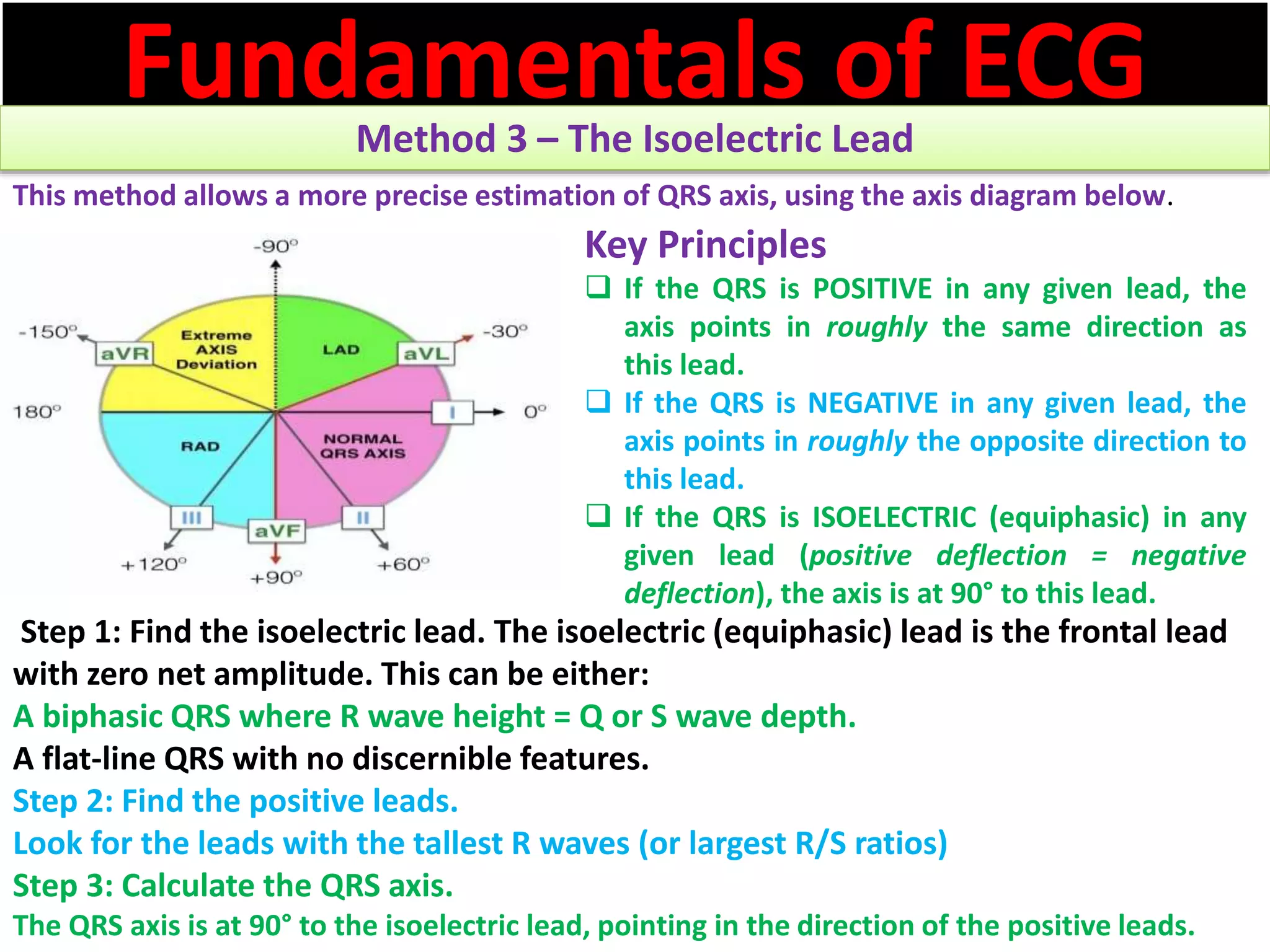
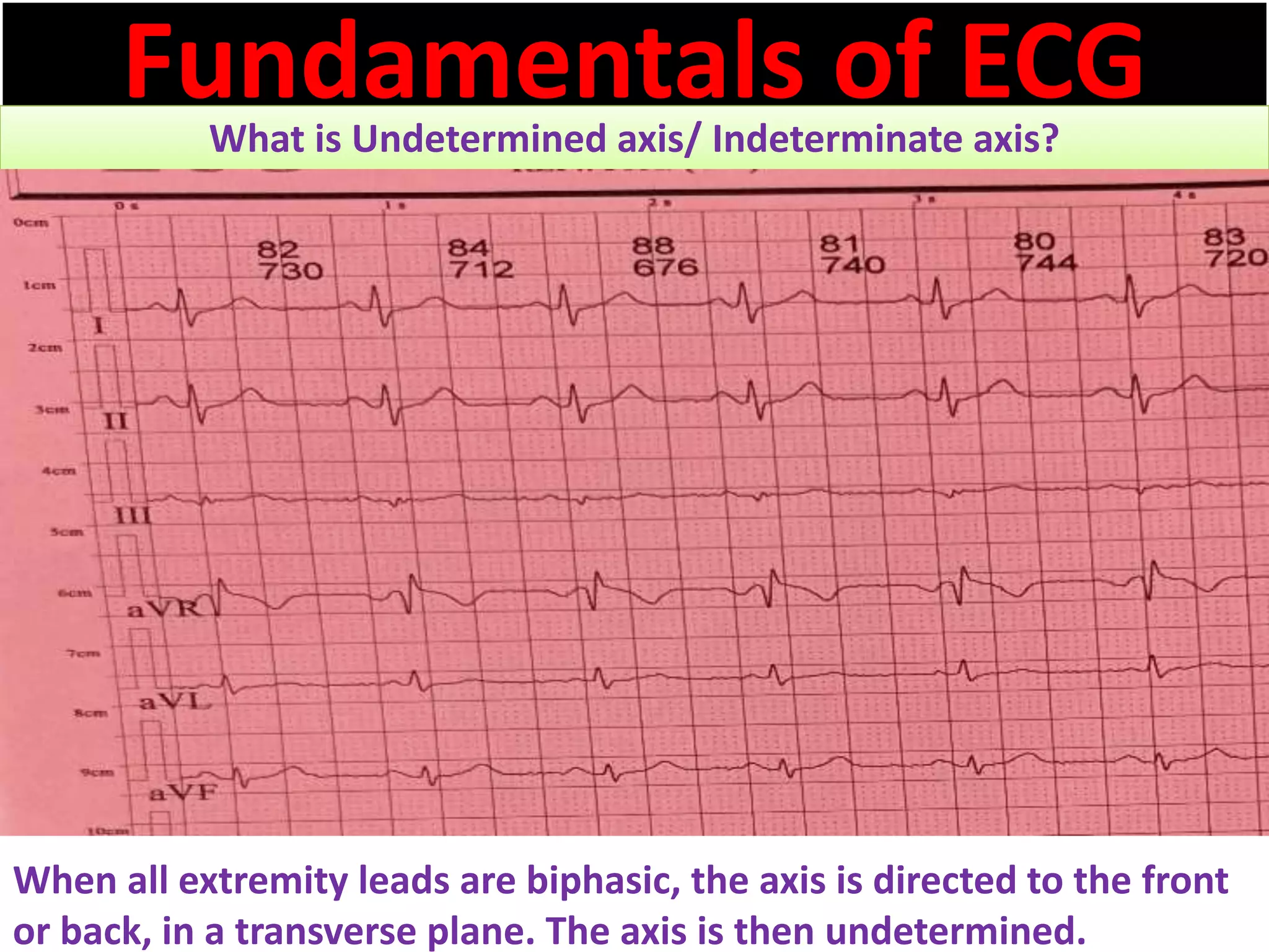
Fundamentals of ECG
Cardiac axis abnormalities in ECG
Dr. Md.Toufiqur Rahman
MBBS, FCPS, MD, FACC, FESC, FRCP, FSCAI,
FCCP,FAPSC, FAPSIC, FAHA,FACP
Professor & head of Cardiology
CMMC, Manikganj
Ex professor of cardiology,
NICVD, Dhaka
drtoufiq1971@gmail.com
Fundamentals of ECG
Cardiac axis abnormalities in ECG
Case-1: ECG with Normal axis ; Case-2: ECG with left axis deviation
Case-3: ECG with extreme right axis deviation
Case-4: ECG with right axis deviation
Clinical significance of cardiac axis
What is Electrical Axis? Types of electrical axis
What are the Methods of ECG Axis Interpretation? How ECG axis can
be determined?
How Ventricular (QRS) Axis is determined in Bundle Branch Blocks ?
What is Undetermined axis/ Indeterminate axis?
What are the causes of abnormal heart axis?
What are the causes of Right Axis Deviation(RAD)?
What are the causes of Left Axis Deviation?
What are the causes of Extreme Axis Deviation (indeterminate axis/
northwest axis)?
Fundamentals of ECG
Cardiac axis abnormalities in ECG
Case1: A 54 years old gentleman presented with palpitations for last 3
months. His pulse 76 beats/min, B.P. 140/90 mm Hg. He had the
following ECG. Comment about QRS complex axis.
Fundamentals of ECG
Cardiac axis abnormalities in ECG
Case2: A 48 years old gentleman presented with vertigo and dizziness
for last 2 months. His pulse beats/min, B.P. 140/90 mm Hg. He had
the following ECG. Comment about QRS complex axis.
Fundamentals of ECG
Cardiac axis abnormalities in ECG
Case3: A 32 years old lady presented with Shortness of Breath(SOB)
for last 6 months. She is a diagnosed case of ASD(secundum) with
Pulmonary hypertension(PASP-102 mmHg) with clubbing. She had the
following ECG. Comment about QRS complex axis.
Fundamentals of ECG
Clinical significance of cardiac axis
The axis of the ECG is the major direction of the overall
electrical activity of the heart.
It can be normal, leftward (left axis deviation, or LAD),
rightward (right axis deviation, or RAD) or indeterminate
(northwest axis).
The QRS axis is the most important to determine.
However, the P wave or T wave axis can also be measured.
To determine the QRS axis, the limb leads (not the
precordial leads) need to be examined.
Being able to interpret the electrical axis may offer insight
into the cardiac pathology and help avoid unnecessary
imaging tests.
Fundamentals of ECG
Types of Electrical axis
There are five main electrical axis:
īļ normal axis
īļ left axis deviation (LAD)
īļ right axis deviation (RAD)
īļ extreme axis deviation, and
īļ indeterminate axis
Fundamentals of ECG
Types of Electrical axis
Normal Axis = QRS axis between -30° and +90°.
Left Axis Deviation = QRS axis less than -30°.
Right Axis Deviation = QRS axis greater than +90°.
Extreme Axis Deviation = QRS axis between -90° and 180°
(AKA “Northwest Axis”).
Fundamentals of ECG
What are the Methods of ECG Axis Interpretation?
How ECG axis can be determined?
There are several complementary approaches to
estimating QRS axis, which are summarized below:
īļ The Quadrant Method – (Lead I and aVF)
īļ Three Lead analysis – (Lead I, Lead II and
aVF)
īļ Isoelectric Lead analysis
īļ Super SAM the Axis Man
Fundamentals of ECG
Method 1 – The Quadrant Method
īļ The most efficient way to estimate axis is to look at LEAD I and LEAD
aVF.
īļ Examine the QRS complex in each lead and determine if it is
Positive, Isoelectric (Equiphasic) or Negative:
īļ A positive QRS in Lead I puts the axis in roughly the same direction as lead I.
īļ A positive QRS in Lead aVF similarly aligns the axis with lead aVF.
īļ Combining both coloured areas – the quadrant of overlap determines the axis.
īļ So If Lead I and aVF are both positive, the axis is between 0° and +90° (i.e. normal
axis).
Fundamentals of ECG
Method 1 – The Quadrant Method
Fundamentals of ECG
Method 1 – The Quadrant Method
Fundamentals of ECG
Cardiac axis abnormalities in ECG
Case1: A 54 years old gentleman presented with palpitations for last 3
months. His pulse 76 beats/min, B.P. 140/90 mm Hg. He had the
following ECG. Comment about QRS complex axis.
Fundamentals of ECG
Cardiac axis abnormalities in ECG
Case2: A 48 years old gentleman presented with vertigo and dizziness
for last 2 months. His pulse beats/min, B.P. 140/90 mm Hg. He had
the following ECG. Comment about QRS complex axis.
Fundamentals of ECG
Cardiac axis abnormalities in ECG
Case3: A 32 years old lady presented with Shortness of Breath(SOB)
for last 6 months. She is a diagnosed case of ASD(secundum) with
Pulmonary hypertension(PASP-102 mmHg) with clubbing. She had the
following ECG. Comment about QRS complex axis.
Fundamentals of ECG
Method 2: Three Lead analysis – (Lead I, Lead II and aVF)
īą Next we add in Lead II to the analysis of Lead I and
aVF
īą A positive QRS in Lead I puts the axis in roughly
the same direction as lead I.
īą A positive QRS in Lead II similarly aligns the axis
with lead II.
īą We can then combine both coloured areas and
the area of overlap determines the axis.
īą So If Lead I and II are both positive, the axis is
between -30° and +90° (i.e. normal axis).
Fundamentals of ECG
Method 2: Three Lead analysis – (Lead I, Lead II and aVF)
īą The combined evaluation of Lead I, Lead II and aVF – allows rapid
and accurate QRS assessment.
īą The addition of Lead II can help determine pathological LAD from
normal axis/physiological LAD
īą Lead III or aVF can both be used in three lead analysis
Fundamentals of ECG
Method 2: Three Lead analysis – (Lead I, Lead II and aVF)
Fundamentals of ECGMethod 3 – The Isoelectric Lead
This method allows a more precise estimation of QRS axis, using the axis diagram below.
Step 1: Find the isoelectric lead. The isoelectric (equiphasic) lead is the frontal lead
with zero net amplitude. This can be either:
A biphasic QRS where R wave height = Q or S wave depth.
A flat-line QRS with no discernible features.
Step 2: Find the positive leads.
Look for the leads with the tallest R waves (or largest R/S ratios)
Step 3: Calculate the QRS axis.
The QRS axis is at 90° to the isoelectric lead, pointing in the direction of the positive leads.
Key Principles
īą If the QRS is POSITIVE in any given lead, the
axis points in roughly the same direction as
this lead.
īą If the QRS is NEGATIVE in any given lead, the
axis points in roughly the opposite direction to
this lead.
īą If the QRS is ISOELECTRIC (equiphasic) in any
given lead (positive deflection = negative
deflection), the axis is at 90° to this lead.
Fundamentals of ECGWhat is Undetermined axis/ Indeterminate axis?
When all extremity leads are biphasic, the axis is directed to the front
or back, in a transverse plane. The axis is then undetermined.
Fundamentals of ECG
What are the causes of abnormal heart axis?
īļ The direction of the vector can changes under different
circumstances:
īļ When the heart itself is rotated (right ventricular overload),
obviously the axis turns with it.
īļ In case of ventricular hypertrophy, the axis will deviate toward the
greater electrical activity and the vector will turn toward the
hypertrophied tissue.
īļ Infarcted tissue is electrically dead. No electrical activity is
registered and the QRS vector turns away from the infarcted tissue
īļ In conduction problems, the axis deviates too. When the right
ventricle depolarizes later than the left ventricle, the axis will turn
to the right (RBBB). This is because the right ventricle will begin the
contraction later and therefore will also finish later. In a normal
situation the vector is influenced by the left ventricle, but in RBBB
only the right ventricle determines it.
Fundamentals of ECG
What are the causes of Right Axis Deviation(RAD)?
 Normal variation (e.g., children, young adults)
 Limb-lead reversal (left- and right-arm electrodes)
 Right ventricular overload syndromes (acute or chronic)
 Right ventricular hypertrophy
 Conduction defects: left posterior fascicular block, right bundle branch
block
 Lateral wall myocardial infarction
 Preexcitation syndromes (e.g., Wolff-Parkinson-White syndrome)
 Ventricular ectopic rhythms (e.g., ventricular tachycardia)
 Congenital heart disease (e.g., secundum atrial septal defect)
 Dextrocardia
 Left pneumothorax
 Mechanical shift, such as with inspiration or emphysema
 Conditions that cause right-ventricular strain (e.g., pulmonary embolism,
pulmonary stenosis, pulmonary hypertension, chronic lung disease, and
resultant cor pulmonale
Fundamentals of ECG
What are the causes of Left Axis Deviation(LAD)?
o Normal variation (physiologic, often age-related change)
o Left ventricular hypertrophy
o Conduction defects: left bundle branch block, left anterior fascicular
block
o Inferior wall myocardial infarction
o Preexcitation syndromes (e.g., Wolff-Parkinson-White syndrome)
o Ventricular ectopic rhythms (e.g., ventricular tachycardia)
o Congenital heart disease (e.g., primum atrial septal defect,
endocardial cushion defect)
o Hyperkalemia
o Emphysema
o Mechanical shift, such as with expiration or raised diaphragm (e.g.,
pregnancy, ascites, abdominal tumor, organomegaly)
o Pacemaker-generated rhythm or paced rhythm
Fundamentals of ECG
What are the causes of Extreme Axis Deviation
(indeterminate axis/ northwest axis)?
īąVentricular rhythms –
e.g.VT, AIVR, ventricular ectopy
īąHyperkalaemia
īąSevere right ventricular
hypertrophy

Cardiac axis abnormalities in ECG

  • 1.
    Fundamentals of ECG Cardiacaxis abnormalities in ECG Dr. Md.Toufiqur Rahman MBBS, FCPS, MD, FACC, FESC, FRCP, FSCAI, FCCP,FAPSC, FAPSIC, FAHA,FACP Professor & head of Cardiology CMMC, Manikganj Ex professor of cardiology, NICVD, Dhaka drtoufiq1971@gmail.com
  • 2.
    Fundamentals of ECG Cardiacaxis abnormalities in ECG Case-1: ECG with Normal axis ; Case-2: ECG with left axis deviation Case-3: ECG with extreme right axis deviation Case-4: ECG with right axis deviation Clinical significance of cardiac axis What is Electrical Axis? Types of electrical axis What are the Methods of ECG Axis Interpretation? How ECG axis can be determined? How Ventricular (QRS) Axis is determined in Bundle Branch Blocks ? What is Undetermined axis/ Indeterminate axis? What are the causes of abnormal heart axis? What are the causes of Right Axis Deviation(RAD)? What are the causes of Left Axis Deviation? What are the causes of Extreme Axis Deviation (indeterminate axis/ northwest axis)?
  • 3.
    Fundamentals of ECG Cardiacaxis abnormalities in ECG Case1: A 54 years old gentleman presented with palpitations for last 3 months. His pulse 76 beats/min, B.P. 140/90 mm Hg. He had the following ECG. Comment about QRS complex axis.
  • 4.
    Fundamentals of ECG Cardiacaxis abnormalities in ECG Case2: A 48 years old gentleman presented with vertigo and dizziness for last 2 months. His pulse beats/min, B.P. 140/90 mm Hg. He had the following ECG. Comment about QRS complex axis.
  • 5.
    Fundamentals of ECG Cardiacaxis abnormalities in ECG Case3: A 32 years old lady presented with Shortness of Breath(SOB) for last 6 months. She is a diagnosed case of ASD(secundum) with Pulmonary hypertension(PASP-102 mmHg) with clubbing. She had the following ECG. Comment about QRS complex axis.
  • 6.
    Fundamentals of ECG Clinicalsignificance of cardiac axis The axis of the ECG is the major direction of the overall electrical activity of the heart. It can be normal, leftward (left axis deviation, or LAD), rightward (right axis deviation, or RAD) or indeterminate (northwest axis). The QRS axis is the most important to determine. However, the P wave or T wave axis can also be measured. To determine the QRS axis, the limb leads (not the precordial leads) need to be examined. Being able to interpret the electrical axis may offer insight into the cardiac pathology and help avoid unnecessary imaging tests.
  • 7.
    Fundamentals of ECG Typesof Electrical axis There are five main electrical axis: īļ normal axis īļ left axis deviation (LAD) īļ right axis deviation (RAD) īļ extreme axis deviation, and īļ indeterminate axis
  • 8.
    Fundamentals of ECG Typesof Electrical axis Normal Axis = QRS axis between -30° and +90°. Left Axis Deviation = QRS axis less than -30°. Right Axis Deviation = QRS axis greater than +90°. Extreme Axis Deviation = QRS axis between -90° and 180° (AKA “Northwest Axis”).
  • 9.
    Fundamentals of ECG Whatare the Methods of ECG Axis Interpretation? How ECG axis can be determined? There are several complementary approaches to estimating QRS axis, which are summarized below: īļ The Quadrant Method – (Lead I and aVF) īļ Three Lead analysis – (Lead I, Lead II and aVF) īļ Isoelectric Lead analysis īļ Super SAM the Axis Man
  • 10.
    Fundamentals of ECG Method1 – The Quadrant Method īļ The most efficient way to estimate axis is to look at LEAD I and LEAD aVF. īļ Examine the QRS complex in each lead and determine if it is Positive, Isoelectric (Equiphasic) or Negative: īļ A positive QRS in Lead I puts the axis in roughly the same direction as lead I. īļ A positive QRS in Lead aVF similarly aligns the axis with lead aVF. īļ Combining both coloured areas – the quadrant of overlap determines the axis. īļ So If Lead I and aVF are both positive, the axis is between 0° and +90° (i.e. normal axis).
  • 11.
    Fundamentals of ECG Method1 – The Quadrant Method
  • 12.
    Fundamentals of ECG Method1 – The Quadrant Method
  • 14.
    Fundamentals of ECG Cardiacaxis abnormalities in ECG Case1: A 54 years old gentleman presented with palpitations for last 3 months. His pulse 76 beats/min, B.P. 140/90 mm Hg. He had the following ECG. Comment about QRS complex axis.
  • 15.
    Fundamentals of ECG Cardiacaxis abnormalities in ECG Case2: A 48 years old gentleman presented with vertigo and dizziness for last 2 months. His pulse beats/min, B.P. 140/90 mm Hg. He had the following ECG. Comment about QRS complex axis.
  • 16.
    Fundamentals of ECG Cardiacaxis abnormalities in ECG Case3: A 32 years old lady presented with Shortness of Breath(SOB) for last 6 months. She is a diagnosed case of ASD(secundum) with Pulmonary hypertension(PASP-102 mmHg) with clubbing. She had the following ECG. Comment about QRS complex axis.
  • 17.
    Fundamentals of ECG Method2: Three Lead analysis – (Lead I, Lead II and aVF) īą Next we add in Lead II to the analysis of Lead I and aVF īą A positive QRS in Lead I puts the axis in roughly the same direction as lead I. īą A positive QRS in Lead II similarly aligns the axis with lead II. īą We can then combine both coloured areas and the area of overlap determines the axis. īą So If Lead I and II are both positive, the axis is between -30° and +90° (i.e. normal axis).
  • 18.
    Fundamentals of ECG Method2: Three Lead analysis – (Lead I, Lead II and aVF) īą The combined evaluation of Lead I, Lead II and aVF – allows rapid and accurate QRS assessment. īą The addition of Lead II can help determine pathological LAD from normal axis/physiological LAD īą Lead III or aVF can both be used in three lead analysis
  • 19.
    Fundamentals of ECG Method2: Three Lead analysis – (Lead I, Lead II and aVF)
  • 20.
    Fundamentals of ECGMethod3 – The Isoelectric Lead This method allows a more precise estimation of QRS axis, using the axis diagram below. Step 1: Find the isoelectric lead. The isoelectric (equiphasic) lead is the frontal lead with zero net amplitude. This can be either: A biphasic QRS where R wave height = Q or S wave depth. A flat-line QRS with no discernible features. Step 2: Find the positive leads. Look for the leads with the tallest R waves (or largest R/S ratios) Step 3: Calculate the QRS axis. The QRS axis is at 90° to the isoelectric lead, pointing in the direction of the positive leads. Key Principles īą If the QRS is POSITIVE in any given lead, the axis points in roughly the same direction as this lead. īą If the QRS is NEGATIVE in any given lead, the axis points in roughly the opposite direction to this lead. īą If the QRS is ISOELECTRIC (equiphasic) in any given lead (positive deflection = negative deflection), the axis is at 90° to this lead.
  • 21.
    Fundamentals of ECGWhatis Undetermined axis/ Indeterminate axis? When all extremity leads are biphasic, the axis is directed to the front or back, in a transverse plane. The axis is then undetermined.
  • 22.
    Fundamentals of ECG Whatare the causes of abnormal heart axis? īļ The direction of the vector can changes under different circumstances: īļ When the heart itself is rotated (right ventricular overload), obviously the axis turns with it. īļ In case of ventricular hypertrophy, the axis will deviate toward the greater electrical activity and the vector will turn toward the hypertrophied tissue. īļ Infarcted tissue is electrically dead. No electrical activity is registered and the QRS vector turns away from the infarcted tissue īļ In conduction problems, the axis deviates too. When the right ventricle depolarizes later than the left ventricle, the axis will turn to the right (RBBB). This is because the right ventricle will begin the contraction later and therefore will also finish later. In a normal situation the vector is influenced by the left ventricle, but in RBBB only the right ventricle determines it.
  • 23.
    Fundamentals of ECG Whatare the causes of Right Axis Deviation(RAD)?  Normal variation (e.g., children, young adults)  Limb-lead reversal (left- and right-arm electrodes)  Right ventricular overload syndromes (acute or chronic)  Right ventricular hypertrophy  Conduction defects: left posterior fascicular block, right bundle branch block  Lateral wall myocardial infarction  Preexcitation syndromes (e.g., Wolff-Parkinson-White syndrome)  Ventricular ectopic rhythms (e.g., ventricular tachycardia)  Congenital heart disease (e.g., secundum atrial septal defect)  Dextrocardia  Left pneumothorax  Mechanical shift, such as with inspiration or emphysema  Conditions that cause right-ventricular strain (e.g., pulmonary embolism, pulmonary stenosis, pulmonary hypertension, chronic lung disease, and resultant cor pulmonale
  • 24.
    Fundamentals of ECG Whatare the causes of Left Axis Deviation(LAD)? o Normal variation (physiologic, often age-related change) o Left ventricular hypertrophy o Conduction defects: left bundle branch block, left anterior fascicular block o Inferior wall myocardial infarction o Preexcitation syndromes (e.g., Wolff-Parkinson-White syndrome) o Ventricular ectopic rhythms (e.g., ventricular tachycardia) o Congenital heart disease (e.g., primum atrial septal defect, endocardial cushion defect) o Hyperkalemia o Emphysema o Mechanical shift, such as with expiration or raised diaphragm (e.g., pregnancy, ascites, abdominal tumor, organomegaly) o Pacemaker-generated rhythm or paced rhythm
  • 25.
    Fundamentals of ECG Whatare the causes of Extreme Axis Deviation (indeterminate axis/ northwest axis)? īąVentricular rhythms – e.g.VT, AIVR, ventricular ectopy īąHyperkalaemia īąSevere right ventricular hypertrophy