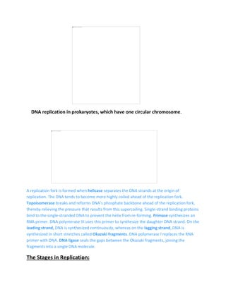
Submitted by:
Name: Sadaf Shaheen
Roll no. 61 ( Evening )
Subject : Molecular Biology
Topic: Replication in Prokaryotes
Submitted To:
Prof.Dr.Farah Rauf
Replication in prokaryotes
DNA replication has been extremely well studied in prokaryotes primarily because
of the small size of the genome and the mutants that are available. E. coli has 4.6
million base pairs in a single circular chromosome and all of it gets replicated in
approximately 42 minutes, starting from a single origin of replication and
proceeding around the circle in both directions. This means that approximately
1000 nucleotides are added per second. The process is quite rapid and occurs
without many mistakes.
DNA replication employs a large number of proteins and enzymes, each of which
plays a critical role during the process. One of the key players is the enzyme DNA
polymerase, also known as DNA pol, which adds nucleotides one by one to the
growing DNA chain that are complementary to the template strand. The addition
of nucleotides requires energy; this energy is obtained from the nucleotides that
have three phosphates attached to them, similar to ATP which has three
phosphate groups attached. When the bond between the phosphates is broken,
the energy released is used to form the phosphodiester bond between the
incoming nucleotide and the growing chain.
In prokaryotes, three main types of polymerases are known: DNA pol I, DNA pol
II, and DNA pol III. It is now known that:
DNA pol III is the enzyme required for "DNA synthesis",
DNA pol I and DNA pol II are primarily required for "repair"
DNA replication in prokaryotes, which have one circular chromosome.
A replication fork is formed when helicase separates the DNA strands at the origin of
replication. The DNA tends to become more highly coiled ahead of the replication fork.
Topoisomerase breaks and reforms DNA’s phosphate backbone ahead of the replication fork,
thereby relieving the pressure that results from this supercoiling. Single-strand binding proteins
bind to the single-stranded DNA to prevent the helix from re-forming. Primase synthesizes an
RNA primer. DNA polymerase III uses this primer to synthesize the daughter DNA strand. On the
leading strand, DNA is synthesized continuously, whereas on the lagging strand, DNA is
synthesized in short stretches called Okazaki fragments. DNA polymerase I replaces the RNA
primer with DNA. DNA ligase seals the gaps between the Okazaki fragments, joining the
fragments into a single DNA molecule.
The Stages in Replication:
The DNA replication in prokaryotes (and eukaryotes) are divided into three stages:
initiation, elongation, and termination.
Initiation:
During initiation the different proteins bind and form complex called orisome at
origin which unwinds the DNA. The different components and their functions are
as follows;
• The origin:
The replication of DNA initiates at a particular sequence of nucleotides in the
genome, known as origin. The prokaryotic cell usually has one origin as the
genome is small and circular. The initial loading of the replication machinery
known as orisome, a nuclear-protein complex, and the initial unwinding takes
place at origin.
In E. coli chromosomal DNA replication initiates at origin of chromosomal DNA,
oriC. The oriC is a 245-bp region containing four 9-bp and are three repeats of a
13-bp A/T-rich sequence.
• DnaA protein: It is also known as the initiator protein. This protein with 4
different domains is the first one to recognize and bind to the origin of replication
at the 9 bp regions. It is the binding of the initiator protein that actually causes
the DNA to stretch and leads to the separation of the strands at the AT-rich
region. As the two strands separate more DnaA proteins bind the unwound DNA.
• Dna C helicase loader: These proteins interact with the ssDNA-bound DnaA
proteins. Dna C helicase loaders help load the Dna B helicase onto the unwound
DNA.
• Dna B helicase: Helicase continues the unwinding of the DNA.
• Single-stranded DNA binding protein (SSB): These proteins bind and stabilize
the unwound DNA. Hence the unwound DNA do not form base pairs again.
• DNA gyrase: DNA gyrase is a topoisomerase that removes the twist resulting
from the unwinding of the DNA, by cleaving a strand of the DNA helix, untwisting
it and then resealing the broken strand again.
As the DNA is unwound, the area appears like a bubble and is known as a
replication bubble.
Circular prokaryotic DNA and the replication bubble.
The replication bubble has two Y shaped ends known as replication forks, (fig 2
shows one of the two replication fork) which is the junction between the wound
ds DNA and ss unwound DNA.
The two unwound strands are complementary and each can serve as the
templates for the synthesis of the new strands of DNA.
Elongation:
During elongation, the DNA pol III extends the daughter strands as the replication
complex known as replisome travels along the length of the chromosome. The
replisome includes a helicase, double-stranded DNA, a DNA polymerase(s) and a
clamp loader.
• DNA Polymerase
The first DNA polymerase enzyme to be characterized, DNA polymerase I (pol I)
from the bacterium Escherichia coli. DNA pol I is not the main DNA replicating
enzyme but is involved in processing of RNA primers during lagging-strand
synthesis and DNA repair mechanisms.
The major polymerase involved in the replication is DNA polymerase III, which is a
dimer, with two similar multisubunit complexes.
DNA pol III
In DNA pol III monomer is made up of core enzyme, γ complex and the β clamp.
DNA pol III core is made up of the αεθ complex, wherein α is the Polymerase
subunit, ε is the exonuclease (proofreading) subunit, and θ is involved with the
stabilizing.
The γ complex is required for the loading and unloading of the β clamps, the ring-
shaped protein complexes that holds and slides along duplex DNA. β clamps allow
the polymerase to remain associated with DNA and still slide along the DNA.
Each pol III monomer of the complex catalyzes the replication of either of the two
DNA strands at the replication fork at the rate of around 1000 bp per second.
DNA polymerase enzymes join the phosphate group at the 5′ carbon of a new
nucleotide to the hydroxyl (OH) group of the 3′ carbon of a nucleotide already in
the chain. Because of this there are two needs that arise;
~ First the DNA pol needs an existing 3’OH end.
This problem is solved by creating RNA primer
• Dna G primase: As the replication bubble is formed, the Dna G primase initiates
the synthesis of RNA primer, a sequence of about 10 RNA nucleotides
complementary to the parent DNA template. RNA primer provides a free 3-OH
end for the addition of the base pairs by the DNA polymerase. The RNA
nucleotides in the primers are then replaced by DNA nucleotides.
~ Second the replication can only proceed in 5′→3′ direction.
Now as DNA polymerase III can add nucleotides only to the 3′ end of a DNA
strand having OH group, the replication always proceeds only in the 5′ → 3′
direction on a growing DNA strand.
However, as the two parent strands of a DNA molecule are antiparallel, one
strand is unwound from the 5′ end and other from the 3′ end at the replication
fork.
In the strand which is unwound from the 5′ end, the primer is synthesized, such
that it’s 3′ end faces towards the unwinding replication fork. Hence the DNA pol III
can continuously add nucleotides to the 3′ end and extend the DNA daughter
strand. This strand which is synthesized continuously is known as the leading
strand, in which the direction of daughter strand elongation is same as the
direction of unwinding.
The replication in the leading strand
The other strand, known as lagging strand, experiences the unwinding from the 3′
end. The primase synthesizes the primer with 5′ end towards the unwinding end
and the and the 3′ end is on the opposite side. Now the DNA pol III extends the
daughter DNA strand at the 3′ end. Hence the unwinding of the parent duplex and
extension of daughter DNA strand takes place in the opposite direction.
In this lagging strand the replication cannot be continuous as in the leading strand.
The primase synthesizes the first primer and moves towards the 3′ end along the
direction of replication fork. The DNA pol III adds nucleotides to the 3′ end of the
primer as the replication fork moves in opposite direction. As the DNA is
unwound, the region to the 5′ end of the primer (P1) remains unreplicated .
To replicate this region primase synthesizes another primer (P2) at the point
closer to the replication fork. Hence the new RNA primer is constructed and the
DNA pol III jumps ahead to it and begin synthesis of the next DNA fragment.
Replication in the lagging strand.
Hence the replication of the lagging strand takes place in discontinuously as a
series of short fragments of DNA. These fragments are called Okazaki fragments,
and are about 1000 to 2000 nucleotides long in prokaryotes.
.
The Okazaki fragments in the lagging strand.
As the Okazaki fragments are completely synthesized, the enzyme DNA
polymerase I removes the RNA primer and fills in the gap. DNA pol I also fills in
any gaps between Okazaki fragments following which the enzyme DNA ligase
joins the adjacent Okazaki fragments to the lagging strand.
“Hence one of the important feature of Replication is that the synthesis of the
leading strand is continuous, while that of the lagging strand is discontinuous.”
Termination:
Replication of the circular chromosome initiates at a origin, with two replication
forks proceeding in opposite directions. The two forks move in the opposite
direction around the circular chromosome and finally move towards the site
opposite to the initiation site. This region is known as the replication terminus.
The Termination sites
The replication terminus contains series of sites called Termination or ‘Ter’ sites. Ter sites sequences are
recognized and bound by the Tus protein monomers.
The Tus–Ter complex is asymmetric and has one face that blocks replisome
progression, the ‘nonpermissive’ face, and a second face that allows replication
to continue, the ‘permissive’ face. The non permissive face of Tus protein blocks
DnaB helicase passage while permissive face permits the passage of the
replisome complex.
When a replisome proceeds towards the nonpermissive face, a cytosine at
position 6 of Ter site binds to the Tus protein. This strengthened association
between Tus and Ter site, causes the replisome to disassemble from the DNA and
single-stranded binding protein (SSB) binds and stabilises the ssDNA.
Later as the second replisome approaches the permissive face of the Tus–Ter
complex, affinity of Tus for Ter site decreases and the Tus protein dissociates.
As the Tus protein dislodges, the replisomes replicates the the two strands,
creating two complete daughter copies.
DNA replication steps
 DNA unwinds at the origin of replication.
 Helicase opens up the DNA-forming replication forks; these are extended
bidirectionally.
 Single-strand binding proteins coat the DNA around the replication fork to
prevent rewinding of the DNA.
 Topoisomerase binds at the region ahead of the replication fork to prevent
supercoiling.
 Primase synthesizes RNA primers complementary to the DNA strand.
 DNA polymerase starts adding nucleotides to the 3'-OH end of the primer.
 Elongation of both the lagging and the leading strand continues.
 RNA primers are removed by exonuclease activity.
 Gaps are filled by DNA pol by adding dNTPs.
 The gap between the two DNA fragments is sealed by DNA ligase, which
helps in the formation of phosphodiester bonds.
Prokaryotic DNA Replication: Enzymes and Their Function
DNA pol I
Exonuclease activity removes RNA primer and replaces with newly synthesized
DNA
DNA pol II
Repair function
DNA pol III
Main enzyme that adds nucleotides in the 5'-3' direction
Helicase
Opens the DNA helix by breaking hydrogen bonds between the nitrogenous bases
Ligase
Seals the gaps between the Okazaki fragments to create one continuous DNA
strand.
Primase
Synthesizes RNA primers needed to start replication
Sliding Clamp
Helps to hold the DNA polymerase in place when nucleotides are being added
Topoisomerase
Helps relieve the stress on DNA when unwinding by causing breaks and then
resealing the DNA
Single-strand binding proteins (SSB)
Binds to single-stranded DNA to avoid DNA rewinding back.
Summary
Replication in prokaryotes starts from a sequence found on the chromosome
called the origin of replication—the point at which the DNA opens up. Helicase
opens up the DNA double helix, resulting in the formation of the replication fork.
Single-strand binding proteins bind to the single-stranded DNA near the
replication fork to keep the fork open. Primase synthesizes an RNA primer to
initiate synthesis by DNA polymerase, which can add nucleotides only in the 5' to
3' direction. One strand is synthesized continuously in the direction of the
replication fork; this is called the leading strand. The other strand is synthesized in
a direction away from the replication fork, in short stretches of DNA known as
Okazaki fragments. This strand is known as the lagging strand. Once replication is
completed, the RNA primers are replaced by DNA nucleotides and the DNA is
sealed with DNA ligase, which creates phosphodiester bonds between the 3'-OH
of one end and the 5' phosphate of the other strand.
Reference:
 https://thebiotechnotes.com/2019/07/13/dna-replication-in-prokaryotes/
 https://openoregon.pressbooks.pub/mhccmajorsbio/chapter/dna-replication-in-
prokaryotes/
 https://bio.libretexts.org/Bookshelves/Introductory_and_General_Biology/General_Biol
ogy_1e_(OpenStax)/3%3A_Genetics/14%3A_DNA_Structure_and_Function/14.4%3A_D
NA_Replication_in_Prokaryotes

molecular biology- Replication in Prokaryotes

  • 1.
    Submitted by: Name: SadafShaheen Roll no. 61 ( Evening ) Subject : Molecular Biology Topic: Replication in Prokaryotes Submitted To: Prof.Dr.Farah Rauf
  • 2.
    Replication in prokaryotes DNAreplication has been extremely well studied in prokaryotes primarily because of the small size of the genome and the mutants that are available. E. coli has 4.6 million base pairs in a single circular chromosome and all of it gets replicated in approximately 42 minutes, starting from a single origin of replication and proceeding around the circle in both directions. This means that approximately 1000 nucleotides are added per second. The process is quite rapid and occurs without many mistakes. DNA replication employs a large number of proteins and enzymes, each of which plays a critical role during the process. One of the key players is the enzyme DNA polymerase, also known as DNA pol, which adds nucleotides one by one to the growing DNA chain that are complementary to the template strand. The addition of nucleotides requires energy; this energy is obtained from the nucleotides that have three phosphates attached to them, similar to ATP which has three phosphate groups attached. When the bond between the phosphates is broken, the energy released is used to form the phosphodiester bond between the incoming nucleotide and the growing chain. In prokaryotes, three main types of polymerases are known: DNA pol I, DNA pol II, and DNA pol III. It is now known that: DNA pol III is the enzyme required for "DNA synthesis", DNA pol I and DNA pol II are primarily required for "repair"
  • 3.
    DNA replication inprokaryotes, which have one circular chromosome. A replication fork is formed when helicase separates the DNA strands at the origin of replication. The DNA tends to become more highly coiled ahead of the replication fork. Topoisomerase breaks and reforms DNA’s phosphate backbone ahead of the replication fork, thereby relieving the pressure that results from this supercoiling. Single-strand binding proteins bind to the single-stranded DNA to prevent the helix from re-forming. Primase synthesizes an RNA primer. DNA polymerase III uses this primer to synthesize the daughter DNA strand. On the leading strand, DNA is synthesized continuously, whereas on the lagging strand, DNA is synthesized in short stretches called Okazaki fragments. DNA polymerase I replaces the RNA primer with DNA. DNA ligase seals the gaps between the Okazaki fragments, joining the fragments into a single DNA molecule. The Stages in Replication:
  • 4.
    The DNA replicationin prokaryotes (and eukaryotes) are divided into three stages: initiation, elongation, and termination. Initiation: During initiation the different proteins bind and form complex called orisome at origin which unwinds the DNA. The different components and their functions are as follows; • The origin: The replication of DNA initiates at a particular sequence of nucleotides in the genome, known as origin. The prokaryotic cell usually has one origin as the genome is small and circular. The initial loading of the replication machinery known as orisome, a nuclear-protein complex, and the initial unwinding takes place at origin. In E. coli chromosomal DNA replication initiates at origin of chromosomal DNA, oriC. The oriC is a 245-bp region containing four 9-bp and are three repeats of a 13-bp A/T-rich sequence. • DnaA protein: It is also known as the initiator protein. This protein with 4 different domains is the first one to recognize and bind to the origin of replication at the 9 bp regions. It is the binding of the initiator protein that actually causes the DNA to stretch and leads to the separation of the strands at the AT-rich region. As the two strands separate more DnaA proteins bind the unwound DNA. • Dna C helicase loader: These proteins interact with the ssDNA-bound DnaA proteins. Dna C helicase loaders help load the Dna B helicase onto the unwound DNA. • Dna B helicase: Helicase continues the unwinding of the DNA. • Single-stranded DNA binding protein (SSB): These proteins bind and stabilize the unwound DNA. Hence the unwound DNA do not form base pairs again.
  • 5.
    • DNA gyrase:DNA gyrase is a topoisomerase that removes the twist resulting from the unwinding of the DNA, by cleaving a strand of the DNA helix, untwisting it and then resealing the broken strand again. As the DNA is unwound, the area appears like a bubble and is known as a replication bubble. Circular prokaryotic DNA and the replication bubble. The replication bubble has two Y shaped ends known as replication forks, (fig 2 shows one of the two replication fork) which is the junction between the wound ds DNA and ss unwound DNA. The two unwound strands are complementary and each can serve as the templates for the synthesis of the new strands of DNA. Elongation:
  • 6.
    During elongation, theDNA pol III extends the daughter strands as the replication complex known as replisome travels along the length of the chromosome. The replisome includes a helicase, double-stranded DNA, a DNA polymerase(s) and a clamp loader. • DNA Polymerase The first DNA polymerase enzyme to be characterized, DNA polymerase I (pol I) from the bacterium Escherichia coli. DNA pol I is not the main DNA replicating enzyme but is involved in processing of RNA primers during lagging-strand synthesis and DNA repair mechanisms. The major polymerase involved in the replication is DNA polymerase III, which is a dimer, with two similar multisubunit complexes. DNA pol III In DNA pol III monomer is made up of core enzyme, γ complex and the β clamp. DNA pol III core is made up of the αεθ complex, wherein α is the Polymerase subunit, ε is the exonuclease (proofreading) subunit, and θ is involved with the stabilizing.
  • 7.
    The γ complexis required for the loading and unloading of the β clamps, the ring- shaped protein complexes that holds and slides along duplex DNA. β clamps allow the polymerase to remain associated with DNA and still slide along the DNA. Each pol III monomer of the complex catalyzes the replication of either of the two DNA strands at the replication fork at the rate of around 1000 bp per second. DNA polymerase enzymes join the phosphate group at the 5′ carbon of a new nucleotide to the hydroxyl (OH) group of the 3′ carbon of a nucleotide already in the chain. Because of this there are two needs that arise; ~ First the DNA pol needs an existing 3’OH end. This problem is solved by creating RNA primer • Dna G primase: As the replication bubble is formed, the Dna G primase initiates the synthesis of RNA primer, a sequence of about 10 RNA nucleotides complementary to the parent DNA template. RNA primer provides a free 3-OH end for the addition of the base pairs by the DNA polymerase. The RNA nucleotides in the primers are then replaced by DNA nucleotides. ~ Second the replication can only proceed in 5′→3′ direction. Now as DNA polymerase III can add nucleotides only to the 3′ end of a DNA strand having OH group, the replication always proceeds only in the 5′ → 3′ direction on a growing DNA strand. However, as the two parent strands of a DNA molecule are antiparallel, one strand is unwound from the 5′ end and other from the 3′ end at the replication fork. In the strand which is unwound from the 5′ end, the primer is synthesized, such that it’s 3′ end faces towards the unwinding replication fork. Hence the DNA pol III can continuously add nucleotides to the 3′ end and extend the DNA daughter
  • 8.
    strand. This strandwhich is synthesized continuously is known as the leading strand, in which the direction of daughter strand elongation is same as the direction of unwinding. The replication in the leading strand The other strand, known as lagging strand, experiences the unwinding from the 3′ end. The primase synthesizes the primer with 5′ end towards the unwinding end and the and the 3′ end is on the opposite side. Now the DNA pol III extends the daughter DNA strand at the 3′ end. Hence the unwinding of the parent duplex and extension of daughter DNA strand takes place in the opposite direction. In this lagging strand the replication cannot be continuous as in the leading strand. The primase synthesizes the first primer and moves towards the 3′ end along the direction of replication fork. The DNA pol III adds nucleotides to the 3′ end of the
  • 9.
    primer as thereplication fork moves in opposite direction. As the DNA is unwound, the region to the 5′ end of the primer (P1) remains unreplicated . To replicate this region primase synthesizes another primer (P2) at the point closer to the replication fork. Hence the new RNA primer is constructed and the DNA pol III jumps ahead to it and begin synthesis of the next DNA fragment. Replication in the lagging strand. Hence the replication of the lagging strand takes place in discontinuously as a series of short fragments of DNA. These fragments are called Okazaki fragments, and are about 1000 to 2000 nucleotides long in prokaryotes.
  • 10.
    . The Okazaki fragmentsin the lagging strand. As the Okazaki fragments are completely synthesized, the enzyme DNA polymerase I removes the RNA primer and fills in the gap. DNA pol I also fills in any gaps between Okazaki fragments following which the enzyme DNA ligase joins the adjacent Okazaki fragments to the lagging strand. “Hence one of the important feature of Replication is that the synthesis of the leading strand is continuous, while that of the lagging strand is discontinuous.” Termination:
  • 11.
    Replication of thecircular chromosome initiates at a origin, with two replication forks proceeding in opposite directions. The two forks move in the opposite direction around the circular chromosome and finally move towards the site opposite to the initiation site. This region is known as the replication terminus. The Termination sites The replication terminus contains series of sites called Termination or ‘Ter’ sites. Ter sites sequences are recognized and bound by the Tus protein monomers.
  • 13.
    The Tus–Ter complexis asymmetric and has one face that blocks replisome progression, the ‘nonpermissive’ face, and a second face that allows replication to continue, the ‘permissive’ face. The non permissive face of Tus protein blocks DnaB helicase passage while permissive face permits the passage of the replisome complex. When a replisome proceeds towards the nonpermissive face, a cytosine at position 6 of Ter site binds to the Tus protein. This strengthened association between Tus and Ter site, causes the replisome to disassemble from the DNA and single-stranded binding protein (SSB) binds and stabilises the ssDNA. Later as the second replisome approaches the permissive face of the Tus–Ter complex, affinity of Tus for Ter site decreases and the Tus protein dissociates. As the Tus protein dislodges, the replisomes replicates the the two strands, creating two complete daughter copies. DNA replication steps  DNA unwinds at the origin of replication.  Helicase opens up the DNA-forming replication forks; these are extended bidirectionally.  Single-strand binding proteins coat the DNA around the replication fork to prevent rewinding of the DNA.  Topoisomerase binds at the region ahead of the replication fork to prevent supercoiling.  Primase synthesizes RNA primers complementary to the DNA strand.  DNA polymerase starts adding nucleotides to the 3'-OH end of the primer.  Elongation of both the lagging and the leading strand continues.  RNA primers are removed by exonuclease activity.  Gaps are filled by DNA pol by adding dNTPs.  The gap between the two DNA fragments is sealed by DNA ligase, which helps in the formation of phosphodiester bonds. Prokaryotic DNA Replication: Enzymes and Their Function
  • 14.
    DNA pol I Exonucleaseactivity removes RNA primer and replaces with newly synthesized DNA DNA pol II Repair function DNA pol III Main enzyme that adds nucleotides in the 5'-3' direction Helicase Opens the DNA helix by breaking hydrogen bonds between the nitrogenous bases Ligase Seals the gaps between the Okazaki fragments to create one continuous DNA strand. Primase Synthesizes RNA primers needed to start replication Sliding Clamp Helps to hold the DNA polymerase in place when nucleotides are being added Topoisomerase Helps relieve the stress on DNA when unwinding by causing breaks and then resealing the DNA Single-strand binding proteins (SSB) Binds to single-stranded DNA to avoid DNA rewinding back. Summary
  • 15.
    Replication in prokaryotesstarts from a sequence found on the chromosome called the origin of replication—the point at which the DNA opens up. Helicase opens up the DNA double helix, resulting in the formation of the replication fork. Single-strand binding proteins bind to the single-stranded DNA near the replication fork to keep the fork open. Primase synthesizes an RNA primer to initiate synthesis by DNA polymerase, which can add nucleotides only in the 5' to 3' direction. One strand is synthesized continuously in the direction of the replication fork; this is called the leading strand. The other strand is synthesized in a direction away from the replication fork, in short stretches of DNA known as Okazaki fragments. This strand is known as the lagging strand. Once replication is completed, the RNA primers are replaced by DNA nucleotides and the DNA is sealed with DNA ligase, which creates phosphodiester bonds between the 3'-OH of one end and the 5' phosphate of the other strand. Reference:  https://thebiotechnotes.com/2019/07/13/dna-replication-in-prokaryotes/  https://openoregon.pressbooks.pub/mhccmajorsbio/chapter/dna-replication-in- prokaryotes/  https://bio.libretexts.org/Bookshelves/Introductory_and_General_Biology/General_Biol ogy_1e_(OpenStax)/3%3A_Genetics/14%3A_DNA_Structure_and_Function/14.4%3A_D NA_Replication_in_Prokaryotes