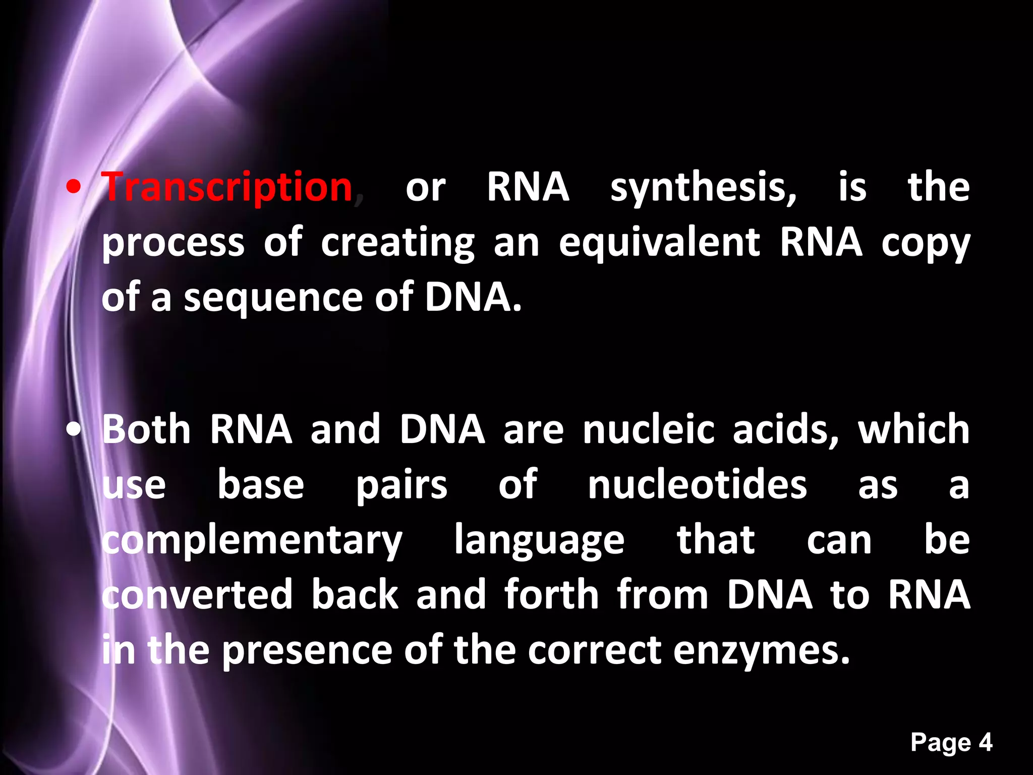
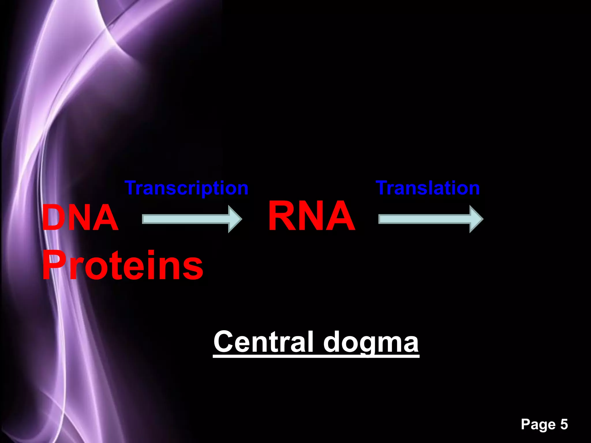
Protein synthesis
           Made By-
       Dr. Nidhi Sharma


                          Page 1
•Transcription
•Genetic code
•tRNA
•Ribosomes
•Translation
•Polyribosomes

                 Page 2
Transcription


                Page 3
• Transcription, or RNA synthesis, is the
  process of creating an equivalent RNA copy
  of a sequence of DNA.

• Both RNA and DNA are nucleic acids, which
  use base pairs of nucleotides as a
  complementary language that can be
  converted back and forth from DNA to RNA
  in the presence of the correct enzymes.
                                         Page 4
Transcription         Translation
DNA                   RNA
Proteins
               Central dogma

                                          Page 5
• During transcription, a DNA sequence is
  read by RNA polymerase, which produces a
  complementary, antiparallel RNA strand.

• As    opposed      to     DNA    replication,
  transcription results in an RNA compliment
  that includes uracil (U) in all instances
  where thymine (T) would have occurred in
  a DNA compliment.


                                          Page 6
• Transcription is the first step leading to gene
  expression.

• The stretch of DNA transcribed into an RNA
  molecule is called a transcription unit and
  encodes at least one gene.

• If the gene transcribed encodes for a protein,
  the result of transcription is messenger RNA
  (mRNA), which will then be used to create
  that protein via the process of translation.
                                             Page 7
• Alternatively, the transcribed gene may
  encode for either ribosomal RNA (rRNA) or
  transfer RNA (tRNA), other components of
  the protein-assembly process, or other
  ribozymes.


                                        Page 8
A DNA transcription unit encoding for a protein contains
not only the sequence that will eventually be directly
translated into the protein (the coding sequence) but
also regulatory sequences that direct and regulate the
synthesis of that protein.


                                                      Page 9
• As in DNA replication, DNA is read from 3' → 5'
  during transcription. Meanwhile, the complementary
  RNA is created from the 5' → 3' direction.

• Only one of the two DNA strands, called the template
  strand, is used for transcription.

• The other DNA strand is called the coding strand,
  because its sequence is the same as the newly
  created RNA transcript (except for the substitution of
  uracil for thymine).


                                                   Page 10
• Transcription is divided into 3
  stages:
1. initiation
2. elongation
3. termination.


                                    Page 11
Initiation
• In bacteria, transcription begins with the binding of RNA
  polymerase to the promoter in DNA.

• RNA polymerase is a core enzyme consisting of five
  subunits: 2 α subunits, 1 β subunit, 1 β' subunit, and 1 ω
  subunit.

• At the start of initiation, the core enzyme is associated
  with a sigma factor that aids in finding the appropriate -35
  and -10 base pairs downstream of promoter sequences.


                                                        Page 12
Elongation
• One strand of DNA, the template strand (or noncoding
  strand), is used as a template for RNA synthesis.

• As transcription proceeds, RNA polymerase traverses
  the template strand and uses base pairing
  complementarity with the DNA template to create an
  RNA copy.

• Although RNA polymerase traverses the template
  strand from 3' → 5', the coding (non-template) strand
  and newly-formed RNA can also be used as reference
  points, so transcription can be described as occurring
  5' → 3'.
                                                   Page 13
• This produces an RNA molecule from 5' → 3', an exact
  copy of the coding strand (except that thymines are
  replaced with uracils, and the nucleotides are
  composed of a ribose (5-carbon) sugar where DNA has
  deoxyribose (one less oxygen atom) in its sugar-
  phosphate backbone).

• Unlike DNA replication, mRNA transcription can
  involve multiple RNA polymerases on a single DNA
  template and multiple rounds of transcription
  (amplification of particular mRNA), so many mRNA
  molecules can be rapidly produced from a single copy
  of a gene.


                                                Page 14
• Elongation also involves a proofreading
  mechanism that can replace incorrectly
  incorporated bases.


                                       Page 15
Termination


• Bacteria use two different strategies for transcription
  termination: Rho-independent and Rho-dependent


                                                     Page 16
In Rho-independent
transcription
termination,      RNA
transcription    stops
when      the   newly
synthesized       RNA
molecule forms a G-C
rich    hairpin   loop
followed by a run of
U's, which makes it
detach from the DNA
                         Fig. Formation of
template.                hairpin


                                             Page 17
In the "Rho-dependent"
type of termination, a
protein factor called
"Rho" destabilizes the
interaction     between
the template and the
mRNA, thus releasing
the newly synthesized
mRNA        from    the
elongation complex.
                      Fig. Rho dependent
                      termination


                                           Page 18
Genetic code

               Page 19
Properties of genetic code
• The code is universal. All prokaryotic and
  eukaryotic organisms use the same codon to
  specify each amino acid.

• The code is triplet. Three nucleotides make one
  codon. 61 of them code for amino acids and 3 viz.,
  UAA, UAG and UGA are nonsense codons or chain
  termination codons.

• The code is degenerate. For a particular amino
  acid more than one word can be used


                                               Page 20
• The code is non overlapping. A base in mRNA is
  not used for two different codons

• The code is commaless. There is no special signal
  or commas between codons.

• The code is non ambiguous. A particular codon
  will always code for the same amino acid,
  wherever it is found.


                                               Page 21
Page 22
tRNA


Fig. Base sequence     Fig. Common features of
of yeast alanyl tRNA   tRNA molecule


                                           Page 23
Fig. 3D structure of tRNA
                            Page 24
Ribosomes
• Bacterial ribosomes consists of two subunits of
  unequal size, the larger having a sedimentation
  coefficient of 50S and the smaller of 30S.

• The two ribosomal subunits have irregular
  shapes which fit together in such a way that a
  cleft is formed through which mRNA passes as
  the ribosome moves along it during the
  translation process and from which the newly
  formed polypeptide chain emerges.


                                            Page 25
Fig. Ribosomes
                 Page 26
Translation


              Page 27
• Translation is the first stage of protein
  biosynthesis (part of the overall process of gene
  expression).

• Translation is the production of proteins by
  decoding mRNA produced in transcription.

• It occurs in the cytoplasm where the ribosomes
  are located.

• Ribosomes are made of a small and large
  subunit which surrounds the mRNA.


                                              Page 28
• In translation, messenger RNA (mRNA) is decoded
  to produce a specific polypeptide according to the
  rules specified by the genetic code.

• This uses an mRNA sequence as a template to
  guide the synthesis of a chain of amino acid that
  form a protein.

• Many types of transcribed RNA, such as transfer
  RNA, ribosomal RNA, and small nuclear RNA are
  not necessarily translated into an amino acid
  sequence.


                                              Page 29
• Translation proceeds in four phases:
  – activation,
  – initiation,
  – elongation and
  – termination
  (all describing the growth of the amino acid
    chain, or polypeptide that is the product
    of translation).
  Amino acids are brought to ribosomes and
    assembled into proteins.


                                        Page 30
Activation

• In activation, the correct amino acid is covalently
  bonded to the correct transfer RNA (tRNA).

• While this is not technically a step in translation, it is
  required for translation to proceed.

• The amino acid is joined by its carboxyl group to the 3'
  OH of the tRNA by an ester bond.

• When the tRNA has an amino acid linked to it, it is
  termed "charged".

                                                       Page 31
Fig. Charged tRNA (N-formyl
  methionyl tRNA f met)


                              Page 32
In Prokaryotes initiation requires the large and small
   ribosome subunits, the mRNA, the initiator tRNA and
   three initiation factors (IF1, IF2, IF3) and GTP. The
   overall sequence of the event is as follows
                  Initiation

• IF3 bind to the free 30S subunit, this helps to prevent
  the large subunit binding to it without an mRNA
  molecule and forming an inactive ribosome

• IF2 complexed with GTP and IF1 then binds to the
  small subunit. It will assist the charged initiator tRNA
  to bind.


                                                     Page 33
• The 30S subunit attached to an mRNA molecule
  making use of the ribosome binding site (RBS) on the
  mRNA

• The initiator tRNA can then bind to the complex by
  base pairing of its anticodon with the AUG codon on
  mRNA.

• At this point, IF3 can be released, as its role in
  keeping the subunits apart and helping the mRNA to
  bind are complete.

• This complex is called 30S initiation complex


                                                  Page 34
• The 50S subunit can now bind, which displace IF1
and IF2, and the GTP is hydrolysed in this energy
consuming step.

•This complex is called as 70S initiation complex.


                                                Page 35
Fig. Formation of the 70S initiation
complex

                                        Page 36
• The assembled ribosome has two tRNA
  binding sites.
• These are called the A and P sites, for amino
  acyl and peptidyl sites.
• The A site is where incoming amino acyl tRNA
  molecules bind, and the P site is where the
  growing polypeptide chain is usually found.
• These sites are in the cleft of small subunit
  and contain adjacent codons that are being
  translated.


                                          Page 37
• One major outcome of initiation is the
  placement of the initiator tRNA in the P
  site.

• It is the only tRNA that does this, as all
  other must enter the A site.


                                        Page 38
Fig. Initiation phase of protein synthesis

                                             Page 39
• With the formation of 70S initiation
              Elongation
  complex the elongation cycle can
  begin.
• In involves three elongation factors EF-
  Tu, EF-Ts and EF-G, GTP, charged tRNA
  and the 70S initiation complex.


                                      Page 40
Elongation is divided into three steps:
1. Amino acyl tRNA delivery.
• EF-Tu is required to deliver the amino acyl tRNA to
   the A site and energy is consumed in this step by
   the hydrolysis of GTP.
• The released EF-Tu. GDP complex is regenerated
   with the help of EF-Ts.
• In the EF-Tu EF-Ts exchange cycle EF-Ts displaces
   the GDP and subsequently is displaced itself by
   GTP.
• The resultant EF-Tu.GTP complex is now able to
   bind another amino acyl tRNA and deliver it to the
   ribosome.
• All amino acyl tRNAs can form this complex with
   EF-Tu except the initiator tRNA.


                                                Page 41
• Fig. Elongation of polypeptide
  chain
                                   Page 42
2. Peptide bond formation.
• After aminoacyl-tRNA delivery, the A- and P- sites are
  both occupied and the two amino acids that are to be
  joined are in close proximity.

• The peptidyl transferase activity of the 50S subunit
  can now form a peptide bond between these two
  amino acids without the input of any more energy,
  since energy in the form of ATP was used to charge
  the tRNA.


                                                   Page 43
• Fig. Peptide bond formation


                                Page 44
3. Translocation.

• A complex of EF-G (translocase) and GTP binds to
  the ribosome and, in an energy consuming step, the
  discharged tRNA is ejected from the P-site, the
  peptidyl-tRNA is moved from the A-site to the P-
  site and the mRNA moves by one codon relative to
  one codon to the ribosome.

• GDP and EF-G are released, the latter being
  reusable. A new codon is now present in the vacant
  A-site.

      The cycle is repeated until one of the
  termination codons (UAA, UAG and UGA) appear in
  the A-site.

                                                Page 45
Fig. Translocation
                     Page 46
• Termination of the polypeptide happens when the A
                  Termination
  site of the ribosome faces a stop codon (UAA, UAG,
  or UGA).

• When this happens, no tRNA can recognize it, but a
  releasing factor can recognize nonsense codons and
  causes the release of the polypeptide chain.

• The 5' end of the mRNA gives rise to the protein's N-
  terminus, and the direction of translation can
  therefore be stated as N->C.


                                                  Page 47
Termination
• Termination of polypeptide synthesis is signalled
  by one of the three termination codons in the
  mRNA (UAA, UAG and UGA) immediately
  following the last amino acid codon.
• In prokaryotes, once a termination codon
  occupies the ribosomal A-site, three termination
  or release factors, viz., the protein RF1, RF2 and
  RF3 contribute to-
 The hydrolysis of the terminal peptidyl-tRNA
   bond.
 Release of the free polypeptide and the last
  uncharged tRNA from the P-site
 The dissociation of the 70S ribosome into its 30S
  and 50S subunits

                                                Page 48
• RF1 recognizes the termination codon UAG
  and UAA and RF2 recognize UGA and UAA.

• Either RF1 or RF2 binds at the termination
  codon and induces peptidyl transferase to
  transfer the growing peptide chain to a
  water molecule rather than to another
  amino acid.

• Function of RF3 is not known.

                                        Page 49
Fig. Termination

                   Page 50
Polyribosomes

• A single strand of mRNA is translated
  simultaneously by many ribosomes, spaced
  closely together.

• Such clusters of ribosomes are called
  polysomes or polyribosomes.

• The simultaneous translation of a single
  mRNA by many ribosomes allow highly
  efficient use of the mRNA
                                      Page 51
Fig. Polyribosomes


                     Page 52
THANK YOU

        Page 53

Protein synthesis

  • 1.
    Protein synthesis Made By- Dr. Nidhi Sharma Page 1
  • 2.
  • 3.
  • 4.
    • Transcription, orRNA synthesis, is the process of creating an equivalent RNA copy of a sequence of DNA. • Both RNA and DNA are nucleic acids, which use base pairs of nucleotides as a complementary language that can be converted back and forth from DNA to RNA in the presence of the correct enzymes. Page 4
  • 5.
    Transcription Translation DNA RNA Proteins Central dogma Page 5
  • 6.
    • During transcription,a DNA sequence is read by RNA polymerase, which produces a complementary, antiparallel RNA strand. • As opposed to DNA replication, transcription results in an RNA compliment that includes uracil (U) in all instances where thymine (T) would have occurred in a DNA compliment. Page 6
  • 7.
    • Transcription isthe first step leading to gene expression. • The stretch of DNA transcribed into an RNA molecule is called a transcription unit and encodes at least one gene. • If the gene transcribed encodes for a protein, the result of transcription is messenger RNA (mRNA), which will then be used to create that protein via the process of translation. Page 7
  • 8.
    • Alternatively, thetranscribed gene may encode for either ribosomal RNA (rRNA) or transfer RNA (tRNA), other components of the protein-assembly process, or other ribozymes. Page 8
  • 9.
    A DNA transcriptionunit encoding for a protein contains not only the sequence that will eventually be directly translated into the protein (the coding sequence) but also regulatory sequences that direct and regulate the synthesis of that protein. Page 9
  • 10.
    • As inDNA replication, DNA is read from 3' → 5' during transcription. Meanwhile, the complementary RNA is created from the 5' → 3' direction. • Only one of the two DNA strands, called the template strand, is used for transcription. • The other DNA strand is called the coding strand, because its sequence is the same as the newly created RNA transcript (except for the substitution of uracil for thymine). Page 10
  • 11.
    • Transcription isdivided into 3 stages: 1. initiation 2. elongation 3. termination. Page 11
  • 12.
    Initiation • In bacteria,transcription begins with the binding of RNA polymerase to the promoter in DNA. • RNA polymerase is a core enzyme consisting of five subunits: 2 α subunits, 1 β subunit, 1 β' subunit, and 1 ω subunit. • At the start of initiation, the core enzyme is associated with a sigma factor that aids in finding the appropriate -35 and -10 base pairs downstream of promoter sequences. Page 12
  • 13.
    Elongation • One strandof DNA, the template strand (or noncoding strand), is used as a template for RNA synthesis. • As transcription proceeds, RNA polymerase traverses the template strand and uses base pairing complementarity with the DNA template to create an RNA copy. • Although RNA polymerase traverses the template strand from 3' → 5', the coding (non-template) strand and newly-formed RNA can also be used as reference points, so transcription can be described as occurring 5' → 3'. Page 13
  • 14.
    • This producesan RNA molecule from 5' → 3', an exact copy of the coding strand (except that thymines are replaced with uracils, and the nucleotides are composed of a ribose (5-carbon) sugar where DNA has deoxyribose (one less oxygen atom) in its sugar- phosphate backbone). • Unlike DNA replication, mRNA transcription can involve multiple RNA polymerases on a single DNA template and multiple rounds of transcription (amplification of particular mRNA), so many mRNA molecules can be rapidly produced from a single copy of a gene. Page 14
  • 15.
    • Elongation alsoinvolves a proofreading mechanism that can replace incorrectly incorporated bases. Page 15
  • 16.
    Termination • Bacteria usetwo different strategies for transcription termination: Rho-independent and Rho-dependent Page 16
  • 17.
    In Rho-independent transcription termination, RNA transcription stops when the newly synthesized RNA molecule forms a G-C rich hairpin loop followed by a run of U's, which makes it detach from the DNA Fig. Formation of template. hairpin Page 17
  • 18.
    In the "Rho-dependent" typeof termination, a protein factor called "Rho" destabilizes the interaction between the template and the mRNA, thus releasing the newly synthesized mRNA from the elongation complex. Fig. Rho dependent termination Page 18
  • 19.
  • 20.
    Properties of geneticcode • The code is universal. All prokaryotic and eukaryotic organisms use the same codon to specify each amino acid. • The code is triplet. Three nucleotides make one codon. 61 of them code for amino acids and 3 viz., UAA, UAG and UGA are nonsense codons or chain termination codons. • The code is degenerate. For a particular amino acid more than one word can be used Page 20
  • 21.
    • The codeis non overlapping. A base in mRNA is not used for two different codons • The code is commaless. There is no special signal or commas between codons. • The code is non ambiguous. A particular codon will always code for the same amino acid, wherever it is found. Page 21
  • 22.
  • 23.
    tRNA Fig. Base sequence Fig. Common features of of yeast alanyl tRNA tRNA molecule Page 23
  • 24.
    Fig. 3D structureof tRNA Page 24
  • 25.
    Ribosomes • Bacterial ribosomesconsists of two subunits of unequal size, the larger having a sedimentation coefficient of 50S and the smaller of 30S. • The two ribosomal subunits have irregular shapes which fit together in such a way that a cleft is formed through which mRNA passes as the ribosome moves along it during the translation process and from which the newly formed polypeptide chain emerges. Page 25
  • 26.
  • 27.
    Translation Page 27
  • 28.
    • Translation isthe first stage of protein biosynthesis (part of the overall process of gene expression). • Translation is the production of proteins by decoding mRNA produced in transcription. • It occurs in the cytoplasm where the ribosomes are located. • Ribosomes are made of a small and large subunit which surrounds the mRNA. Page 28
  • 29.
    • In translation,messenger RNA (mRNA) is decoded to produce a specific polypeptide according to the rules specified by the genetic code. • This uses an mRNA sequence as a template to guide the synthesis of a chain of amino acid that form a protein. • Many types of transcribed RNA, such as transfer RNA, ribosomal RNA, and small nuclear RNA are not necessarily translated into an amino acid sequence. Page 29
  • 30.
    • Translation proceedsin four phases: – activation, – initiation, – elongation and – termination (all describing the growth of the amino acid chain, or polypeptide that is the product of translation). Amino acids are brought to ribosomes and assembled into proteins. Page 30
  • 31.
    Activation • In activation,the correct amino acid is covalently bonded to the correct transfer RNA (tRNA). • While this is not technically a step in translation, it is required for translation to proceed. • The amino acid is joined by its carboxyl group to the 3' OH of the tRNA by an ester bond. • When the tRNA has an amino acid linked to it, it is termed "charged". Page 31
  • 32.
    Fig. Charged tRNA(N-formyl methionyl tRNA f met) Page 32
  • 33.
    In Prokaryotes initiationrequires the large and small ribosome subunits, the mRNA, the initiator tRNA and three initiation factors (IF1, IF2, IF3) and GTP. The overall sequence of the event is as follows Initiation • IF3 bind to the free 30S subunit, this helps to prevent the large subunit binding to it without an mRNA molecule and forming an inactive ribosome • IF2 complexed with GTP and IF1 then binds to the small subunit. It will assist the charged initiator tRNA to bind. Page 33
  • 34.
    • The 30Ssubunit attached to an mRNA molecule making use of the ribosome binding site (RBS) on the mRNA • The initiator tRNA can then bind to the complex by base pairing of its anticodon with the AUG codon on mRNA. • At this point, IF3 can be released, as its role in keeping the subunits apart and helping the mRNA to bind are complete. • This complex is called 30S initiation complex Page 34
  • 35.
    • The 50Ssubunit can now bind, which displace IF1 and IF2, and the GTP is hydrolysed in this energy consuming step. •This complex is called as 70S initiation complex. Page 35
  • 36.
    Fig. Formation ofthe 70S initiation complex Page 36
  • 37.
    • The assembledribosome has two tRNA binding sites. • These are called the A and P sites, for amino acyl and peptidyl sites. • The A site is where incoming amino acyl tRNA molecules bind, and the P site is where the growing polypeptide chain is usually found. • These sites are in the cleft of small subunit and contain adjacent codons that are being translated. Page 37
  • 38.
    • One majoroutcome of initiation is the placement of the initiator tRNA in the P site. • It is the only tRNA that does this, as all other must enter the A site. Page 38
  • 39.
    Fig. Initiation phaseof protein synthesis Page 39
  • 40.
    • With theformation of 70S initiation Elongation complex the elongation cycle can begin. • In involves three elongation factors EF- Tu, EF-Ts and EF-G, GTP, charged tRNA and the 70S initiation complex. Page 40
  • 41.
    Elongation is dividedinto three steps: 1. Amino acyl tRNA delivery. • EF-Tu is required to deliver the amino acyl tRNA to the A site and energy is consumed in this step by the hydrolysis of GTP. • The released EF-Tu. GDP complex is regenerated with the help of EF-Ts. • In the EF-Tu EF-Ts exchange cycle EF-Ts displaces the GDP and subsequently is displaced itself by GTP. • The resultant EF-Tu.GTP complex is now able to bind another amino acyl tRNA and deliver it to the ribosome. • All amino acyl tRNAs can form this complex with EF-Tu except the initiator tRNA. Page 41
  • 42.
    • Fig. Elongationof polypeptide chain Page 42
  • 43.
    2. Peptide bondformation. • After aminoacyl-tRNA delivery, the A- and P- sites are both occupied and the two amino acids that are to be joined are in close proximity. • The peptidyl transferase activity of the 50S subunit can now form a peptide bond between these two amino acids without the input of any more energy, since energy in the form of ATP was used to charge the tRNA. Page 43
  • 44.
    • Fig. Peptidebond formation Page 44
  • 45.
    3. Translocation. • Acomplex of EF-G (translocase) and GTP binds to the ribosome and, in an energy consuming step, the discharged tRNA is ejected from the P-site, the peptidyl-tRNA is moved from the A-site to the P- site and the mRNA moves by one codon relative to one codon to the ribosome. • GDP and EF-G are released, the latter being reusable. A new codon is now present in the vacant A-site. The cycle is repeated until one of the termination codons (UAA, UAG and UGA) appear in the A-site. Page 45
  • 46.
  • 47.
    • Termination ofthe polypeptide happens when the A Termination site of the ribosome faces a stop codon (UAA, UAG, or UGA). • When this happens, no tRNA can recognize it, but a releasing factor can recognize nonsense codons and causes the release of the polypeptide chain. • The 5' end of the mRNA gives rise to the protein's N- terminus, and the direction of translation can therefore be stated as N->C. Page 47
  • 48.
    Termination • Termination ofpolypeptide synthesis is signalled by one of the three termination codons in the mRNA (UAA, UAG and UGA) immediately following the last amino acid codon. • In prokaryotes, once a termination codon occupies the ribosomal A-site, three termination or release factors, viz., the protein RF1, RF2 and RF3 contribute to-  The hydrolysis of the terminal peptidyl-tRNA bond.  Release of the free polypeptide and the last uncharged tRNA from the P-site  The dissociation of the 70S ribosome into its 30S and 50S subunits Page 48
  • 49.
    • RF1 recognizesthe termination codon UAG and UAA and RF2 recognize UGA and UAA. • Either RF1 or RF2 binds at the termination codon and induces peptidyl transferase to transfer the growing peptide chain to a water molecule rather than to another amino acid. • Function of RF3 is not known. Page 49
  • 50.
  • 51.
    Polyribosomes • A singlestrand of mRNA is translated simultaneously by many ribosomes, spaced closely together. • Such clusters of ribosomes are called polysomes or polyribosomes. • The simultaneous translation of a single mRNA by many ribosomes allow highly efficient use of the mRNA Page 51
  • 52.
  • 53.
    THANK YOU Page 53