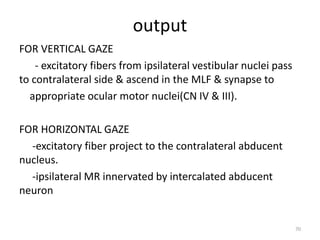
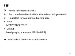
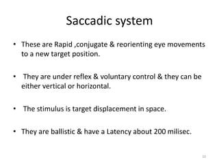
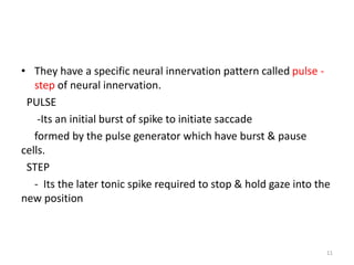
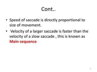
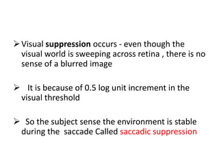
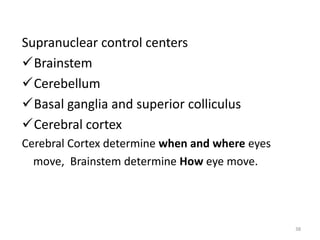
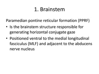
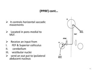
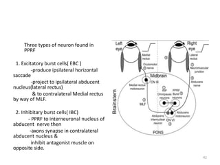
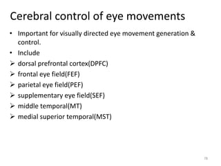
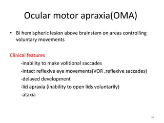
Supranuclear Control of
Eye Movements
By Dr.Desta G (Opthalmology resident l)
Advisor Dr.Sadik(Assistant Prof. of Opthalmology,
CHS,AAU)
June / 2018GC.
OUTLINE
• INTRODUCTION
• SUPRANUCLEAR CONTROL SYSTEM
• SUPRANUCLEAR DISORDERS
• REFERENCE
2
Introduction
• Successful use of vision is because of properly aligned gaze
which result from the sum of body, head & eye movements.
• To achieve these there are different oculomotor systems or
eye movement systems each with their own characteristics.
3
 There are six extraocular muscles involed in moving the eye
Extraocular muscles are supplied by 3,4,6th
 cranial nerves which have their nuclei in the
Midbrain and Brainstem.
Intro…
The different types of eye movement can be either gaze shifting
or holding;
6
GAZE SHIFTING
- Its moving the eye to a new position & its
also an eye
movement which follows a moving
target.
- Its acquisition of an image on the fovea
and maintaining foveal fixation while the object
is moving.
Cont..
• It includes
- saccadic system
- smooth pursuit
- vergence eye movements
8
Gaze holding
• It’s a type of eye movements that fixate on a stationary
objects & maintain that fixation despite our head
movements.
These Include;
 Visual fixation
 Vestibuloocular reflex(VOR)
 Optokinetic reflex(OKR)
9
Saccadic system
• These are Rapid ,conjugate & reorienting eye movements
to a new target position.
• They are under reflex & voluntary control & they can be
either vertical or horizontal.
• The stimulus is target displacement in space.
• They are ballistic & have a Latency about 200 milisec.
10
• They have a specific neural innervation pattern called pulse -
step of neural innervation.
PULSE
-Its an initial burst of spike to initiate saccade
formed by the pulse generator which have burst & pause
cells.
STEP
- Its the later tonic spike required to stop & hold gaze into the
new position
11
Cont..
• Speed of saccade is directly proportional to
size of movement.
• Velocity of a larger saccade is faster than the
velocity of a slow saccade , this is known as
Main sequence
12
Visual suppression occurs - even though the
visual world is sweeping across retina , there is no
sense of a blurred image
 It is because of 0.5 log unit increment in the
visual threshold
 So the subject sense the environment is stable
during the saccade Called saccadic suppression
Purposes of saccades
1. Fast phases of nystagmus reset the
eyes following vestibular or optokinetic slow-
phase movements.
2. Spontaneous saccades, at a frequency of
about 20 per minute, allow visual search,
although they also occur in darkness.
Purposes of saccades…
3. Reflexive saccades are made in response to
new visual, auditory, or tactile cues at short
latency.
4. Voluntary saccades point the eyes at a
predetermined location or goal.
Clinical examination
The fixation objects should be presented at an angular separation
of about 20 to 30°.
• SPEED - slowing of
saccades can be seen in
 AIDS dementia complex
 Lipid storage disorders ,
 PSP
 Drug intoxications
• SMOOTHNESS –
Affected in cerebellar
diseases
• ACCURACY –
Hypometric or
Hypermetric ,
affected in cerebellar
diseases
• Saccades mainly assessed by their accuracy;
i.e the final position of the eye in relation to the
target
1.Orthometric saccades.
- normal saccades
2.Hypometric saccades.
-undergo corrective saccade to refixate the
target
3.Hypermetric saccades.
-undergo dynamic overshoot to refixate the
target
18
Smooth pursuit movements
• It’s a smooth ,conjugate movement of the eye in response to a
slow moving stimuli.
• It has a latency of about 125 milisec
• Its accuracy assessed by pursuit gain( ratio of eye velocity to
target velocity) which is normally 1.
• If less than one ,slow eye velocity so eye lags behind the target
so it need catch up saccades for refoveation called saccadic
pursuit.
19
• Have the capacity for compensation unlike
saccades - when speed of target is varied after
initiation of the movement , speed of pursuit
can be varied.
Cont..
• The type of innervation in smooth pursuit is
called a step – ramp innervation.
step
- its an instantaneous jump in firing
frequency.
ramp
- its the linear increase in frequency
which occur after the step.
22
Vergence system
• Its a disjugate movement of the eye to maintain
the target on the two fovea's.
• Important to obtain Depth cues ( the relative
location of the target either away or toward the
observer).
• It can be either divergent or convergent
movement
23
• Its one component of the triadic response which
include accommodation & pupillary changes.
• They are cross coupled in that vergence drive
accommodation and vice versa.
• Image blur & retinal disparity are the main
stimulus.
24
Retinal blur – accomodative vergence
Disparity of location of images- fusional
vergence
Visual fixation
• It maintain a stationary target of interest with
in the fovea.
Persistent foveation of target attenuates the
neural response in the retina to the same
target.
• These Degrade the quality of image of the
stationary target on the retina
26
• These effect counter acted by Micro Saccade
refixation movements
These are small amplitude saccades which are;
1 Small enough so that the image is
maintained on the retina
2 But large enough to provide a change in
image to the photoreceptors so they increase
perceptional quality.
27
Vestibuloocular reflex(VOR)
• It’s a reflexive compensatory eye movement in
response to brief high frequency head rotation.
• It produce eye movement in a direction opposite to
head acceleration.
• It produce fast response with short latency (15
milisec)
• Response is better when its combined with visual
cues & the reflexive response is called visually
enhanced VOR(VVOR)
28
• It respond to all the three natural head
movements(head rotation ,translation & tilt)
• Semicircular canal & otolith organs sense
angular & linear acceleration of the head
respectively.
• They change the head acceleration to velocity
signal by the hair cells.
29
 The velocity signal reach
vestibular nuclei & changed
to positional signal which
project to ocular motor
neurons
 Stimulation of Ampulla of
horizontal semicircular
canal conjuate movement
towards contralateral side
 Information from anterior
and posterior semicircular
canals - combination of
vertical and torsional eye
movements
30
• VOR accuracy assessed by its VOR GAIN which
is the ratio of the magnitude of eye velocity to
head velocity.
• Perfect VOR is when eye moves with same
amount but in opposite direction as the head
movements.
• VOR attenuated quickly & is not responsible for
very slow head movements.
31
VOR cancellation test…cerebellar
function
• When a target moves with that of the head
movement VOR SUPRESSION is important for
precise foveation of the target
33
Optokinetic response
A combination of slow and fast phase eye
movements.
Seen when an individual follows an object
with their eyes, which then moves out of the
field of vision at which point their eyes moves
back to position it was when it first saw object
The reflex starts at 6th month of age
34
Optokinetic nystagmus OKN occurs in
response to a rotation movement.
Its latency is ≈100milisec
With sustained head rotation at a constant
velocity , vestibular response fades and
optokinetic response takes over
 OKN prevents a continuous blur from relative
motion of the moving visual field
Clinical Examination
• But most widely used
tests is the rotating
drum with black and
white stripes.
• It actually subtend
portion of visual field &
its really a test of
saccadic system &
smooth pursuit system.
36
37
SUPRA NUCLEAR CONTROL OF EYE
MOVEMENTS & THEIR
DISORDERS
Supranuclear control centers
Brainstem
Cerebellum
Basal ganglia and superior colliculus
Cerebral cortex
Cerebral Cortex determine when and where eyes
move, Brainstem determine How eye move.
38
1. Brainstem
Paramedian pontine reticular formation (PPRF)
• Is the brainstem structure responsible for
generating horizontal conjugate gaze
• Positioned ventral to the medial longitudinal
fasciculus (MLF) and adjacent to the abducens
nerve nucleus
40
(PPRF) cont…
 It controls horizontal saccadic
movements
 Located in pons medial to
MLF.
 Receive an input from
I. FEF & Superior colliculus
II. cerebellum
III. vestibular nuclei
 send an out put to ipsilateral
abducent nucleus
41
Three types of neuron found in
PPRF
1. Excitatory burst cells( EBC )
-produce ipsilateral horizontal
saccade
-project to ipsilateral abducent
nucleus(lateral rectus)
& to contralateral Medial rectus
by way of MLF.
2. Inhibitary burst cells( IBC)
- PPRF to interneuronal nucleus of
abducent nerve then
-axons synapse in contralateral
abducent nucleus &
inhibit antagonist muscle on
opposite side.
42
3. Pause cells
-They tonically discharge to inhibit the EBC
except during saccades.
Lesion in PPRF
-Inability to make ipsilateral saccades
-but pursuit system & VOR are intact.
 Parkinson , Huntington, & Alzheimer
diseases.
43
Medial longitudinal fasciculus(MLF)
• It’s a fiber tract which extend from the spinal
cord to oculomotor nuclei.
• Majority of them are ascending fibers arising
from the superior & medial vestibular nuclei.
Most importantly they link contralateral
abducent nucleus with ipsilateral medial
rectus sub nuclei.
44
Internuclear ophthalmoplegia (INO)
lesion on the MLF If unilateral;
 loss of adduction ipsilateral to lesion
 abduction overshoot contralateral to lesion
• If bilateral
 impaired vertical pursuit & gaze evoked nystagmus
 multiple sclerosis , brain stem tumors & infarction,
syphilis, drug intoxications, Wernicke's encephalopathy
are common causes.
45
Unilateral INO
Straight eyes in the primary
position.
• Defective adduction of the eye
on the side of the lesion and
nystagmus of the contralateral
eye on abduction; note that the
side of the lesion is named for
the side of the adduction
deficit. ○
• Gaze to the side of the lesion
is normal. ○ Convergence is
intact if the lesion is anterior
and discrete but impaired if the
lesion is posterior or extensive.
Bilateral INO
• Limitation of left
adduction and
Dissociated Nystagmus of
the right eye on right
gaze.
• Limitation of right
adduction and
Dissociated Nystagmus of
the left eye on left gaze.
• Convergence may be
intact Or impaired
One-and-a-half syndrome
• PPRF and MLF lesions combined on the
same side give rise to the ‘one-and-a-half
syndrome’
• characterized by a combination of
ipsilateral gaze palsy and INO.
• The only residual movement is abduction
of the contralateral eye, which exhibits
abduction nystagmus.
Nucleus prepositus hypoglossus (NPH)
• It’s the major neural integrator for horizontal conjugate gazes
which is found in medulla.
• Combination of medial vestibular nuclei & CN XII nucleus
• Important for gaze holding mechanism in horizontal eye
movement.(hold eyes in eccentric positions after horizontal
movements)
49
Lesion in NPH;
 abolish gaze holding mechanism for horizontal gaze
 eyes not able to hold an eccentric position so they drift back
to primary position.
 which produce corrective quick phases
 gives gaze evoked nystagmus.
50
Dorsolateral pontine nucleus(DLPN)
 Receive signals from middle temporal & middle superior
temporal area
 responsible for smooth pursuit generation.
51
Brain stem control for vertical &
torsional eye movements
52
 Found in midbrain at rostral
termination of MLF.
 Connect to motor neurons of
oculomotor nucleus &
trochlear nucleus as well as to
PPRF.
 Important for vertical &
torsional saccade generation.
 Receive Input from vestibular
nuclei via the MLF
53
ROSTRAL INTERESTITIAL NUCLEUS OF MEDIAL LONGTUIDNAL
FASCULUS(riMLF)
 It Project to motor neurons
of elevators bilaterally & to
motor neurons of
depressors ipsilateraly so
neurons burst
for both upward & down ward
movements.
54
55
 Lesion in riMLF
-unilateral
-mild defect in vertical saccades
-torsional quick phase defect is specific
-bilateral
-clinically apparent vertical eye movement abnormality.
- gaze holding ,horizontal movements & vestibular
movements are intact.
56
Interstitial nucleus of cajal (INC)
 Found in rostral mid brain
 Receive input from riMLF & vestibular nuclei from
contralateral INC
 Project to vertical motor neuron nucleus to motor
neurons of the neck & trunk( to coordinate eye & head
movement )
 Important for torsional & vertical gaze holding function.
• Lesion in INC
-impair gaze holding function in the vertical plane
-skew deviation occur.
57
VERTICAL SMOOTH PURSUIT & VESTIBULA
CONTROL
 MST to ipsilateral DLPN to ipsilateral folucullus,parafolluculus,
to contralateral cerebellar nuclei to ipsilateral vestibular
nuclei to contralateral ocular motor nuclei.
58
Brain stem control of vergence eye
movement
• Centers for vergence control include
-Mesencephalic reticular formation(1 to 2 mm dorsal and
dorsolateral to the oculomotor nucleus)
• Three main types of neurons can be found;
 cells that discharge in relation to vergence angle,
 vergence velocity, and
 both vergence angle and velocity.
60
Nuclear reticularis tegmenti pontis(NRTP)
• important for coordinating pursuit and
saccadic movements with vergence, and
• also for holding the eyes steady at a specified
vergence angle
DISORDERS OF VERGENCE
include -convergence insufficiency
-divergence insufficiency
-convergence spasm
Convergence insufficiency
• Its impairment of convergence eye movement
• Occur in midbrain reticular formation lesions
• Accommodation & pupillary constriction occur in an
attempt to converge)
• Multiple sclerosis ,encephalitis ,Parkinson disease, closed
head trauma ,old age & lack of effort are some of the
causes.
63
Convergence spasm
 Excess convergence tone because of In born abnormalities of
convergence resulting high accommodative convergence to
accommodation ratio giving early onset esotropia.
 The near reflex is uncomfortable for the patient.
 Mimic unilateral or bilateral abducent paresis
 Dorsal midbrain syndrome, seizure , head trauma, thalamic
hemorrhage, psychogenic factors are some of the causes.
64
Divergence insufficiency
• Its insufficiency of divergence so patient is esotropic which is
greater at distance than near.
• Normal abduction with normal abducting saccades rule out
bilateral 6th nerve palsy.
• Seen in increased ICP, midbrain tumor , spinocerebellar ataxia
65
Vestibular control system
• Consists of two systems
-peripheral (semicircular canal
& otolith organs)
-central (vestibular nuclei)
SEMICIRCULAR CANAL
-responds to angular acceleration
produced by head rotation
-Change acceleration/deceleration
signal to velocity signal
send it to ipsilateral vestibular
nuclei
66
Composed of three canals.
Each send signal to move the eye conjugatly to opposite side
during head rotation by supplying the yolk muscles.
 horizontal(lateral)canal….supply ipsilateral MR &
contralateral LR
 anterior (superior)canal ..supply ipsilateral SR &
contralateral IO
 posterior (vertical )canal ..supply ipsilateral SO &
contralateral IR.
67
OTOLITH ORGANS
• Include utricle & saccule
• They respond to linear acceleration of the head & head tilt.
• Keep the eye fixed in position to a change in head position.
 Disruption of peripheral vestibular system results;
-skew deviation
-ocular tilt reaction(head tilt & cyclotorsion rotation)
-eye movement to same side of lesion(unopposed CL
action)
68
Vestibular nuclei
• In lateral medulla beneath the floor of the 4th ventricle.
• Important for VOR & OKN.
INPUT
-vestibular information from ipsilateral labyrinth.
-visual input from Visual cortex(for OKN)
-proprioceptive information from the neck
-cerebellum
-frontal & parietal cortex
69
output
FOR VERTICAL GAZE
- excitatory fibers from ipsilateral vestibular nuclei pass
to contralateral side & ascend in the MLF & synapse to
appropriate ocular motor nuclei(CN IV & III).
FOR HORIZONTAL GAZE
-excitatory fiber project to the contralateral abducent
nucleus.
-ipsilateral MR innervated by intercalated abducent
neuron
70
71
• Each vestibular system exert a continuous tonic pressure to
turn eye to opposite side.
• Lesion on the right -----eye moves to the right(unopposed
action from the left vestibular system)
72
Cerebellar influence on eye
movement
• Two parts of cerebellum
majorly involved in eye
movements.
-vestibular
cerebellum(flocculus ,
paraflocculus, nodulus)
-dorsal vermis of the
posterior lobe.
Function of cerebellum
-modulate on going eye
movement & maintain
accuracy ocular motor
performance. 73
• The overall role of the cerebellum in the
control of eye movements is to optimize them
so that they can provide clear and stable
vision.
• Repair shop for eye movements
Vestibular cerebellum
• Found inferior to cerebellar peduncle
 INPUT
- from vestibular nuclei & NPH
 OUTPUT
-to ipsilateral vestibular nuclei
 FUNCTION
--for smooth pursuit & combined eye –head tracking
--for gaze holding & VOR
75
Dorsal vermis of the posterior lobe
 INPUT
-PPRF & NRTP
-vestibular nuclei
 OUTPUT
-PPRF & riMLF
 FUNCTION
-important for saccadic eye movement
76
 Lesion on vestibular cerebellum
-impair contralateral smooth pursuit
-impair gaze holding mechanisms
-loss of adaptation to VOR
 Lesion on the dorsal vermis
-ipsilateral saccadic dysmetria (hypometria)
77
Cerebral control of eye movements
• Important for visually directed eye movement generation &
control.
• Include
 dorsal prefrontal cortex(DPFC)
 frontal eye field(FEF)
 parietal eye field(PEF)
 supplementary eye field(SEF)
 middle temporal(MT)
 medial superior temporal(MST)
78
79
FEF
 Found in broadman area 8
 For contralateral vertical & horizontal saccadic generation
 Important for voluntary redirecting gaze
• Input
-MT,MST,PFC,PEF,SEF
• Output
-basal ganglia, brainstem(PPRF & riMLF)
 Lesion in FEF…increase saccadic latency
80
PFC
 to make accurate saccades to remembered spatial location of
targets.
 If lesion occurs it disrupts our working memory.
SEF
 Found in dorsomedial frontal lobe
 Important to generate saccades & to remember chronologic
sequence of multiple targets to make sequential saccades
81
MT & MST
• Found at junction of temporal , parietal & occipital lobe
• Neurons record speed & direction of moving targets
• Important for smooth pursuit movement
Input
-FEF ,PEF ,visual cortex
Output
-cerebellum, FEF, brain stem(PPRF ,riMLF )
-dorsolateral pontine nuclei (DLPN)
 Lesion in these area…ipsilateral slow pursuit & akinetopsia
82
 Because of existence of multiple parallel pathways for each
type of eye movements.
 inability to move eyes completely should involve multiple
areas of cortex bilaterally.
83
Ocular motor apraxia(OMA)
• Bi hemispheric lesion above brainstem on areas controlling
voluntary movements
Clinical features
-inability to make volitional saccades
-intact reflexive eye movements(VOR ,reflexive saccades)
-delayed development
-lid apraxia (inability to open lids voluntarily)
-ataxia
84
• Congenital or acquired
Congenital OMA
-characterized by horizontal head thrust to fixate on targets
 patient make extreme contra version of head( employ its VOR )
Until foveation on target occur
 While maintaining fixation patient slowly turn head until eye
see straight to target ( primary position)
85
86
Acquired OMA
• Occur In bilateral frontoparieto occipital lesion
-bilateral stroke
-anoxic encephalopathy
Clinical features
-impaired volitional saccades
-inaccurate arm pointing
-simultagnosia (difficult to perceive major visual
scenes at once)
Its also called ballient syndrome
87
References
• Leigh RJ, Zee DS. The Neurology of Eye
Movements. 5th ed
• Duane clinical Ophthalmology, 2012
• BCSC section 2,6
• Adlers physiology of the eye,11th edition
• Online sources
Supra nuclear eye movements

Supra nuclear eye movements

  • 1.
    Supranuclear Control of EyeMovements By Dr.Desta G (Opthalmology resident l) Advisor Dr.Sadik(Assistant Prof. of Opthalmology, CHS,AAU) June / 2018GC.
  • 2.
    OUTLINE • INTRODUCTION • SUPRANUCLEARCONTROL SYSTEM • SUPRANUCLEAR DISORDERS • REFERENCE 2
  • 3.
    Introduction • Successful useof vision is because of properly aligned gaze which result from the sum of body, head & eye movements. • To achieve these there are different oculomotor systems or eye movement systems each with their own characteristics. 3
  • 4.
     There aresix extraocular muscles involed in moving the eye Extraocular muscles are supplied by 3,4,6th  cranial nerves which have their nuclei in the Midbrain and Brainstem.
  • 6.
    Intro… The different typesof eye movement can be either gaze shifting or holding; 6
  • 7.
    GAZE SHIFTING - Itsmoving the eye to a new position & its also an eye movement which follows a moving target. - Its acquisition of an image on the fovea and maintaining foveal fixation while the object is moving.
  • 8.
    Cont.. • It includes -saccadic system - smooth pursuit - vergence eye movements 8
  • 9.
    Gaze holding • It’sa type of eye movements that fixate on a stationary objects & maintain that fixation despite our head movements. These Include;  Visual fixation  Vestibuloocular reflex(VOR)  Optokinetic reflex(OKR) 9
  • 10.
    Saccadic system • Theseare Rapid ,conjugate & reorienting eye movements to a new target position. • They are under reflex & voluntary control & they can be either vertical or horizontal. • The stimulus is target displacement in space. • They are ballistic & have a Latency about 200 milisec. 10
  • 11.
    • They havea specific neural innervation pattern called pulse - step of neural innervation. PULSE -Its an initial burst of spike to initiate saccade formed by the pulse generator which have burst & pause cells. STEP - Its the later tonic spike required to stop & hold gaze into the new position 11
  • 12.
    Cont.. • Speed ofsaccade is directly proportional to size of movement. • Velocity of a larger saccade is faster than the velocity of a slow saccade , this is known as Main sequence 12
  • 13.
    Visual suppression occurs- even though the visual world is sweeping across retina , there is no sense of a blurred image  It is because of 0.5 log unit increment in the visual threshold  So the subject sense the environment is stable during the saccade Called saccadic suppression
  • 14.
    Purposes of saccades 1.Fast phases of nystagmus reset the eyes following vestibular or optokinetic slow- phase movements. 2. Spontaneous saccades, at a frequency of about 20 per minute, allow visual search, although they also occur in darkness.
  • 15.
    Purposes of saccades… 3.Reflexive saccades are made in response to new visual, auditory, or tactile cues at short latency. 4. Voluntary saccades point the eyes at a predetermined location or goal.
  • 16.
    Clinical examination The fixationobjects should be presented at an angular separation of about 20 to 30°.
  • 17.
    • SPEED -slowing of saccades can be seen in  AIDS dementia complex  Lipid storage disorders ,  PSP  Drug intoxications • SMOOTHNESS – Affected in cerebellar diseases • ACCURACY – Hypometric or Hypermetric , affected in cerebellar diseases
  • 18.
    • Saccades mainlyassessed by their accuracy; i.e the final position of the eye in relation to the target 1.Orthometric saccades. - normal saccades 2.Hypometric saccades. -undergo corrective saccade to refixate the target 3.Hypermetric saccades. -undergo dynamic overshoot to refixate the target 18
  • 19.
    Smooth pursuit movements •It’s a smooth ,conjugate movement of the eye in response to a slow moving stimuli. • It has a latency of about 125 milisec • Its accuracy assessed by pursuit gain( ratio of eye velocity to target velocity) which is normally 1. • If less than one ,slow eye velocity so eye lags behind the target so it need catch up saccades for refoveation called saccadic pursuit. 19
  • 20.
    • Have thecapacity for compensation unlike saccades - when speed of target is varied after initiation of the movement , speed of pursuit can be varied.
  • 22.
    Cont.. • The typeof innervation in smooth pursuit is called a step – ramp innervation. step - its an instantaneous jump in firing frequency. ramp - its the linear increase in frequency which occur after the step. 22
  • 23.
    Vergence system • Itsa disjugate movement of the eye to maintain the target on the two fovea's. • Important to obtain Depth cues ( the relative location of the target either away or toward the observer). • It can be either divergent or convergent movement 23
  • 24.
    • Its onecomponent of the triadic response which include accommodation & pupillary changes. • They are cross coupled in that vergence drive accommodation and vice versa. • Image blur & retinal disparity are the main stimulus. 24
  • 25.
    Retinal blur –accomodative vergence Disparity of location of images- fusional vergence
  • 26.
    Visual fixation • Itmaintain a stationary target of interest with in the fovea. Persistent foveation of target attenuates the neural response in the retina to the same target. • These Degrade the quality of image of the stationary target on the retina 26
  • 27.
    • These effectcounter acted by Micro Saccade refixation movements These are small amplitude saccades which are; 1 Small enough so that the image is maintained on the retina 2 But large enough to provide a change in image to the photoreceptors so they increase perceptional quality. 27
  • 28.
    Vestibuloocular reflex(VOR) • It’sa reflexive compensatory eye movement in response to brief high frequency head rotation. • It produce eye movement in a direction opposite to head acceleration. • It produce fast response with short latency (15 milisec) • Response is better when its combined with visual cues & the reflexive response is called visually enhanced VOR(VVOR) 28
  • 29.
    • It respondto all the three natural head movements(head rotation ,translation & tilt) • Semicircular canal & otolith organs sense angular & linear acceleration of the head respectively. • They change the head acceleration to velocity signal by the hair cells. 29
  • 30.
     The velocitysignal reach vestibular nuclei & changed to positional signal which project to ocular motor neurons  Stimulation of Ampulla of horizontal semicircular canal conjuate movement towards contralateral side  Information from anterior and posterior semicircular canals - combination of vertical and torsional eye movements 30
  • 31.
    • VOR accuracyassessed by its VOR GAIN which is the ratio of the magnitude of eye velocity to head velocity. • Perfect VOR is when eye moves with same amount but in opposite direction as the head movements. • VOR attenuated quickly & is not responsible for very slow head movements. 31
  • 33.
    VOR cancellation test…cerebellar function •When a target moves with that of the head movement VOR SUPRESSION is important for precise foveation of the target 33
  • 34.
    Optokinetic response A combinationof slow and fast phase eye movements. Seen when an individual follows an object with their eyes, which then moves out of the field of vision at which point their eyes moves back to position it was when it first saw object The reflex starts at 6th month of age 34
  • 35.
    Optokinetic nystagmus OKNoccurs in response to a rotation movement. Its latency is ≈100milisec With sustained head rotation at a constant velocity , vestibular response fades and optokinetic response takes over  OKN prevents a continuous blur from relative motion of the moving visual field
  • 36.
    Clinical Examination • Butmost widely used tests is the rotating drum with black and white stripes. • It actually subtend portion of visual field & its really a test of saccadic system & smooth pursuit system. 36
  • 37.
    37 SUPRA NUCLEAR CONTROLOF EYE MOVEMENTS & THEIR DISORDERS
  • 38.
    Supranuclear control centers Brainstem Cerebellum Basalganglia and superior colliculus Cerebral cortex Cerebral Cortex determine when and where eyes move, Brainstem determine How eye move. 38
  • 39.
    1. Brainstem Paramedian pontinereticular formation (PPRF) • Is the brainstem structure responsible for generating horizontal conjugate gaze • Positioned ventral to the medial longitudinal fasciculus (MLF) and adjacent to the abducens nerve nucleus
  • 40.
  • 41.
    (PPRF) cont…  Itcontrols horizontal saccadic movements  Located in pons medial to MLF.  Receive an input from I. FEF & Superior colliculus II. cerebellum III. vestibular nuclei  send an out put to ipsilateral abducent nucleus 41
  • 42.
    Three types ofneuron found in PPRF 1. Excitatory burst cells( EBC ) -produce ipsilateral horizontal saccade -project to ipsilateral abducent nucleus(lateral rectus) & to contralateral Medial rectus by way of MLF. 2. Inhibitary burst cells( IBC) - PPRF to interneuronal nucleus of abducent nerve then -axons synapse in contralateral abducent nucleus & inhibit antagonist muscle on opposite side. 42
  • 43.
    3. Pause cells -Theytonically discharge to inhibit the EBC except during saccades. Lesion in PPRF -Inability to make ipsilateral saccades -but pursuit system & VOR are intact.  Parkinson , Huntington, & Alzheimer diseases. 43
  • 44.
    Medial longitudinal fasciculus(MLF) •It’s a fiber tract which extend from the spinal cord to oculomotor nuclei. • Majority of them are ascending fibers arising from the superior & medial vestibular nuclei. Most importantly they link contralateral abducent nucleus with ipsilateral medial rectus sub nuclei. 44
  • 45.
    Internuclear ophthalmoplegia (INO) lesionon the MLF If unilateral;  loss of adduction ipsilateral to lesion  abduction overshoot contralateral to lesion • If bilateral  impaired vertical pursuit & gaze evoked nystagmus  multiple sclerosis , brain stem tumors & infarction, syphilis, drug intoxications, Wernicke's encephalopathy are common causes. 45
  • 46.
    Unilateral INO Straight eyesin the primary position. • Defective adduction of the eye on the side of the lesion and nystagmus of the contralateral eye on abduction; note that the side of the lesion is named for the side of the adduction deficit. ○ • Gaze to the side of the lesion is normal. ○ Convergence is intact if the lesion is anterior and discrete but impaired if the lesion is posterior or extensive.
  • 47.
    Bilateral INO • Limitationof left adduction and Dissociated Nystagmus of the right eye on right gaze. • Limitation of right adduction and Dissociated Nystagmus of the left eye on left gaze. • Convergence may be intact Or impaired
  • 48.
    One-and-a-half syndrome • PPRFand MLF lesions combined on the same side give rise to the ‘one-and-a-half syndrome’ • characterized by a combination of ipsilateral gaze palsy and INO. • The only residual movement is abduction of the contralateral eye, which exhibits abduction nystagmus.
  • 49.
    Nucleus prepositus hypoglossus(NPH) • It’s the major neural integrator for horizontal conjugate gazes which is found in medulla. • Combination of medial vestibular nuclei & CN XII nucleus • Important for gaze holding mechanism in horizontal eye movement.(hold eyes in eccentric positions after horizontal movements) 49
  • 50.
    Lesion in NPH; abolish gaze holding mechanism for horizontal gaze  eyes not able to hold an eccentric position so they drift back to primary position.  which produce corrective quick phases  gives gaze evoked nystagmus. 50
  • 51.
    Dorsolateral pontine nucleus(DLPN) Receive signals from middle temporal & middle superior temporal area  responsible for smooth pursuit generation. 51
  • 52.
    Brain stem controlfor vertical & torsional eye movements 52
  • 53.
     Found inmidbrain at rostral termination of MLF.  Connect to motor neurons of oculomotor nucleus & trochlear nucleus as well as to PPRF.  Important for vertical & torsional saccade generation.  Receive Input from vestibular nuclei via the MLF 53 ROSTRAL INTERESTITIAL NUCLEUS OF MEDIAL LONGTUIDNAL FASCULUS(riMLF)
  • 54.
     It Projectto motor neurons of elevators bilaterally & to motor neurons of depressors ipsilateraly so neurons burst for both upward & down ward movements. 54
  • 55.
  • 56.
     Lesion inriMLF -unilateral -mild defect in vertical saccades -torsional quick phase defect is specific -bilateral -clinically apparent vertical eye movement abnormality. - gaze holding ,horizontal movements & vestibular movements are intact. 56
  • 57.
    Interstitial nucleus ofcajal (INC)  Found in rostral mid brain  Receive input from riMLF & vestibular nuclei from contralateral INC  Project to vertical motor neuron nucleus to motor neurons of the neck & trunk( to coordinate eye & head movement )  Important for torsional & vertical gaze holding function. • Lesion in INC -impair gaze holding function in the vertical plane -skew deviation occur. 57
  • 58.
    VERTICAL SMOOTH PURSUIT& VESTIBULA CONTROL  MST to ipsilateral DLPN to ipsilateral folucullus,parafolluculus, to contralateral cerebellar nuclei to ipsilateral vestibular nuclei to contralateral ocular motor nuclei. 58
  • 60.
    Brain stem controlof vergence eye movement • Centers for vergence control include -Mesencephalic reticular formation(1 to 2 mm dorsal and dorsolateral to the oculomotor nucleus) • Three main types of neurons can be found;  cells that discharge in relation to vergence angle,  vergence velocity, and  both vergence angle and velocity. 60
  • 61.
    Nuclear reticularis tegmentipontis(NRTP) • important for coordinating pursuit and saccadic movements with vergence, and • also for holding the eyes steady at a specified vergence angle
  • 62.
    DISORDERS OF VERGENCE include-convergence insufficiency -divergence insufficiency -convergence spasm
  • 63.
    Convergence insufficiency • Itsimpairment of convergence eye movement • Occur in midbrain reticular formation lesions • Accommodation & pupillary constriction occur in an attempt to converge) • Multiple sclerosis ,encephalitis ,Parkinson disease, closed head trauma ,old age & lack of effort are some of the causes. 63
  • 64.
    Convergence spasm  Excessconvergence tone because of In born abnormalities of convergence resulting high accommodative convergence to accommodation ratio giving early onset esotropia.  The near reflex is uncomfortable for the patient.  Mimic unilateral or bilateral abducent paresis  Dorsal midbrain syndrome, seizure , head trauma, thalamic hemorrhage, psychogenic factors are some of the causes. 64
  • 65.
    Divergence insufficiency • Itsinsufficiency of divergence so patient is esotropic which is greater at distance than near. • Normal abduction with normal abducting saccades rule out bilateral 6th nerve palsy. • Seen in increased ICP, midbrain tumor , spinocerebellar ataxia 65
  • 66.
    Vestibular control system •Consists of two systems -peripheral (semicircular canal & otolith organs) -central (vestibular nuclei) SEMICIRCULAR CANAL -responds to angular acceleration produced by head rotation -Change acceleration/deceleration signal to velocity signal send it to ipsilateral vestibular nuclei 66
  • 67.
    Composed of threecanals. Each send signal to move the eye conjugatly to opposite side during head rotation by supplying the yolk muscles.  horizontal(lateral)canal….supply ipsilateral MR & contralateral LR  anterior (superior)canal ..supply ipsilateral SR & contralateral IO  posterior (vertical )canal ..supply ipsilateral SO & contralateral IR. 67
  • 68.
    OTOLITH ORGANS • Includeutricle & saccule • They respond to linear acceleration of the head & head tilt. • Keep the eye fixed in position to a change in head position.  Disruption of peripheral vestibular system results; -skew deviation -ocular tilt reaction(head tilt & cyclotorsion rotation) -eye movement to same side of lesion(unopposed CL action) 68
  • 69.
    Vestibular nuclei • Inlateral medulla beneath the floor of the 4th ventricle. • Important for VOR & OKN. INPUT -vestibular information from ipsilateral labyrinth. -visual input from Visual cortex(for OKN) -proprioceptive information from the neck -cerebellum -frontal & parietal cortex 69
  • 70.
    output FOR VERTICAL GAZE -excitatory fibers from ipsilateral vestibular nuclei pass to contralateral side & ascend in the MLF & synapse to appropriate ocular motor nuclei(CN IV & III). FOR HORIZONTAL GAZE -excitatory fiber project to the contralateral abducent nucleus. -ipsilateral MR innervated by intercalated abducent neuron 70
  • 71.
  • 72.
    • Each vestibularsystem exert a continuous tonic pressure to turn eye to opposite side. • Lesion on the right -----eye moves to the right(unopposed action from the left vestibular system) 72
  • 73.
    Cerebellar influence oneye movement • Two parts of cerebellum majorly involved in eye movements. -vestibular cerebellum(flocculus , paraflocculus, nodulus) -dorsal vermis of the posterior lobe. Function of cerebellum -modulate on going eye movement & maintain accuracy ocular motor performance. 73
  • 74.
    • The overallrole of the cerebellum in the control of eye movements is to optimize them so that they can provide clear and stable vision. • Repair shop for eye movements
  • 75.
    Vestibular cerebellum • Foundinferior to cerebellar peduncle  INPUT - from vestibular nuclei & NPH  OUTPUT -to ipsilateral vestibular nuclei  FUNCTION --for smooth pursuit & combined eye –head tracking --for gaze holding & VOR 75
  • 76.
    Dorsal vermis ofthe posterior lobe  INPUT -PPRF & NRTP -vestibular nuclei  OUTPUT -PPRF & riMLF  FUNCTION -important for saccadic eye movement 76
  • 77.
     Lesion onvestibular cerebellum -impair contralateral smooth pursuit -impair gaze holding mechanisms -loss of adaptation to VOR  Lesion on the dorsal vermis -ipsilateral saccadic dysmetria (hypometria) 77
  • 78.
    Cerebral control ofeye movements • Important for visually directed eye movement generation & control. • Include  dorsal prefrontal cortex(DPFC)  frontal eye field(FEF)  parietal eye field(PEF)  supplementary eye field(SEF)  middle temporal(MT)  medial superior temporal(MST) 78
  • 79.
  • 80.
    FEF  Found inbroadman area 8  For contralateral vertical & horizontal saccadic generation  Important for voluntary redirecting gaze • Input -MT,MST,PFC,PEF,SEF • Output -basal ganglia, brainstem(PPRF & riMLF)  Lesion in FEF…increase saccadic latency 80
  • 81.
    PFC  to makeaccurate saccades to remembered spatial location of targets.  If lesion occurs it disrupts our working memory. SEF  Found in dorsomedial frontal lobe  Important to generate saccades & to remember chronologic sequence of multiple targets to make sequential saccades 81
  • 82.
    MT & MST •Found at junction of temporal , parietal & occipital lobe • Neurons record speed & direction of moving targets • Important for smooth pursuit movement Input -FEF ,PEF ,visual cortex Output -cerebellum, FEF, brain stem(PPRF ,riMLF ) -dorsolateral pontine nuclei (DLPN)  Lesion in these area…ipsilateral slow pursuit & akinetopsia 82
  • 83.
     Because ofexistence of multiple parallel pathways for each type of eye movements.  inability to move eyes completely should involve multiple areas of cortex bilaterally. 83
  • 84.
    Ocular motor apraxia(OMA) •Bi hemispheric lesion above brainstem on areas controlling voluntary movements Clinical features -inability to make volitional saccades -intact reflexive eye movements(VOR ,reflexive saccades) -delayed development -lid apraxia (inability to open lids voluntarily) -ataxia 84
  • 85.
    • Congenital oracquired Congenital OMA -characterized by horizontal head thrust to fixate on targets  patient make extreme contra version of head( employ its VOR ) Until foveation on target occur  While maintaining fixation patient slowly turn head until eye see straight to target ( primary position) 85
  • 86.
  • 87.
    Acquired OMA • OccurIn bilateral frontoparieto occipital lesion -bilateral stroke -anoxic encephalopathy Clinical features -impaired volitional saccades -inaccurate arm pointing -simultagnosia (difficult to perceive major visual scenes at once) Its also called ballient syndrome 87
  • 88.
    References • Leigh RJ,Zee DS. The Neurology of Eye Movements. 5th ed • Duane clinical Ophthalmology, 2012 • BCSC section 2,6 • Adlers physiology of the eye,11th edition • Online sources

Editor's Notes

  • #11 For voluntary latency 180-200msec…for involuntary its up to0 msec
  • #42 Supranuclear inputs converge on the paramedian pontine reticular formation (PPRF), The innervation for a horizontal eye movement flows from the ipsilateral PPRF to both an abducens motor neuron and an internuclear neuron in the abducens nucleus. The latter internuclear neuron decussates to the contralateral medial longitudinal fasciculus, where it ascends to reach the contralateral medial rectus subnucleus.
  • #56 Anatomic schemes for the synthesis of upward, downward, and torsional eye movements. From the vertical semicircular canals, primary afferents on the vestibular nerve (vn) synapse in the vestibular nuclei (VN) and ascend in the medial longitudinal fasciculus (MLF) and brachium conjunctivum (not shown) to contact neurons in the trochlear nucleus (CN IV), oculomotor nucleus (CN III), and the interstitial nucleus of Cajal (INC). The rostral interstitial nucleus of the medial longitudinal fasciculus (riMLF), which lies in the prerubral fields, contains saccadic burst neurons. It receives an inhibitory input from omnipause neurons of the nucleus raphe interpositus (rip), which lie in the pons (for clarity, this projection is only shown for upward movements). Excitatory burst neurons in riMLF project to the motoneurons of CN III and CN IV, and also send an axon collateral to INC. Each riMLF neuron sends axon collaterals to yoke-pair muscles (Hering's law). Projections to the elevator subnuclei (innervating the superior rectus and inferior oblique muscles) may be bilateral due to axon collaterals crossing at the level of the CN III nucleus. Projections of inhibitory burst neurons are less well understood, and are not shown. The INC provides a gaze-holding signal, and projects to vertical motoneurons via the posterior commissure. Signals contributing to vertical smooth pursuit and eye–head tracking reach CN III from the y-group via the brachium conjunctivum and a crossing ventral tegmental tract. Neurotransmitters: asp, aspartate; glu, glutamate; gly, glycine. (Modified from Leigh RJ, Zee DS. The Neurology of Eye Movements, 3rd ed. New York: Oxford University Press, 1999) 13436 In the prerubral fields at the junction of midbrain and diencephalon, lies the rostral interstitial nucleus of the medial longitudinal fasciculus (riMLF),73 which has also been called the nucleus of the prerubral fields and the nucleus of the fields of Forel. It contains burst neurons for vertical saccades and quick phases, and for torsional quick phases. Each riMLF contains neurons that burst for both upward and downward eye movements, but for torsional quick phases in only one direction. For example, the right riMLF discharges for quick phases that are directed clockwise with respect to the subject.74 Each riMLF is connected to its counterpart commissural projections. The riMLF projects to motoneurons innervating elevator muscles bilaterally, but to motoneurons innervating depressor muscles ipsilaterally; therefore each riMLF is responsible for innervation of the contralateral superior oblique and ipsilateral inferior rectus muscles for saccades.75,76 Furthermore, each burst neuron in the riMLF sends axon collaterals to motoneurons supplying yoke muscle pairs; this appears to be part of the neural substrate for Hering's law in the vertical plane.77