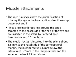
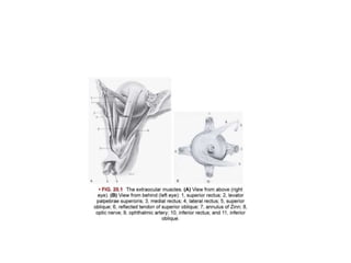
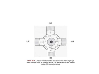
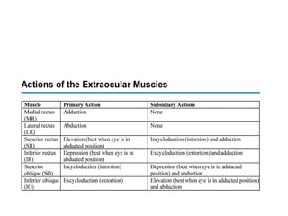
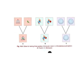
STRABISMUS
Dr Ritu Chaturvedi
HOD,Department of Ophthalmology
SRVS Medical College
The motor apparatus of the eye
• The extraocular muscles and their central nervous
control comprise the motor apparatus of the eye.
• The motility and coordination of the two eyes are
subserved by an unusually accurate and responsive
neuromuscular apparatus.
• This is required in order to ensure the following:
• • Each eye must rapidly and accurately fix upon the
object being viewed.
• • The image of the object must fall upon the fovea
of each eye.
• • Both eyes, in their every movement, must move
in unison so that binocular vision is attained.
Position of eyes in orbit and in
relation to each other
• The position of the eye is judged with respect to its
position in the orbit, its position with respect to the
object being viewed and its position relative to the
other eye. Normally, the two eyes are aligned with
each other when looking at an object, such that the
visual axes of the two eyes meet at the object of
regard
• • The visual axis passes from the object being
viewed, through the nodal point, to the fovea
centralis. • The optic axis marks the centre of the
cornea and the lens and passes through the centre
of rotation of the eye and approximately through
• • The visual axis and the optic axis do not coincide
and angle formed between them is called angle
kappa. If the angle is large, it gives the appearance
of a pseudosquint.
• • If the angle kappa is positive, it gives the
appearance of pseudoexotropia or
pseudodivergent squint.
• • If the angle kappa is negative, it gives the
appearance of pseudoesotropia or
pseudoconvergent squint.
The extraocular muscles
• A set of six muscles controls the movements of
each eye.
• • Four rectus muscles—superior, inferior, lateral
and medial rectus
• • Two obliques—superior and inferior oblique
Muscle attachments
• The rectus muscles have the primary action of
rotating the eye in the four cardinal directions—up,
down, out and in
• They arise in a fibrous ring around the optic
foramen to the nasal side of the axis of the eye and
are inserted in the sclera by flat tendinous
insertions about 10 mm broad.
• The medial rectus is inserted into the sclera about
5.5 mm to the nasal side of the corneoscleral
margin, the inferior rectus 6.6 mm below, the
lateral rectus 7 mm to the temporal side and the
superior rectus 7.75 mm above
• The oblique muscles, the primary function of which is
rotation of the globe, are differently arranged.
• The superior oblique arises from the common origin at
the apex of the orbit, runs forwards to the trochlea, a
cartilaginous ring at the upper and inner angle of the
orbit and, having threaded through this, becomes
tendinous.
• The tendon changes its direction completely and runs
backwards and sideways over the globe under the
superior rectus to attach itself above and lateral to the
posterior pole . The action of the muscle is thus
determined by the oblique direction of its tendon after
it has left the trochlea.
• The inferior oblique maintains a similar direction
throughout its course and is the only muscle not
arising from the apex of the orbit.
• It arises anteriorly from the lower and inner orbital
walls near the lacrimal fossa and, running below
the inferior rectus (i.e. the inferior rectus lies
between the globe and inferior oblique muscle),
finds an insertion in the sclera below and lateral to
the posterior pole of the globe.
The action of the extraocular
muscles
• These rotate the eye around a centre of rotation,
which lies in the horizontal plane approximately 12
or 13 mm behind the cornea, and in every
movement of the globe each muscle is involved to
some degree, by either contraction or inhibition
(Table 20.1). Three types of rotation or ‘degrees of
freedom’ are possible around the centre of
rotation: 1. Rotation around the vertical axis
whereby the globe is turned from side to side 2.
Rotation around the horizontal axis whereby the
globe is turned upwards and downwards
• 3. Rotation around the anteroposterior axis—an
involuntary movement of torsion; intorsion when
the upper pole of the cornea rotates
• n The medial and lateral rectus muscles rotate the
eye horizontally inwards (adduction) and outwards
(abduction), respectively.asally, extorsion when
temporally
• • The superior and inferior rectus muscles rotate
the eye vertically upwards (elevation) and
downwards (depression), respectively, but due to
the obliquity of their course, contraction of the
superior and inferior recti also involves some
torsion .
• Thus, when the superior rectus acts upon the globe
in the primary position, it pulls the eye not only
upwards but also inwards and intorts it. Similarly,
when the inferior rectus acts, the eye is pulled
downwards and inwards and extorted.
• • The superior and inferior oblique muscles rotate
the eye nasally (intorsion) and temporally
(extorsion). Since the obliques are inserted behind
the centre of rotation, their effective action is to
pull the back of the eye forwards and inwards.
• Therefore, when the superior oblique contracts,if
the globe is in the primary position, the main effect
is intorsion but it also rotates the eye downwards
and outwards; the inferior oblique primarily causes
extorsion but also rotates the globe upwards and
outwards.
• • The superior rectus and inferior oblique act
simultaneously to move the eye directly upwards,
the upward movement caused by each muscle
being summated, while the inward movement and
torsion of the superior rectus is exactly
compensated by the outward movement and
contrary torsion of the inferior oblique.
Coordination Between the Various
Extraocular Muscles in Binocular
Movements (Versions)
• Every movement of the eyeball is thus a synkinesis.
Not only is there uniocular synkinesis but also in
normal circumstances there is always binocular
synkinesis. Abduction of one eye is accompanied by
adduction of the other—which is known as a
conjugate movement. The only exception to this
rule is the bilateral adduction of the eyes in
convergence and abduction of both eyes in
divergence (dysconjugate movements). Elevation or
depression of one eye is always accompanied by
elevation or depression, respectively, of the other.
• Elevation of both eyes is accompanied by slight
abduction (divergence), depression by slight adduction
(convergence). In these movements, the muscles which
contract together are called synergists, and those which
suffer inhibition are called antagonists.
• Muscles contracting together to move both the eyes in
the direction of any of the arrows in Fig. 20.5B are
synergists and the muscles that would work in a
directly opposite direction are relaxed and called
antagonists for that particular action. Thus, in rotation
to the right (dextroversion), the synergists are the right
lateral rectus and left medial rectus, while the
antagonists are the right medial rectus and left lateral
rectus.
• In rotation upwards, synergists are the right and
left superior recti primarily and the right and left
inferior obliques secondarily. The antagonists are
the right and left inferior recti and right and left
superior obliques. The testing of eye movements is
complete only if the examination of all types of eye
movements is done systematically.
Laws governing the neural control of
ocular movements are as follows:
• Hering law of equal innervation applies to both
eyes. Equal and simultaneous innervation flows
from the brain to a pair of synergistic (yoke)
muscles which contract simultaneously in
conjugate binocular movements. For example, in
laevoversion, the lateral rectus of the left eye and
medial rectus of the right eye receive an equal and
simultaneous flow of innervation; during
convergence, both medial recti; and so on. In the
case of a paretic squint, the amount of innervation
to both eyes is always determined by the fixating
eye so that the angle of deviation will vary
according to which eye is used for fixation
• Sherrington law of reciprocal innervation: During
the initiation of an eye movement, increased
innervation to an extraocular muscle is
accompanied by simultaneous inhibition (a
reciprocal decrease in innervation) of the direct
antagonist of the contracting muscle of the same
eye. If the left medial rectus muscle receives
innervational flow to initiate adduction of the left
eye, there is simultaneous decreased and inhibitory
flow to the left lateral rectus muscle to make it
relax and enable the eyeball to move medially.
Nerves and centres
• The nervous control of ocular movements is
complicated. The muscles are supplied by nerves
arising from nuclei in the mid-brain. Their action is
coordinated by intermediate ‘centres’ situated in this
region by which reflex activities are governed. Finally,
these intermediate centres are linked with the
vestibular apparatus whereby they become
associated with the equilibration reflexes and the
cerebral cortex so that voluntary movements and
participation in the higher reflexes involving
perception become possible.
• The oculomotor, or third cranial nerve, supplies all
the extrinsic muscles except the lateral rectus and
superior oblique. It also supplies the sphincter
pupillae and ciliary muscle.
• The superior oblique is supplied by the trochlear
(fourth) nerve and the lateral rectus by the
abducens (sixth) nerve
Fixation, projection, correspondence
and binocular vision
• Fixation and projection
• We have already seen that the location of the
image of an external object on the retina is
determined by a line passing from the object
through the nodal point of the eye. Conversely, an
object is projected in space along the line passing
through the retinal image and the nodal point.
Fixation is the ability of the eye to steadily look at
the object of regard. When a distant object is
looked at, the visual axes are practically parallel;
the object forms an image upon each fovea
centralis.
• Correspondence Any object to one side of the fixation
target forms its retinal images upon the temporal side
of one retina and upon the nasal side of the other;
these retinal areas are coordinated visually in the
occipital cortex so that such an object is seen with both
eyes as a single object. These are known as
corresponding points, the most important pair of
which, of course, is the foveae. Points on the two
retinae, which are not corresponding points in this
sense of the term, are called disparate points, and if an
object forms its retinal images on these, it will be seen
double (binocular diplopia). If the disparity is slight,
there is a tendency to move the eyes so that the images
may be fused by means of the fusion reflexes from the
occipital cortex.
• Fixation, fusion and reflex movements
• Since the most accurate vision is attained by the
foveae, it is necessary that the eyes be rapidly
orientated so that the image of an object of
interest falls upon them or that of a moving object
be retained on them. This ascendancy of the foveae
is maintained by the fixation reflex (Fig. 20.8) and
whenever the image leaves the foveae, the eyes are
at once reoriented so that it falls upon them. This
continues till the object moves outside the
binocular field of vision and the eyes then refixate
on another object.
• The activity of this reflex is demonstrated by the
rapid to-and-fro movements of the eyes of a person
watching passing objects such as trees or electric
poles while looking out of the window of a moving
train, and can be demonstrated clinically by
regarding a revolving drum or a moving tape on
which black and white stripes are painted
(optokinetic nystagmus). The latter phenomenon
can be used as a test to demonstrate the integrity
of the reflex paths.
• The same reflex produces involuntary fusional
movements of the eyes to maintain single binocular
vision. This may be demonstrated clinically by
placing a small prism in front of one eye while the
patient regards a distant light. The eye will at once
turn away from the primary position to allow the
deflected rays to fall again upon the fovea. The
strongest prism whose deviating effect can be
tolerated without developing diplopia or double
vision is a measure of the reflex fusional capacity
(Fig. 20.11). A prism bar consists of a battery of
prisms of increasing strength and is a convenient
instrument in clinical testing
Binocular vision
• In view of the distance between the two eyes, it is
obvious that the retinal images of both eyes cannot
be identical, since each eye regards a slightly
different aspect of any object observed. If the
object is a solid body, the right eye sees a little
more of the right side of the object, and vice versa.
The two images are fused psychologically, and this
fusion of the slightly diverse images, combined with
other facts derived from experience, enables the
person to appreciate the solidity of objects or
perceive the sensation of depth or stereopsis.
• Even with one eye, a person can appreciate depth
by monocular clues such as contour overlay, distant
objects appearing smaller and motion parallax with
far objects moving faster
• . Moreover, it is obvious from that if both eyes are
fixing upon a particular object, the images of other
objects nearer or further away cannot fall upon
corresponding points.
• If their projection through the nodal point is
continued to the retina, it is seen that the images
of objects nearer than the object of fixation fall on
the temporal side, and those farther away to the
• This can be easily demonstrated by holding a pencil
in front of the eyes: if the pencil is fixated, more
distant objects appear doubled; if a distant object is
fixated, the pencil appears doubled.
• It will thus be found that near objects suffer a
crossed (heteronymous) diplopia, and distant
objects an uncrossed (homonymous) diplopia.
• This diplopia is physiological and is perceptually
suppressed in actual vision, but produces a
psychological impression, which is translated into
appreciation of distance. It follows that accuracy of
stereoscopic vision depends on good sight with
GRADES OF BINOCULAR SINGLE
VISION
Grade I-Simultaneous macular perception.
It is the power to see two dissimilar objects
simultaneously. It is tested by projecting two
dissimilar objects ( which can be joined or
superimposed to form a complete picture) in front of
the two eyes. For example, when a picture of a bird is
projected onto the right eye and that of a cage on to
the left eye, an individual with presence of
simultaneous perception will see the bird in the cage
• Grade 2- Fusion. It consists of the power to
superimpose two incomplete but similar images to
form one complete image (Fig. 14.8B). The ability of
the subject to continue to see one complete picture
when his eyes are made to converge or diverge a
few degrees, gives the positive and negative fusion
range, respectively.
• Grade lll- Stereopsis. lt consists of the ability to
perceive the third dimension (depth perception). lt
can be tested with stereopsis slides in
synoptophore (Fig. l 4.8C).
Convergence and accommodation
• When a distant object is observed by an emmetropic
person, the visual axes are parallel and no effort of
accommodation is made. If, however, a near object is
regarded, the eyes converge upon it and an effort of
accommodation corresponding to the distance of the
object is made.
• These movements are reflex and are controlled, as we
have seen, by a centre in the occipital cortex , the afferent
path being the visual pathways, the efferent path running
to the Perlia nucleus region of the Edinger–Westphal
nucleus. The associated pupillary contraction is a purely
low-level reflex arc, the afferent path running from the
medial recti to the Edinger–Westphal nucleus and the
efferent by the parasympathetic fibres in the third nerve
• An instrument called the Royal Air Force (RAF) ruler
is used to measure the near point of
accommodation and convergence by measuring the
distance at which the target appears ‘blurred’ or
‘double’, respectively.
STRABISMUS new-WPS Office.pptx

STRABISMUS new-WPS Office.pptx

  • 1.
    STRABISMUS Dr Ritu Chaturvedi HOD,Departmentof Ophthalmology SRVS Medical College
  • 2.
    The motor apparatusof the eye • The extraocular muscles and their central nervous control comprise the motor apparatus of the eye. • The motility and coordination of the two eyes are subserved by an unusually accurate and responsive neuromuscular apparatus. • This is required in order to ensure the following:
  • 3.
    • • Eacheye must rapidly and accurately fix upon the object being viewed. • • The image of the object must fall upon the fovea of each eye. • • Both eyes, in their every movement, must move in unison so that binocular vision is attained.
  • 4.
    Position of eyesin orbit and in relation to each other • The position of the eye is judged with respect to its position in the orbit, its position with respect to the object being viewed and its position relative to the other eye. Normally, the two eyes are aligned with each other when looking at an object, such that the visual axes of the two eyes meet at the object of regard • • The visual axis passes from the object being viewed, through the nodal point, to the fovea centralis. • The optic axis marks the centre of the cornea and the lens and passes through the centre of rotation of the eye and approximately through
  • 5.
    • • Thevisual axis and the optic axis do not coincide and angle formed between them is called angle kappa. If the angle is large, it gives the appearance of a pseudosquint. • • If the angle kappa is positive, it gives the appearance of pseudoexotropia or pseudodivergent squint. • • If the angle kappa is negative, it gives the appearance of pseudoesotropia or pseudoconvergent squint.
  • 6.
    The extraocular muscles •A set of six muscles controls the movements of each eye. • • Four rectus muscles—superior, inferior, lateral and medial rectus • • Two obliques—superior and inferior oblique
  • 7.
    Muscle attachments • Therectus muscles have the primary action of rotating the eye in the four cardinal directions—up, down, out and in • They arise in a fibrous ring around the optic foramen to the nasal side of the axis of the eye and are inserted in the sclera by flat tendinous insertions about 10 mm broad. • The medial rectus is inserted into the sclera about 5.5 mm to the nasal side of the corneoscleral margin, the inferior rectus 6.6 mm below, the lateral rectus 7 mm to the temporal side and the superior rectus 7.75 mm above
  • 10.
    • The obliquemuscles, the primary function of which is rotation of the globe, are differently arranged. • The superior oblique arises from the common origin at the apex of the orbit, runs forwards to the trochlea, a cartilaginous ring at the upper and inner angle of the orbit and, having threaded through this, becomes tendinous. • The tendon changes its direction completely and runs backwards and sideways over the globe under the superior rectus to attach itself above and lateral to the posterior pole . The action of the muscle is thus determined by the oblique direction of its tendon after it has left the trochlea.
  • 11.
    • The inferioroblique maintains a similar direction throughout its course and is the only muscle not arising from the apex of the orbit. • It arises anteriorly from the lower and inner orbital walls near the lacrimal fossa and, running below the inferior rectus (i.e. the inferior rectus lies between the globe and inferior oblique muscle), finds an insertion in the sclera below and lateral to the posterior pole of the globe.
  • 12.
    The action ofthe extraocular muscles • These rotate the eye around a centre of rotation, which lies in the horizontal plane approximately 12 or 13 mm behind the cornea, and in every movement of the globe each muscle is involved to some degree, by either contraction or inhibition (Table 20.1). Three types of rotation or ‘degrees of freedom’ are possible around the centre of rotation: 1. Rotation around the vertical axis whereby the globe is turned from side to side 2. Rotation around the horizontal axis whereby the globe is turned upwards and downwards
  • 13.
    • 3. Rotationaround the anteroposterior axis—an involuntary movement of torsion; intorsion when the upper pole of the cornea rotates • n The medial and lateral rectus muscles rotate the eye horizontally inwards (adduction) and outwards (abduction), respectively.asally, extorsion when temporally
  • 14.
    • • Thesuperior and inferior rectus muscles rotate the eye vertically upwards (elevation) and downwards (depression), respectively, but due to the obliquity of their course, contraction of the superior and inferior recti also involves some torsion . • Thus, when the superior rectus acts upon the globe in the primary position, it pulls the eye not only upwards but also inwards and intorts it. Similarly, when the inferior rectus acts, the eye is pulled downwards and inwards and extorted.
  • 16.
    • • Thesuperior and inferior oblique muscles rotate the eye nasally (intorsion) and temporally (extorsion). Since the obliques are inserted behind the centre of rotation, their effective action is to pull the back of the eye forwards and inwards. • Therefore, when the superior oblique contracts,if the globe is in the primary position, the main effect is intorsion but it also rotates the eye downwards and outwards; the inferior oblique primarily causes extorsion but also rotates the globe upwards and outwards.
  • 17.
    • • Thesuperior rectus and inferior oblique act simultaneously to move the eye directly upwards, the upward movement caused by each muscle being summated, while the inward movement and torsion of the superior rectus is exactly compensated by the outward movement and contrary torsion of the inferior oblique.
  • 19.
    Coordination Between theVarious Extraocular Muscles in Binocular Movements (Versions) • Every movement of the eyeball is thus a synkinesis. Not only is there uniocular synkinesis but also in normal circumstances there is always binocular synkinesis. Abduction of one eye is accompanied by adduction of the other—which is known as a conjugate movement. The only exception to this rule is the bilateral adduction of the eyes in convergence and abduction of both eyes in divergence (dysconjugate movements). Elevation or depression of one eye is always accompanied by elevation or depression, respectively, of the other.
  • 20.
    • Elevation ofboth eyes is accompanied by slight abduction (divergence), depression by slight adduction (convergence). In these movements, the muscles which contract together are called synergists, and those which suffer inhibition are called antagonists. • Muscles contracting together to move both the eyes in the direction of any of the arrows in Fig. 20.5B are synergists and the muscles that would work in a directly opposite direction are relaxed and called antagonists for that particular action. Thus, in rotation to the right (dextroversion), the synergists are the right lateral rectus and left medial rectus, while the antagonists are the right medial rectus and left lateral rectus.
  • 22.
    • In rotationupwards, synergists are the right and left superior recti primarily and the right and left inferior obliques secondarily. The antagonists are the right and left inferior recti and right and left superior obliques. The testing of eye movements is complete only if the examination of all types of eye movements is done systematically.
  • 23.
    Laws governing theneural control of ocular movements are as follows: • Hering law of equal innervation applies to both eyes. Equal and simultaneous innervation flows from the brain to a pair of synergistic (yoke) muscles which contract simultaneously in conjugate binocular movements. For example, in laevoversion, the lateral rectus of the left eye and medial rectus of the right eye receive an equal and simultaneous flow of innervation; during convergence, both medial recti; and so on. In the case of a paretic squint, the amount of innervation to both eyes is always determined by the fixating eye so that the angle of deviation will vary according to which eye is used for fixation
  • 24.
    • Sherrington lawof reciprocal innervation: During the initiation of an eye movement, increased innervation to an extraocular muscle is accompanied by simultaneous inhibition (a reciprocal decrease in innervation) of the direct antagonist of the contracting muscle of the same eye. If the left medial rectus muscle receives innervational flow to initiate adduction of the left eye, there is simultaneous decreased and inhibitory flow to the left lateral rectus muscle to make it relax and enable the eyeball to move medially.
  • 25.
    Nerves and centres •The nervous control of ocular movements is complicated. The muscles are supplied by nerves arising from nuclei in the mid-brain. Their action is coordinated by intermediate ‘centres’ situated in this region by which reflex activities are governed. Finally, these intermediate centres are linked with the vestibular apparatus whereby they become associated with the equilibration reflexes and the cerebral cortex so that voluntary movements and participation in the higher reflexes involving perception become possible.
  • 26.
    • The oculomotor,or third cranial nerve, supplies all the extrinsic muscles except the lateral rectus and superior oblique. It also supplies the sphincter pupillae and ciliary muscle. • The superior oblique is supplied by the trochlear (fourth) nerve and the lateral rectus by the abducens (sixth) nerve
  • 29.
    Fixation, projection, correspondence andbinocular vision • Fixation and projection • We have already seen that the location of the image of an external object on the retina is determined by a line passing from the object through the nodal point of the eye. Conversely, an object is projected in space along the line passing through the retinal image and the nodal point. Fixation is the ability of the eye to steadily look at the object of regard. When a distant object is looked at, the visual axes are practically parallel; the object forms an image upon each fovea centralis.
  • 30.
    • Correspondence Anyobject to one side of the fixation target forms its retinal images upon the temporal side of one retina and upon the nasal side of the other; these retinal areas are coordinated visually in the occipital cortex so that such an object is seen with both eyes as a single object. These are known as corresponding points, the most important pair of which, of course, is the foveae. Points on the two retinae, which are not corresponding points in this sense of the term, are called disparate points, and if an object forms its retinal images on these, it will be seen double (binocular diplopia). If the disparity is slight, there is a tendency to move the eyes so that the images may be fused by means of the fusion reflexes from the occipital cortex.
  • 31.
    • Fixation, fusionand reflex movements • Since the most accurate vision is attained by the foveae, it is necessary that the eyes be rapidly orientated so that the image of an object of interest falls upon them or that of a moving object be retained on them. This ascendancy of the foveae is maintained by the fixation reflex (Fig. 20.8) and whenever the image leaves the foveae, the eyes are at once reoriented so that it falls upon them. This continues till the object moves outside the binocular field of vision and the eyes then refixate on another object.
  • 32.
    • The activityof this reflex is demonstrated by the rapid to-and-fro movements of the eyes of a person watching passing objects such as trees or electric poles while looking out of the window of a moving train, and can be demonstrated clinically by regarding a revolving drum or a moving tape on which black and white stripes are painted (optokinetic nystagmus). The latter phenomenon can be used as a test to demonstrate the integrity of the reflex paths.
  • 33.
    • The samereflex produces involuntary fusional movements of the eyes to maintain single binocular vision. This may be demonstrated clinically by placing a small prism in front of one eye while the patient regards a distant light. The eye will at once turn away from the primary position to allow the deflected rays to fall again upon the fovea. The strongest prism whose deviating effect can be tolerated without developing diplopia or double vision is a measure of the reflex fusional capacity (Fig. 20.11). A prism bar consists of a battery of prisms of increasing strength and is a convenient instrument in clinical testing
  • 35.
    Binocular vision • Inview of the distance between the two eyes, it is obvious that the retinal images of both eyes cannot be identical, since each eye regards a slightly different aspect of any object observed. If the object is a solid body, the right eye sees a little more of the right side of the object, and vice versa. The two images are fused psychologically, and this fusion of the slightly diverse images, combined with other facts derived from experience, enables the person to appreciate the solidity of objects or perceive the sensation of depth or stereopsis.
  • 36.
    • Even withone eye, a person can appreciate depth by monocular clues such as contour overlay, distant objects appearing smaller and motion parallax with far objects moving faster • . Moreover, it is obvious from that if both eyes are fixing upon a particular object, the images of other objects nearer or further away cannot fall upon corresponding points. • If their projection through the nodal point is continued to the retina, it is seen that the images of objects nearer than the object of fixation fall on the temporal side, and those farther away to the
  • 37.
    • This canbe easily demonstrated by holding a pencil in front of the eyes: if the pencil is fixated, more distant objects appear doubled; if a distant object is fixated, the pencil appears doubled. • It will thus be found that near objects suffer a crossed (heteronymous) diplopia, and distant objects an uncrossed (homonymous) diplopia. • This diplopia is physiological and is perceptually suppressed in actual vision, but produces a psychological impression, which is translated into appreciation of distance. It follows that accuracy of stereoscopic vision depends on good sight with
  • 38.
    GRADES OF BINOCULARSINGLE VISION Grade I-Simultaneous macular perception. It is the power to see two dissimilar objects simultaneously. It is tested by projecting two dissimilar objects ( which can be joined or superimposed to form a complete picture) in front of the two eyes. For example, when a picture of a bird is projected onto the right eye and that of a cage on to the left eye, an individual with presence of simultaneous perception will see the bird in the cage
  • 39.
    • Grade 2-Fusion. It consists of the power to superimpose two incomplete but similar images to form one complete image (Fig. 14.8B). The ability of the subject to continue to see one complete picture when his eyes are made to converge or diverge a few degrees, gives the positive and negative fusion range, respectively.
  • 40.
    • Grade lll-Stereopsis. lt consists of the ability to perceive the third dimension (depth perception). lt can be tested with stereopsis slides in synoptophore (Fig. l 4.8C).
  • 42.
    Convergence and accommodation •When a distant object is observed by an emmetropic person, the visual axes are parallel and no effort of accommodation is made. If, however, a near object is regarded, the eyes converge upon it and an effort of accommodation corresponding to the distance of the object is made. • These movements are reflex and are controlled, as we have seen, by a centre in the occipital cortex , the afferent path being the visual pathways, the efferent path running to the Perlia nucleus region of the Edinger–Westphal nucleus. The associated pupillary contraction is a purely low-level reflex arc, the afferent path running from the medial recti to the Edinger–Westphal nucleus and the efferent by the parasympathetic fibres in the third nerve
  • 43.
    • An instrumentcalled the Royal Air Force (RAF) ruler is used to measure the near point of accommodation and convergence by measuring the distance at which the target appears ‘blurred’ or ‘double’, respectively.