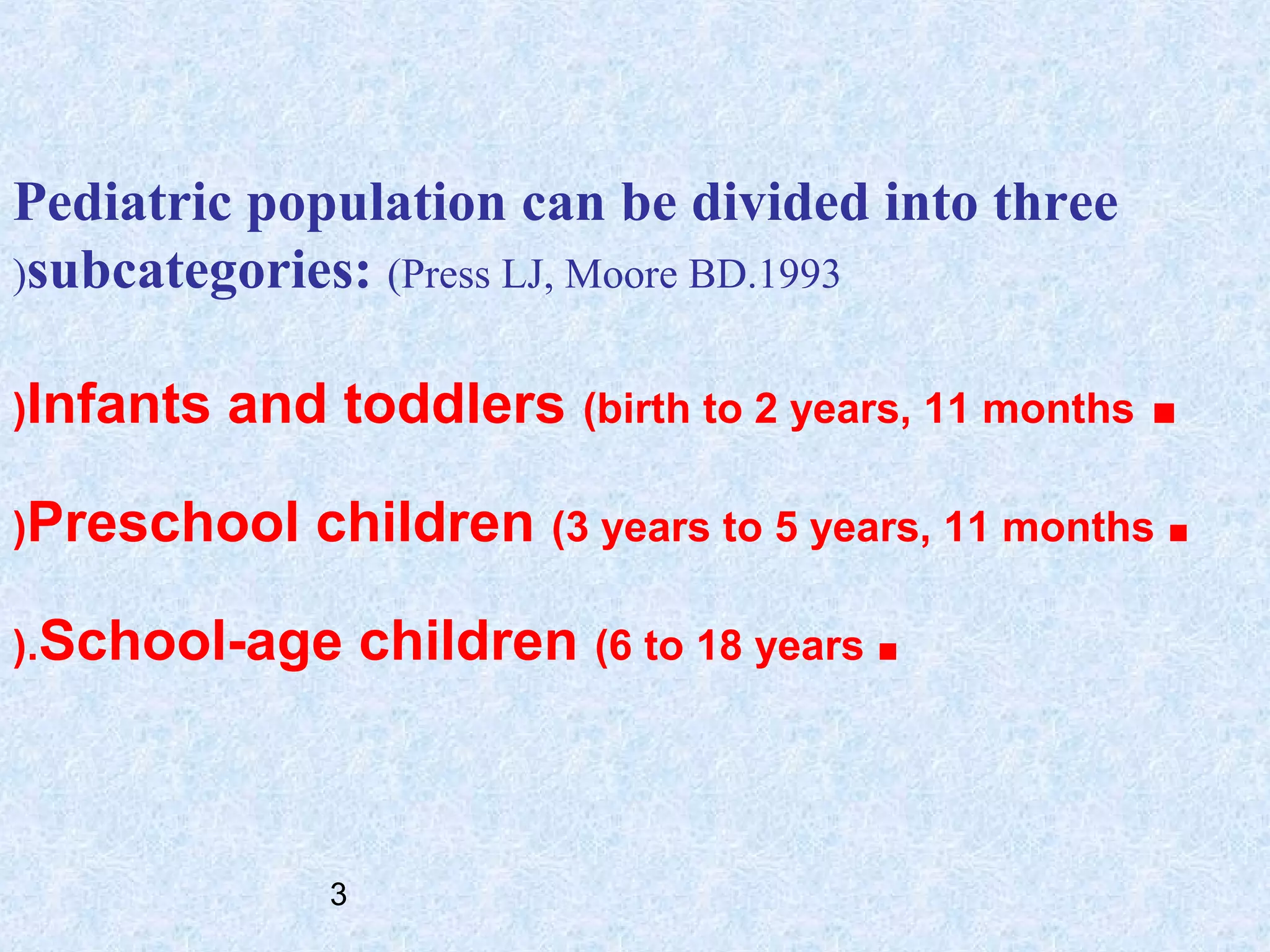
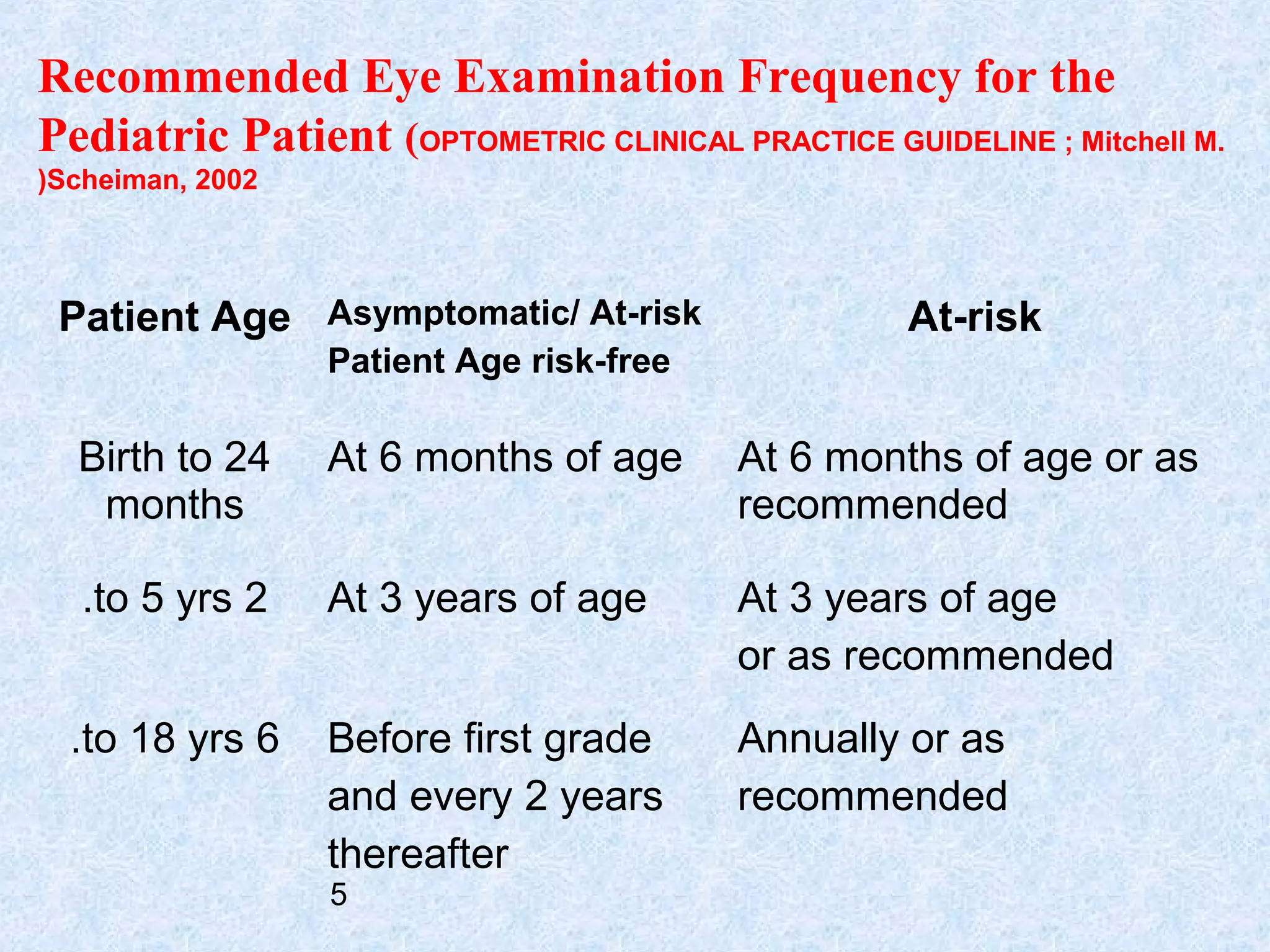
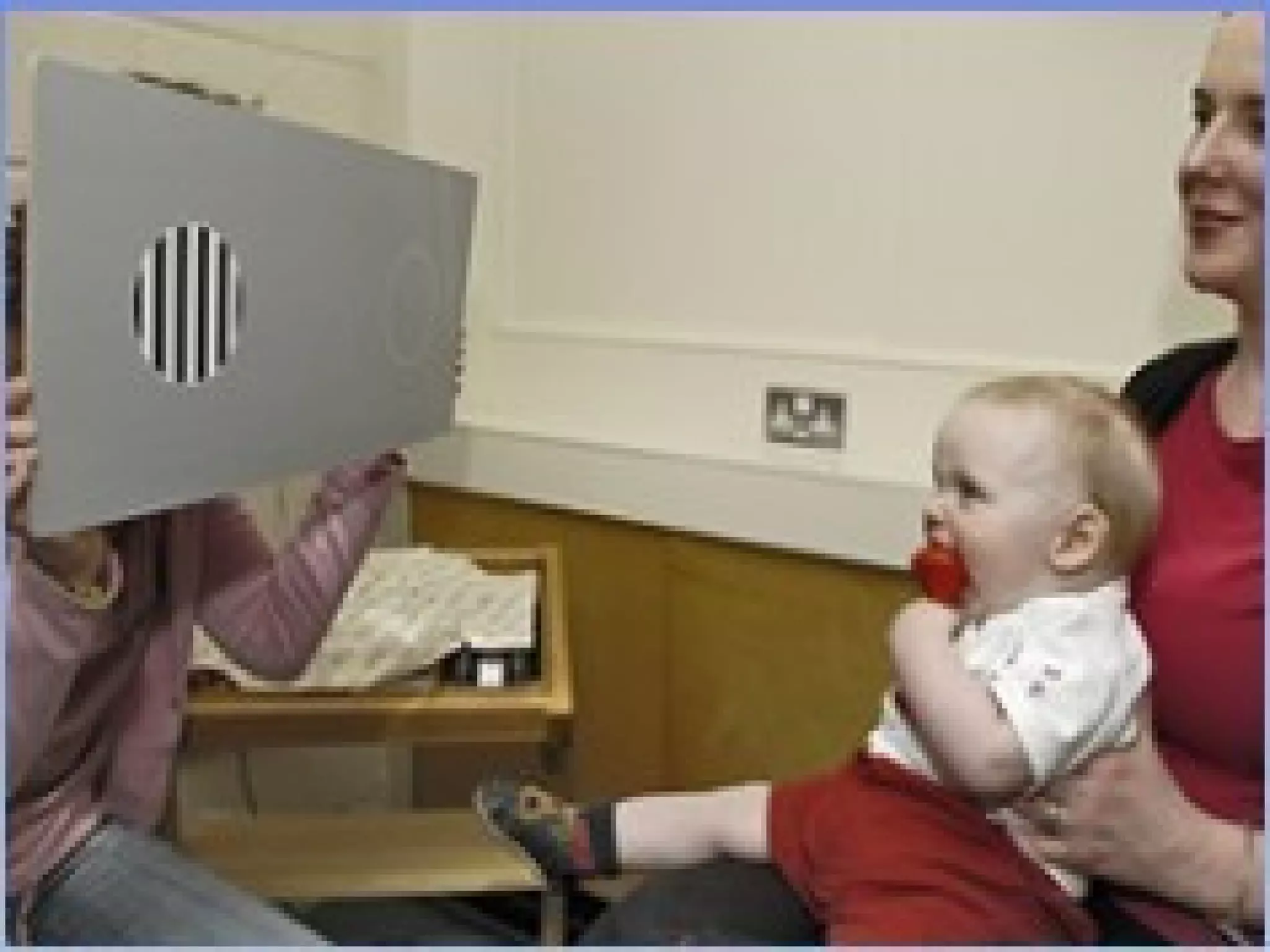
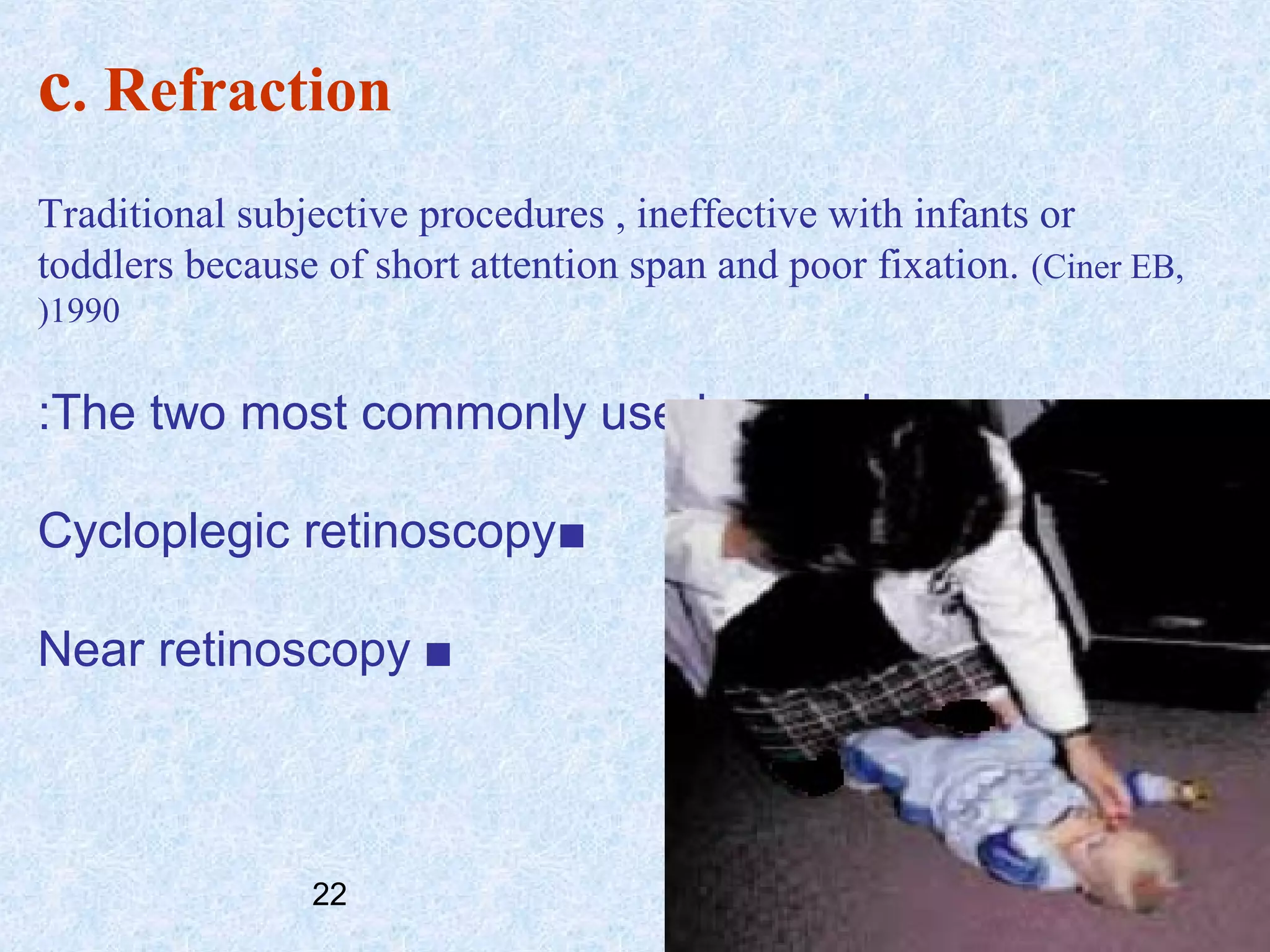
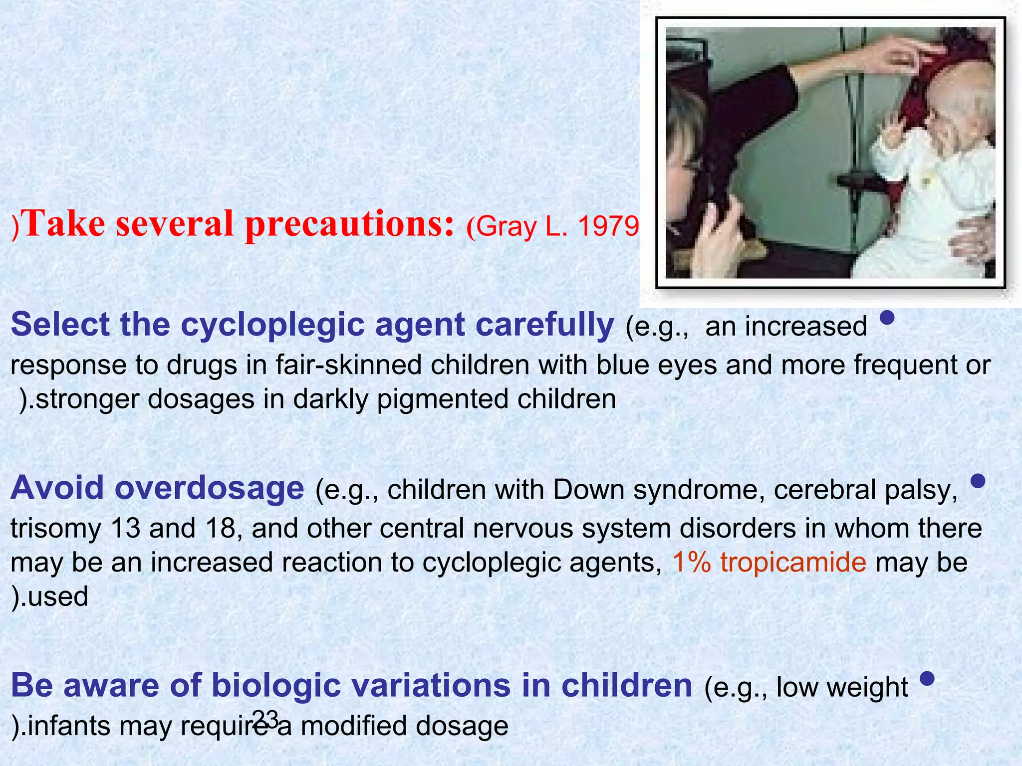
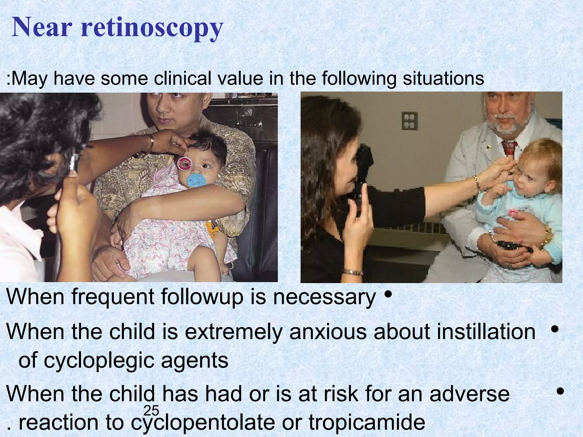
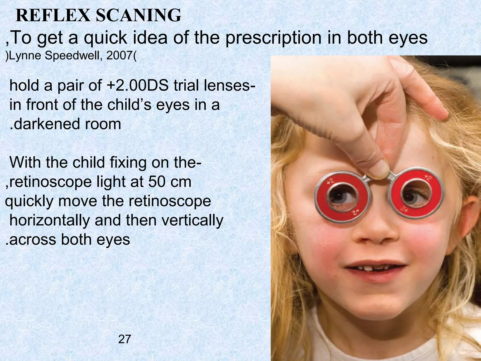
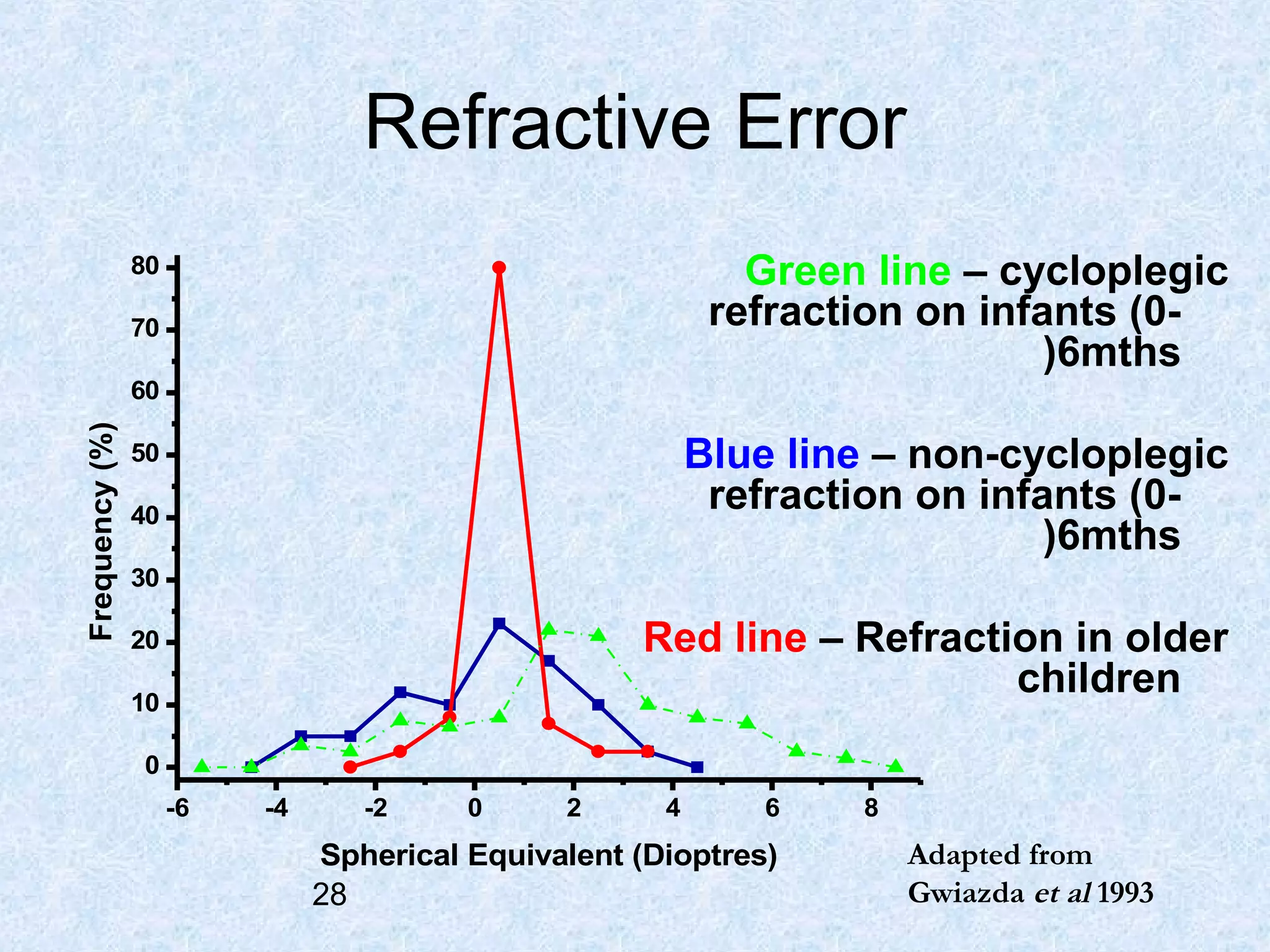
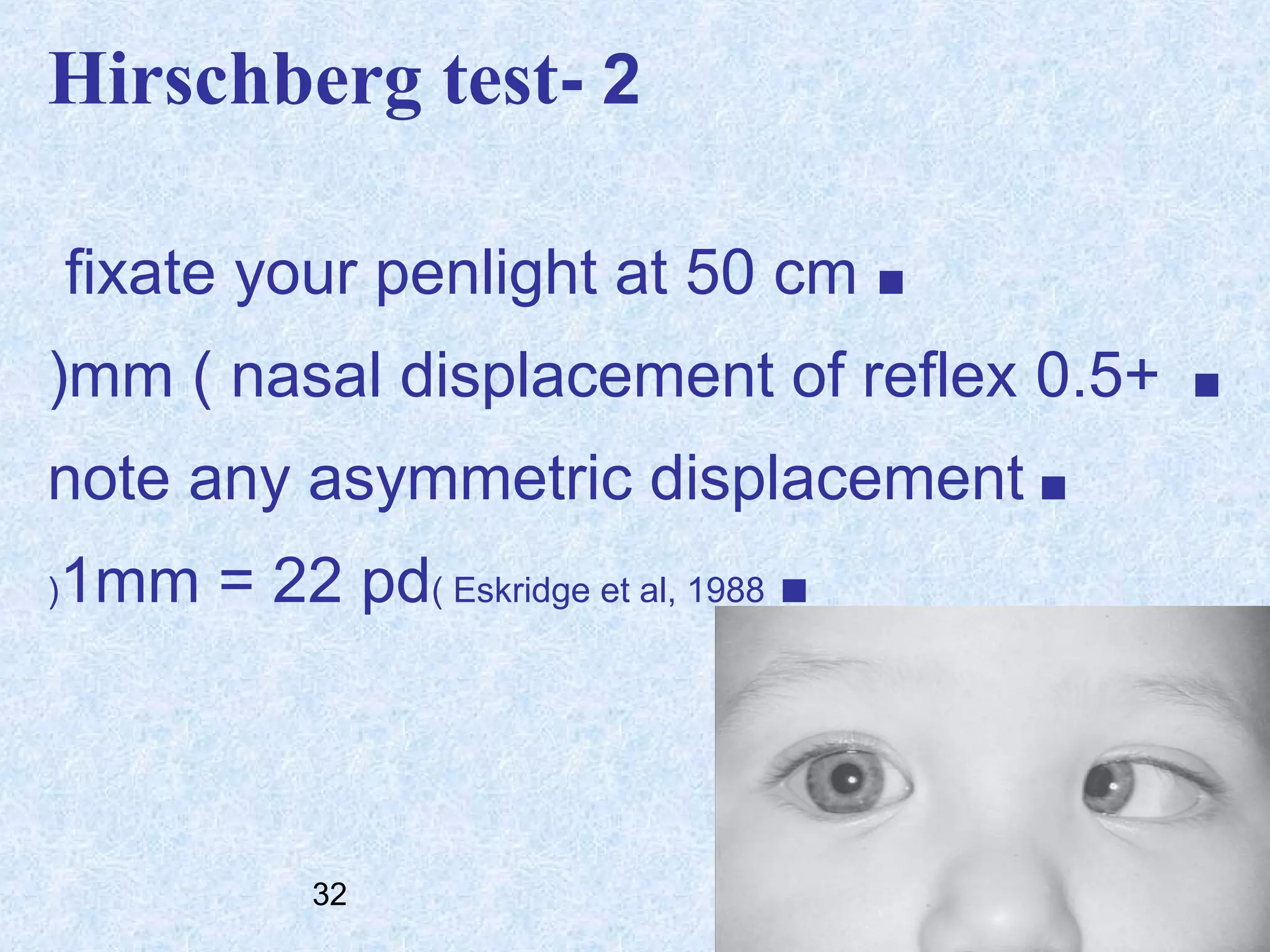
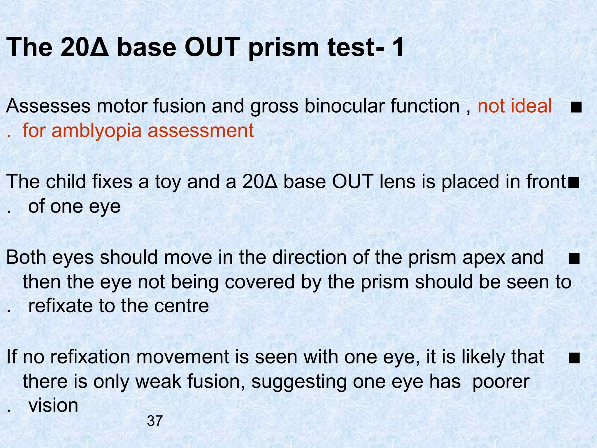
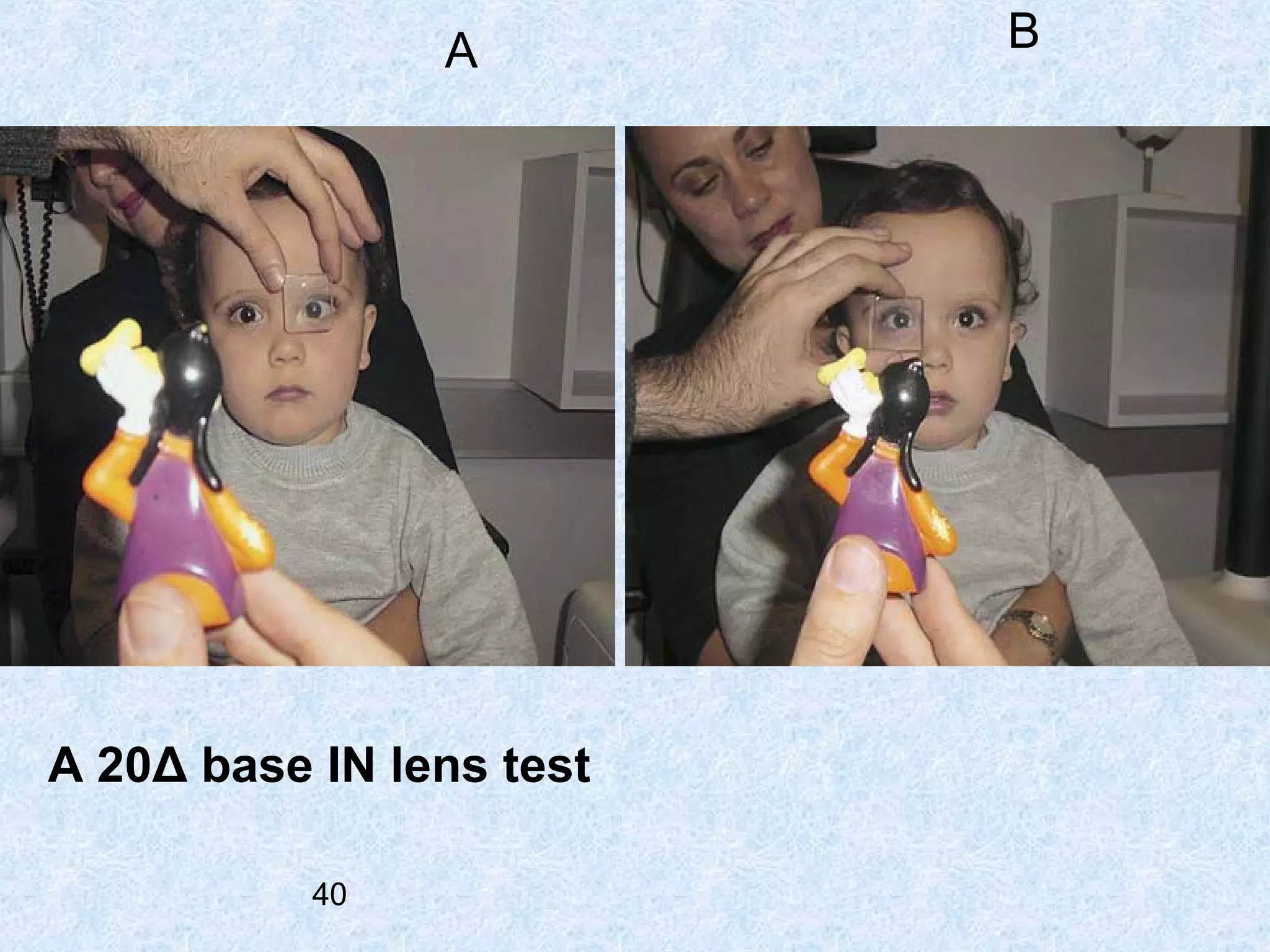
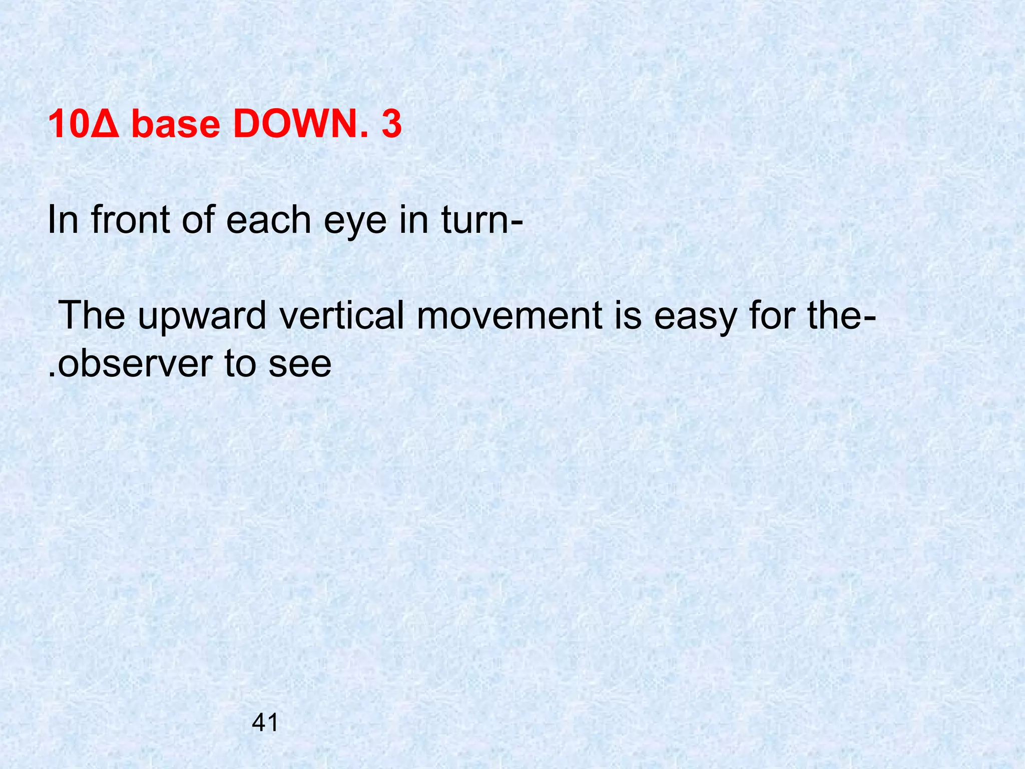
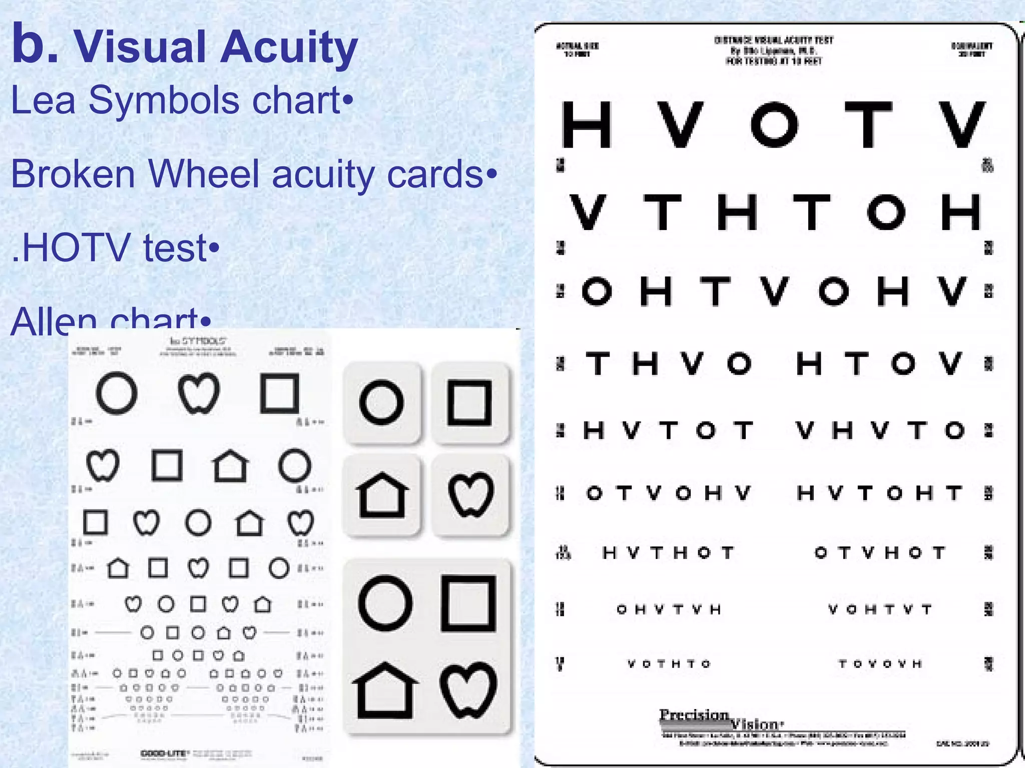
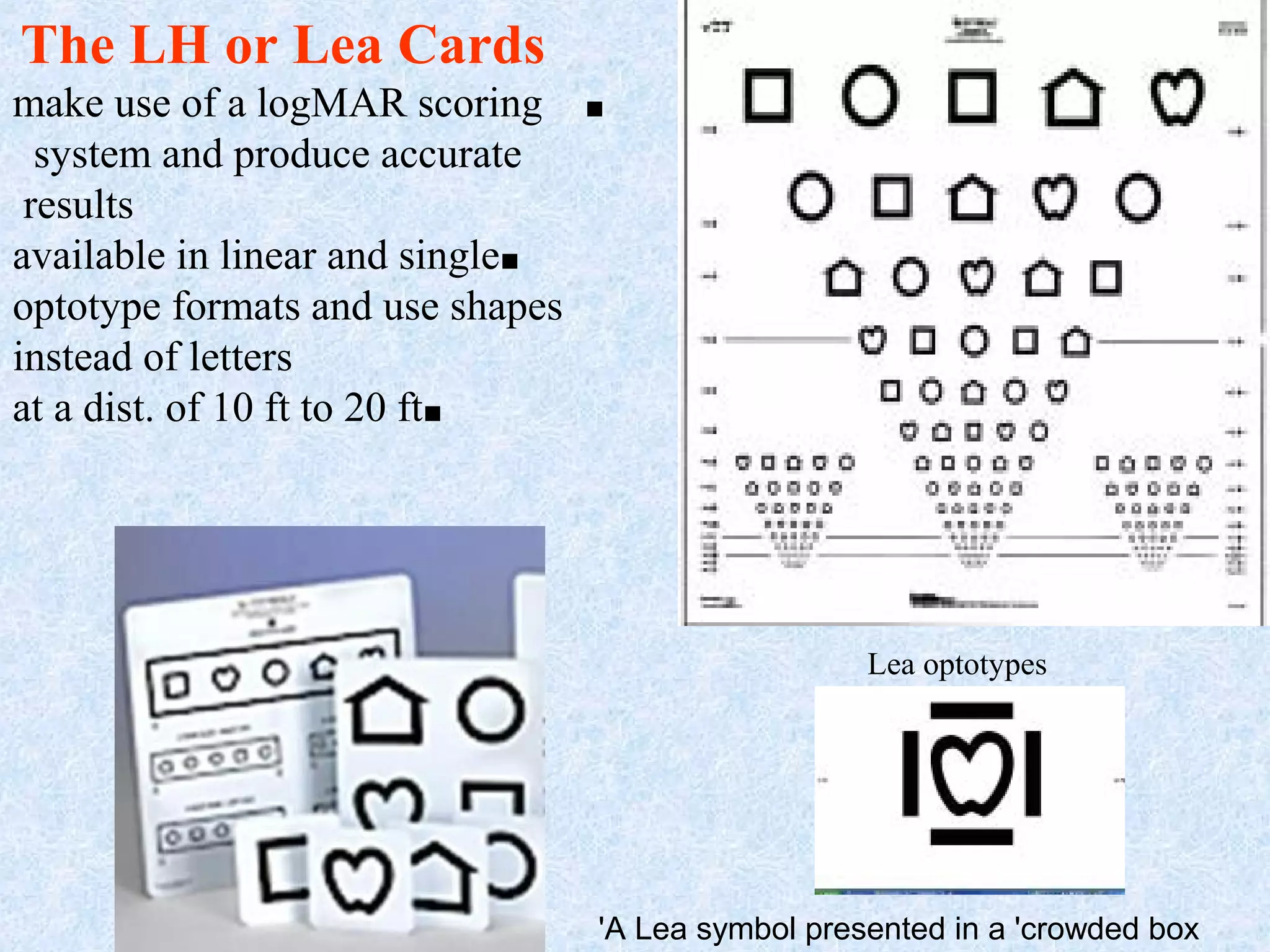
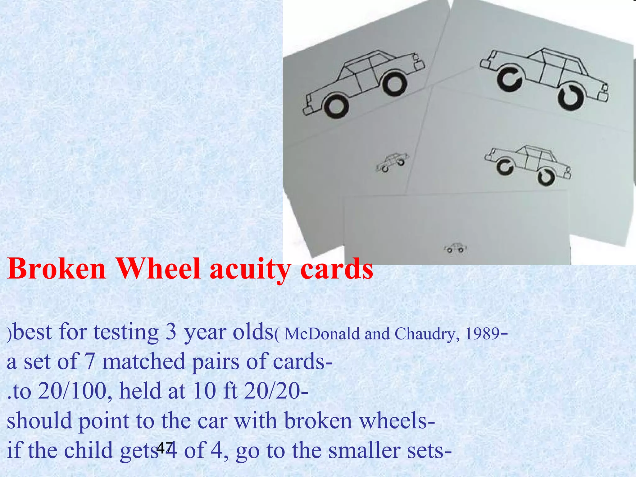
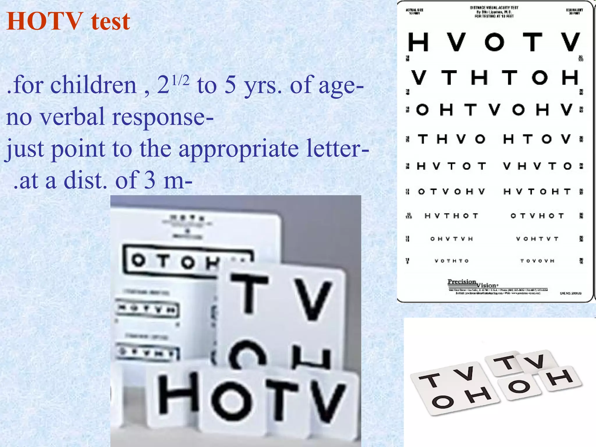
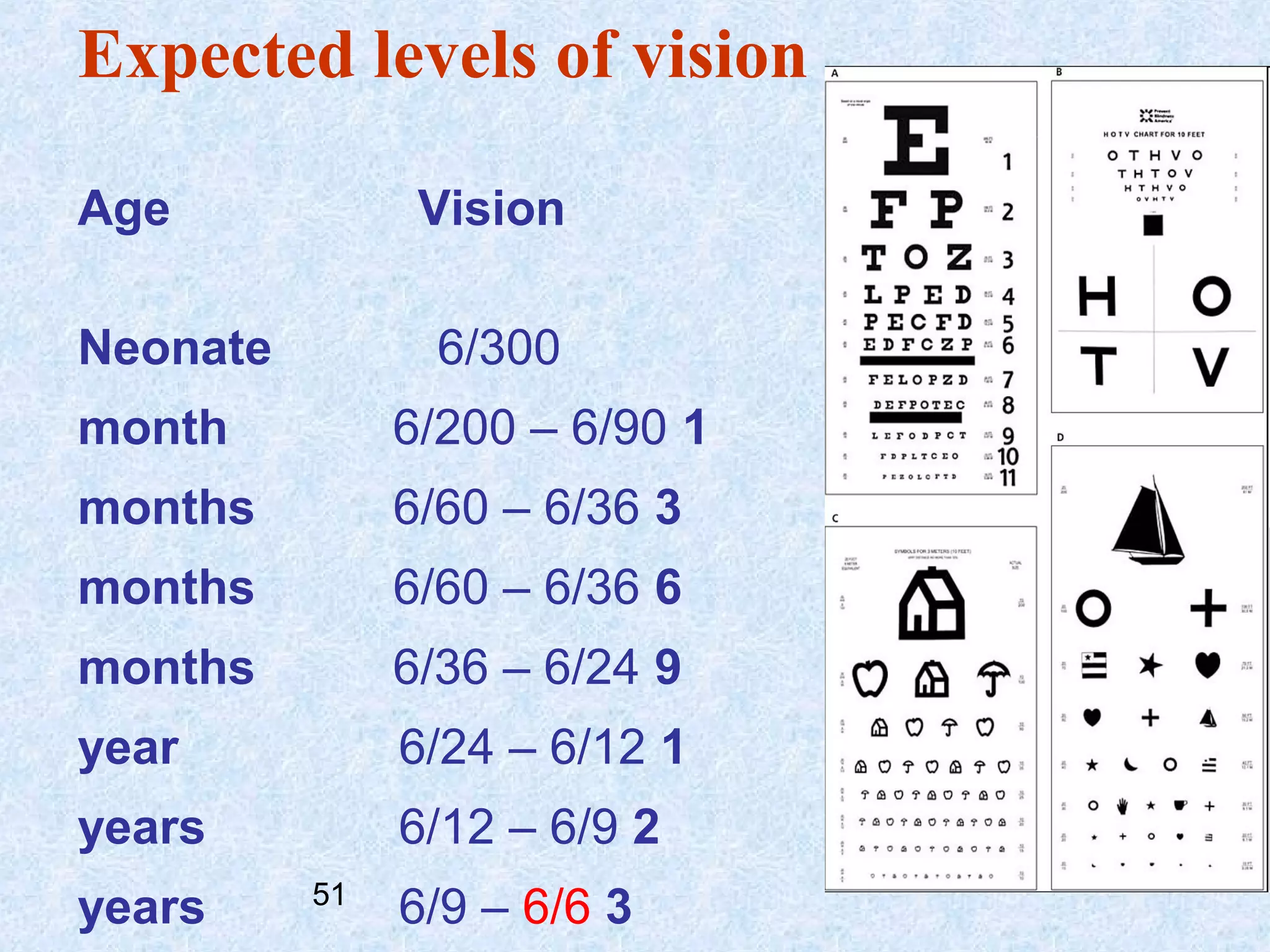
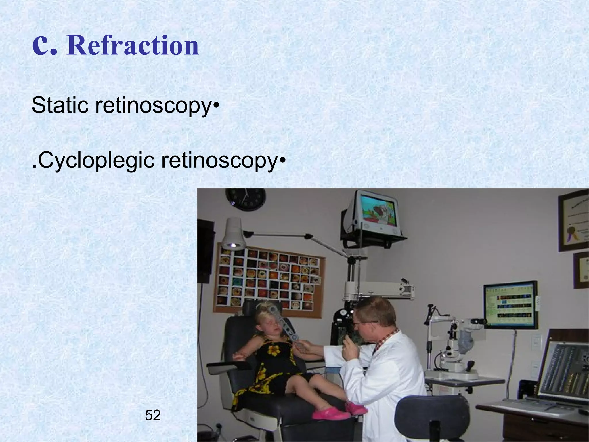
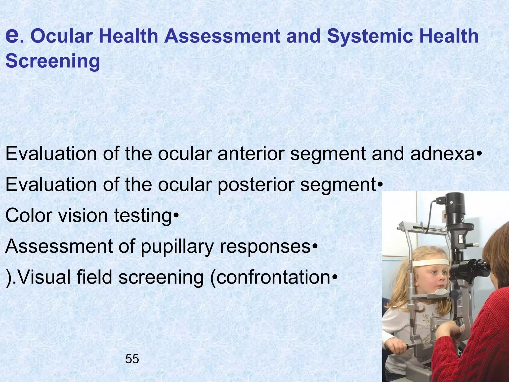
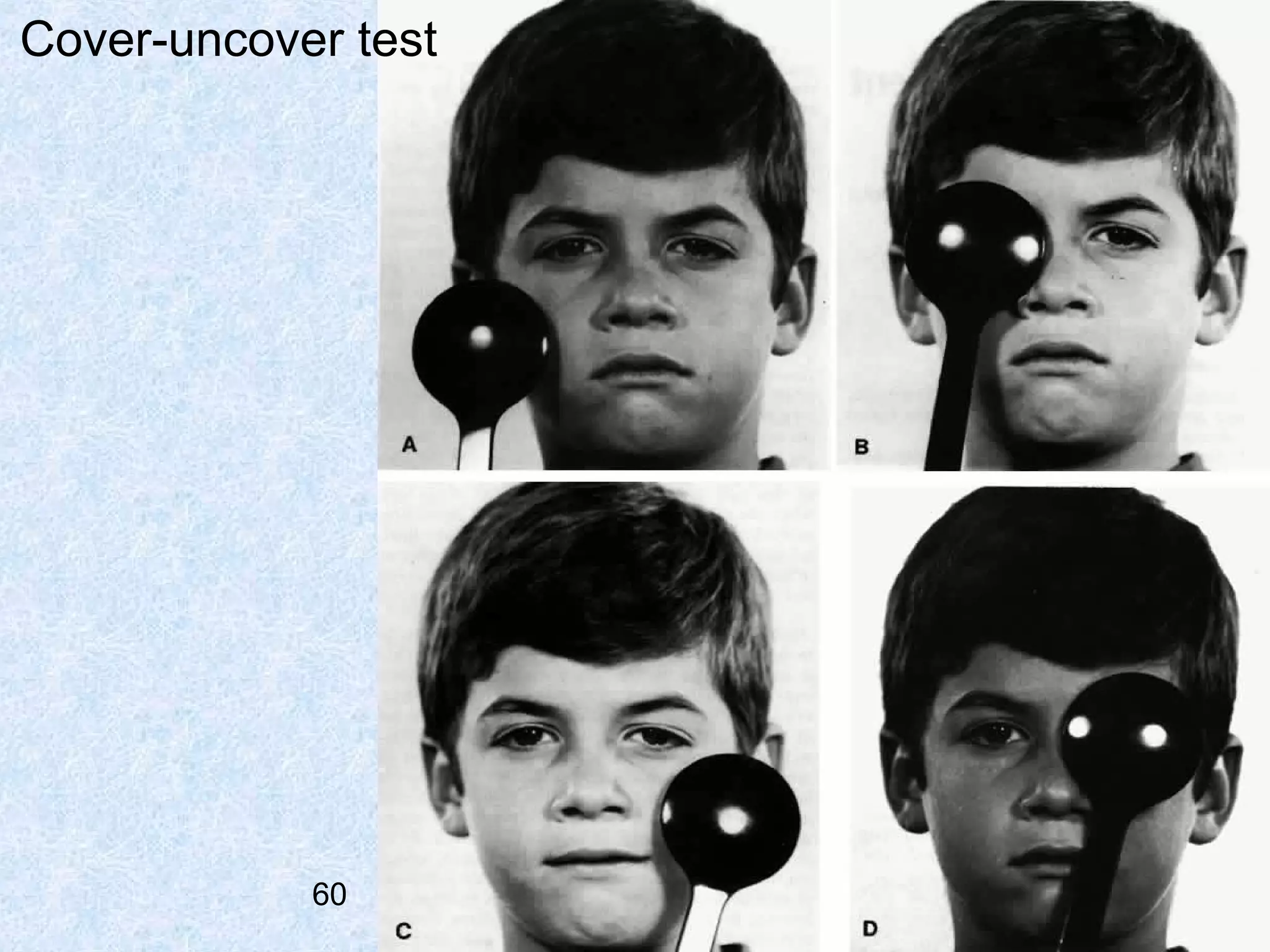
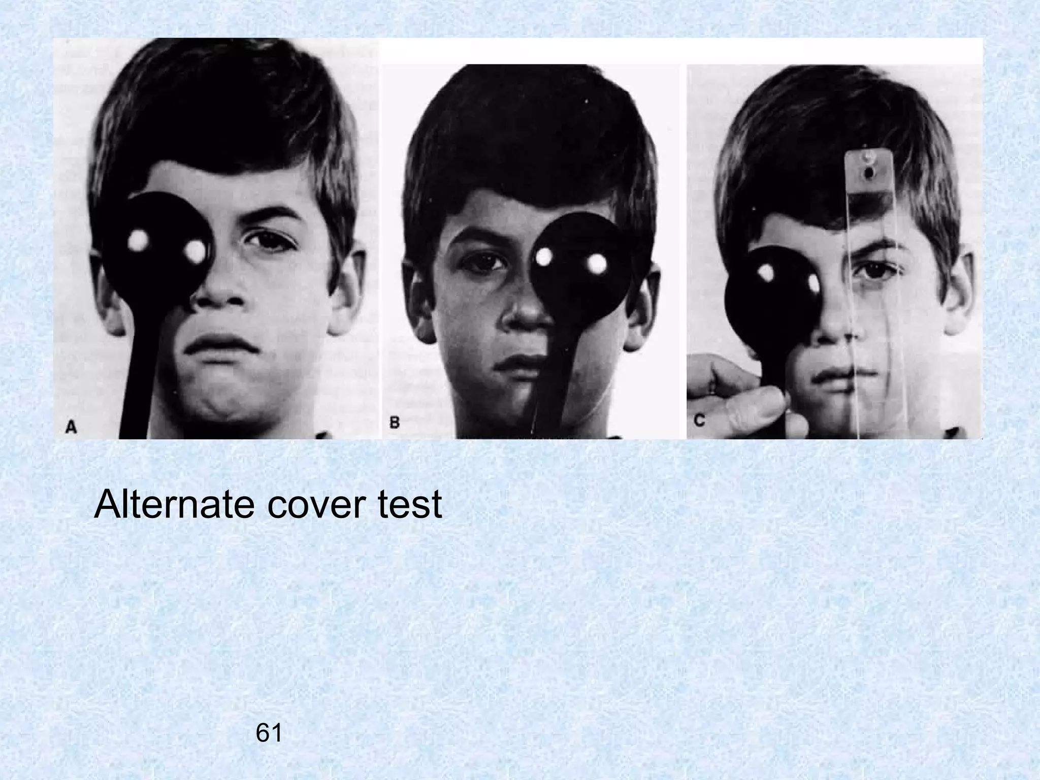
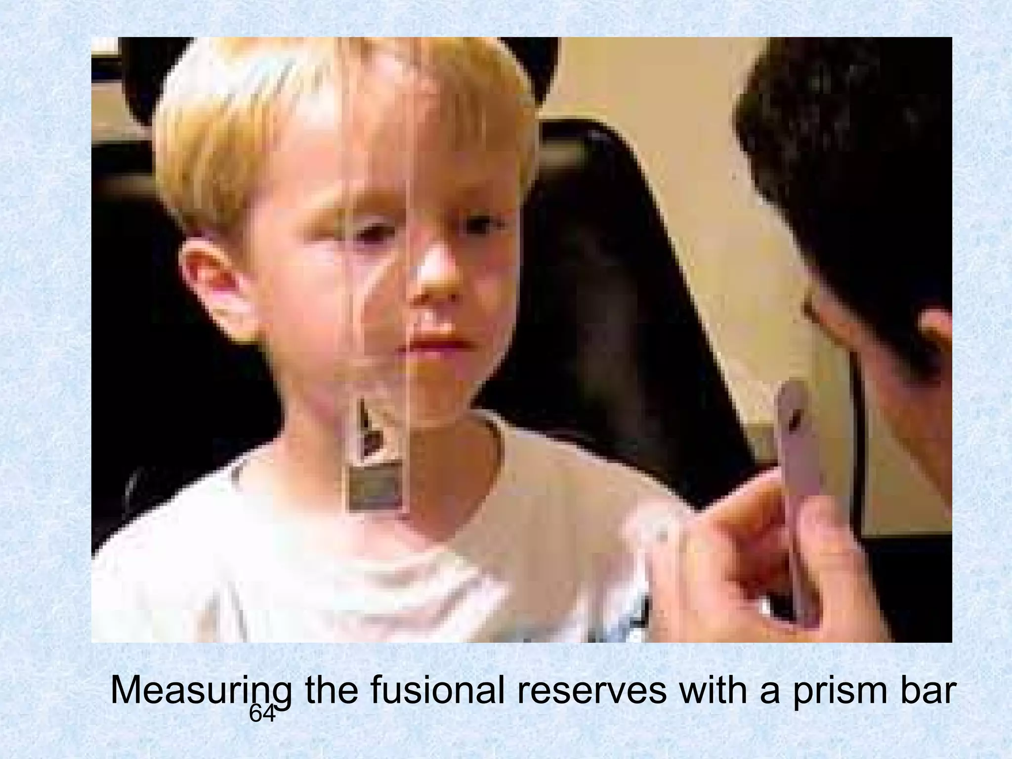
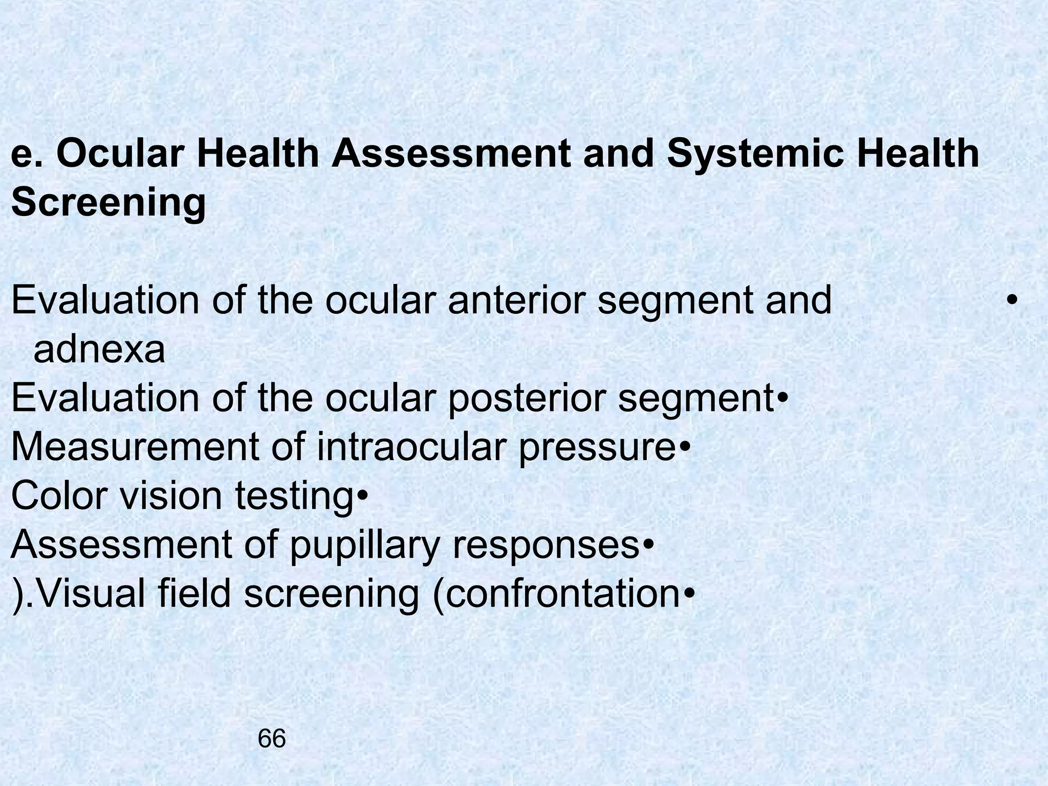
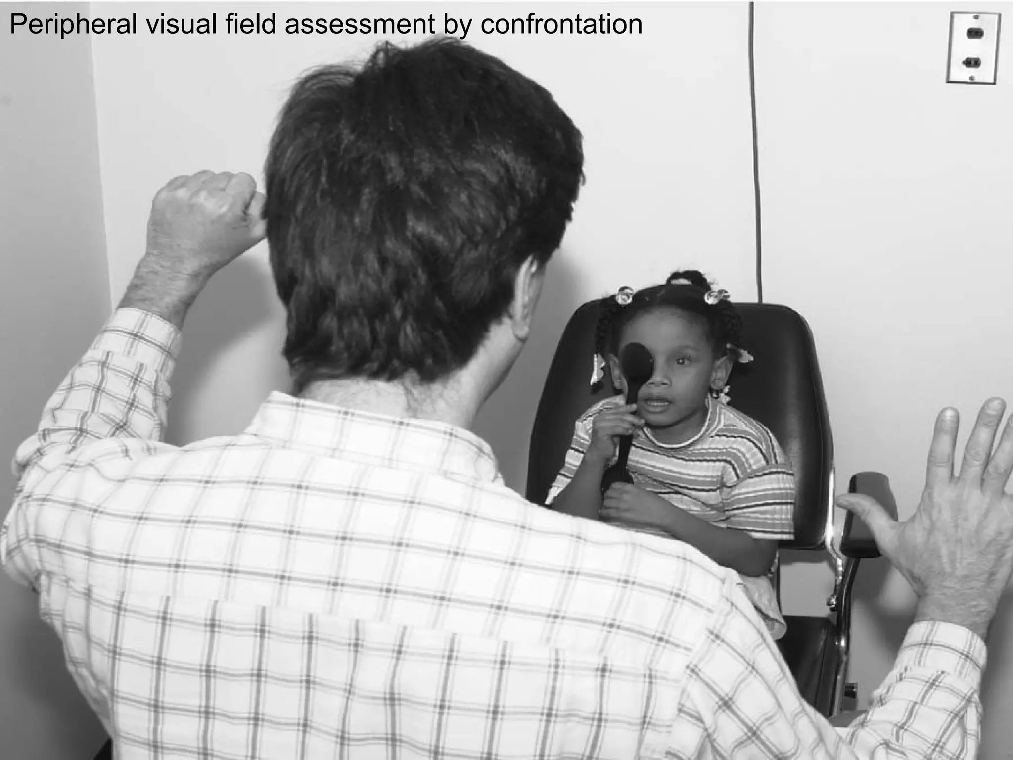
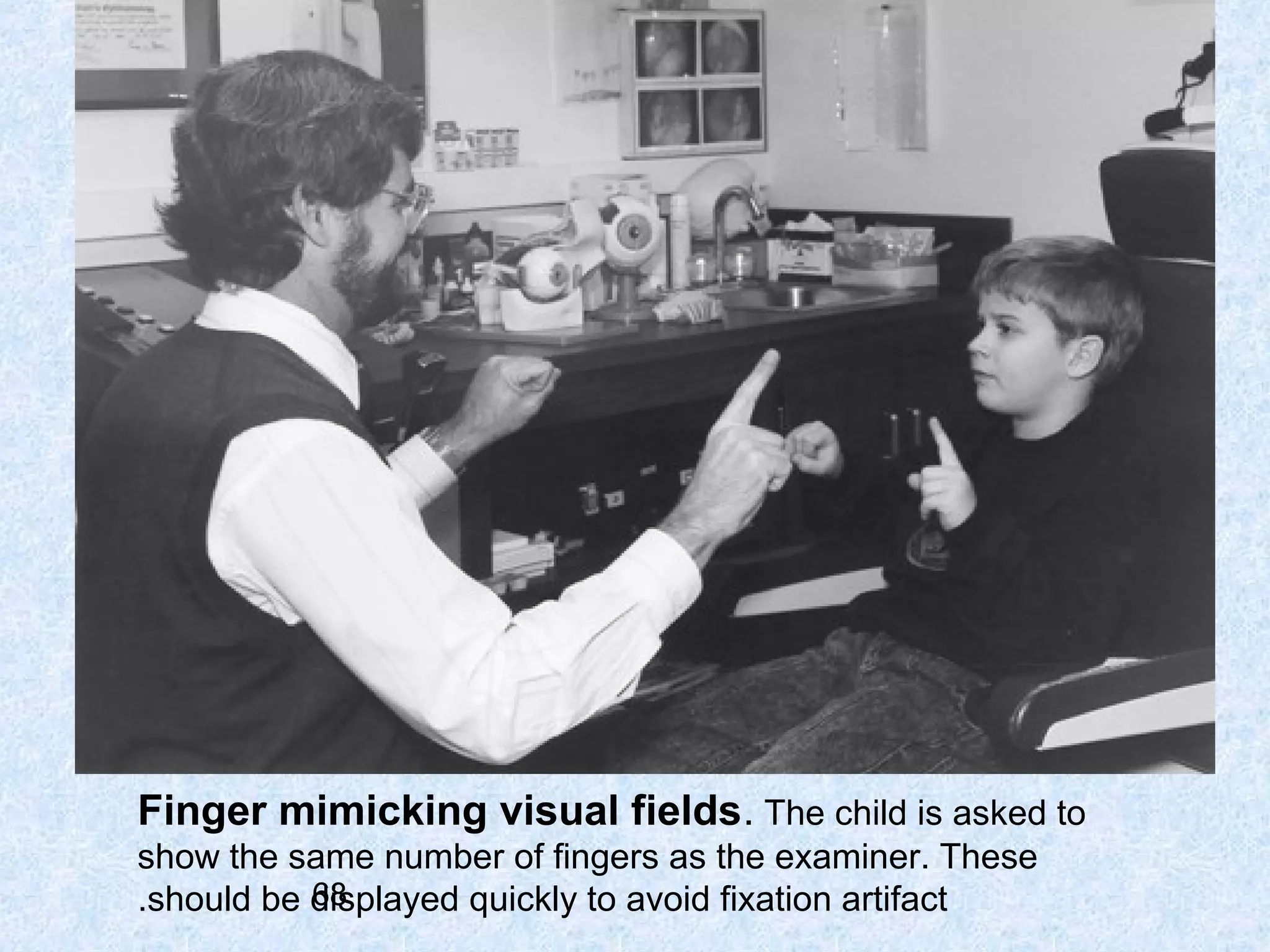
1
Pediatric Eye And Vision
Examination
‫شکرا شفائی کارشناس ارشد‬
‫اپتومتری‬

2
Pediatric population can be divided into three
(subcategories: (Press LJ, Moore BD.1993
(Infants

and toddlers (birth to 2 years, 11 months ■

(Preschool

children (3 years to 5 years, 11 months ■

(.School-age

3

children (6 to 18 years ■
4
Recommended Eye Examination Frequency for the
Pediatric Patient (OPTOMETRIC CLINICAL PRACTICE GUIDELINE ; Mitchell M.
(Scheiman, 2002

Patient Age Asymptomatic/ At-risk

At-risk

Patient Age risk-free

Birth to 24
months

At 6 months of age

At 6 months of age or as
recommended

.to 5 yrs 2

At 3 years of age

At 3 years of age
or as recommended

.to 18 yrs 6

Before first grade
and every 2 years
thereafter

Annually or as
recommended

5
A. Examination of Infants and Toddlers
General Considerations. 1
time of examination; morning visit types of examination; objective fast but exact; age appropriate -

Early Detection and Prevention. 2
Examination Sequence. 3
a. Patient History
Nature of the presenting problem, including chief complaint■
Visual and ocular history■
General health history, including prenatal, perinatal, and postnatal
history and review of systems
Family eye and medical histories■
.Developmental history of the child■
6

■
b. Visual Acuity
Assessment of visual acuity for infants and
:toddlers may include these procedures
Gross estimate of visual function at near•
Fixation maintenance & preference tests•
prism diopter (PD) lens 10-

OKN•
Preferential looking visual acuity test•
VEP•

7
(.pincer grasp to pick up cake decoration ( or raisin
(Candy

beads at 8 cm = 20/285 ( Frenkel & Evans, 1980
40
.lights on

lights turned down

Note marked retraction of the upper lids. indicating some
9
.visual function
Fixation maintenance test
To test central & steady fixation■
Shine a light at uncovered eye■
Estimate the position of the light reflexes (or angle Kappa(;
displaced +0.5 mm nasally
Any deviation = eccentric fixation■

10

■
Fixation preference test
can be judged when obvious strabismus is present■
cover and uncover the fixing eye to force fixation to the
nonpreferred eye
observe the fixation pattern ■
good vision in case of alternate fixation 11

■
Teller cards
pack of 16 cardsfrom 38.0 to 0.32 cycles/cmtest dist. 38 cm for infants , 55 cmfor toddlers

12
Spatial frequency paddles
)calibrated for a specific distance( 1 m-

.paddles and 6 different sp. fr 4. hold the gray over one of sp. frseparate the paddles and notice the fixation 13
Preferential looking tests of vision
Keeler/Teller cards for infants

14
15
Normal levels of vision
(Preferential

looking tests

(Mayer et al, 1995

month

24/400 – 20/1600 1

month

20/100 – 20/400 4

month

20/80 – 20/300 12
16
Vertical optokinetic nystagmus testing using an OKN
drum. If a vertical nystagmus can be elicited, vision is
.20/400 or better
17
vestibular-ocular reflex, which stimulates the semicircular
.canals, causing the eyes to deviate in the direction of rotation
18
The early decreased visual acuity in the infant is due
:to
foveal cone immaturities; small and stumpy- 1
at 4 years, the length of an adult cone at 3.5 – 4 years, adult level of cone density cortical immaturities- 2
incomplete myelination of the optic pathways- 3

19
Cardiff acuity cards for toddlers; vanishing optotype
in children from 1 to 3 years of age■
sets of cards are available ; 20/400 to 20/20 11 ■
test dis. 0.5 m and 1.0 m ■
present one set of cards and notice the fixation ■
(better done binocularly first( Adoh and Woodhouse,1994 ■
20
(

Cardiff acuity test (Monocular; Adoh & Woodhouse 1994

+0.4 to +0.8 L.MAR

months(20/50-20/120) 12-18

+0.1 to +0.7 L.MAR

months( 20/25- 20/100) 24- 18

+0.1 to +0.5

L.MAR

months( 20/25-20/60) 24-30

+0.0 to +0.3

LMAR

months(20/20-20/40) 30-36

21
c. Refraction
Traditional subjective procedures , ineffective with infants or
toddlers because of short attention span and poor fixation. (Ciner EB,
(1990

:The two most commonly used procedures are
Cycloplegic retinoscopy■
Near retinoscopy ■

22
(Take

several precautions: (Gray L. 1979

Select the cycloplegic agent carefully (e.g., an increased •
response to drugs in fair-skinned children with blue eyes and more frequent or
(.stronger dosages in darkly pigmented children

Avoid overdosage (e.g., children with Down syndrome, cerebral palsy,

•

trisomy 13 and 18, and other central nervous system disorders in whom there
may be an increased reaction to cycloplegic agents, 1% tropicamide may be
(.used

Be aware of biologic variations in children (e.g., low weight •
23
(.infants may require a modified dosage
Cyclopentolate hydrochloride , the cycloplegic
agent of choice
, One drop , twice, 5 minutes apart, in each eye
for children from birth to 1 year 0.5% (for older children. (Amos JF, 2001 1% Spray administration of the drug , a viable alternative.

(Amos JF,

)2001
The child has less of an avoidance response) 1(
A single application can achieve both cycloplegia and pupillary dilation ) 2(
.a mixture of 0.5% cyclopentolate, 0.5% tropicamide, and 2.5% phenylephrine is used) 3(

Retinoscopy may be performed 20-30 minutes after instillation.
((Bartlett JD, 1993
24
Near retinoscopy
:May have some clinical value in the following situations

When frequent followup is necessary •
When the child is extremely anxious about instillation
of cycloplegic agents
When the child has had or is at risk for an adverse
25
. reaction to cyclopentolate or tropicamide

•
•
Near retinoscopy , an alternative to cycloplegic refraction in ■
(. children
and infants (Mohindra 1977
A dim retinoscope light is used as a fixation target and seen in
complete darkness and perform retinoscopy using a lens rack or
. individual trial case lenses

■

Working at 50 cm, -1.25 DS rather than the standard -2.00 DS is
(.
added to the final retinoscopy result .(Mohindra 1977

■

The test is comparable to cycloplegic retinoscopy (e.g. Saunders &

■

(.

Westall1992

Add - 0.75 DS for infants (2 years( ; -1.00 DS for children (over 2
( years( rather than the original 1.25 DS. (Saunders & Westall (1992
26

■
REFLEX SCANING
,To get a quick idea of the prescription in both eyes
(Lynne Speedwell, 2007(

hold a pair of +2.00DS trial lensesin front of the child’s eyes in a
.darkened room
With the child fixing on the,retinoscope light at 50 cm
quickly move the retinoscope
horizontally and then vertically
.across both eyes

27
Refractive Error
Green line – cycloplegic
refraction on infants (0)6mths

80
70

Frequency (%)

60

Blue line – non-cycloplegic
refraction on infants (0)6mths

50
40
30

Red line – Refraction in older
children

20
10
0
-6

-4

-2

0

2

4

6

Spherical Equivalent (Dioptres)
28

8

Adapted from
Gwiazda et al 1993
d. Binocular Vision and Ocular Motility
Observation•
Hirschberg test•
Krimsky test•
Brückner test•
Cover test•

29
30
General observation- 1
size of head■
IPD ■
nose bridge ■
lid asymmetries ■
pupil or orbit position ■
epicanthal folds ■
31

lid asymmetries
Hirschberg test- 2
fixate your penlight at 50 cm ■
(mm ( nasal displacement of reflex 0.5+
note any asymmetric displacement ■
(

1mm = 22 pd( Eskridge et al, 1988 ■

32

■
Krimsky test- 3
A( The light reflection in the(
deviated right eye is
temporal to the pupillary
. center

B( Using a prism, the(
Krimsky test measures the
amplitude of the esotropia
by centering the light
.reflection in the right pupil
33
Brückner test- 4
fixate an ophthalmoscope light at one meter ■
illuminate both eyes at the same time ■
observe the relative whiteness & brightness of ■
each pupillary reflex
strabismus in case of whiter & brighter reflex ■
it can help in the diagnosis of small angle
) strabismus (Miller et al. 1995
34

■
Cover test- 5
Targets; brightly colored with sound & fine detail Use your thumb to avoiddistraction
For distance, moving targets-

35
Binocular vision testing
intermittent esotropia should stop by the age of two months ■
(.■ exotropia by six months (Sondhi et al, 1988

There are three types of prism test that are useful on young
:children
The 20Δ base out prism test- 1
A 20Δ base in lens- 2
A 10Δ base down- 3
36
The 20Δ base OUT prism test- 1
Assesses motor fusion and gross binocular function , not ideal ■
. for amblyopia assessment
The child fixes a toy and a 20Δ base OUT lens is placed in front ■
. of one eye
Both eyes should move in the direction of the prism apex and ■
then the eye not being covered by the prism should be seen to
. refixate to the centre
If no refixation movement is seen with one eye, it is likely that
there is only weak fusion, suggesting one eye has poorer
. vision
37

■
A

B

.The 20 Δ base out test
A- fixation toy is used as a target and corrective eye
B - movements are observed in response to a 20
. diopters base out prism challenge
38
A 20Δ base IN lens. 2
will assess fixation preference and will therefore ■
.demonstrate amblyopia
The prism is held in front of first one eye then the
. other, whilst the child fixes a light

■

The eye underneath the prism should move
(. outwards (in the direction of the prism apex

■

If the eye under the prism is amblyopic, it will ■
. not move when the prism is placed in front of it
39
A

A 20Δ base IN lens test
40

B
10Δ base DOWN. 3
In front of each eye in turnThe upward vertical movement is easy for the .observer to see

41
e. Ocular Health Assessment and Systemic Health
Screening
:An evaluation of ocular health may include

Evaluation of the ocular anterior segment and adnexa •
Evaluation of the ocular posterior segment •
Assessment of pupillary responses•
(.Visual field screening (confrontation•
42
Testing visual fields
in a young child.
Begin by attracting
the child’s attention
(. straight ahead (top

Then move an object
in from the side. A
head movement to the
side of the target
(bottom( indicates
.intact peripheral field
43
B. Examination of Preschool
Children

44
b. Visual Acuity
Lea Symbols chart•
Broken Wheel acuity cards•
.HOTV test•
Allen chart•

45
The LH or Lea Cards
make use of a logMAR scoring
system and produce accurate
results
available in linear and single■
optotype formats and use shapes
instead of letters
at a dist. of 10 ft to 20 ft■

■

Lea optotypes

46

'A Lea symbol presented in a 'crowded box
Broken Wheel acuity cards
(best

for testing 3 year olds( McDonald and Chaudry, 1989a set of 7 matched pairs of cards.to 20/100, held at 10 ft 20/20should point to the car with broken wheelsif the child gets47 of 4, go to the smaller sets4
Kay picture cards
Having vision checked using
the crowded 3 meter LogMAR

48
HOTV test
.for children , 21/2 to 5 yrs. of ageno verbal responsejust point to the appropriate letter .at a dist. of 3 m-

49
The Allen Preschool Vision Test

ALLEN CARDS
optotypes for 2.5 to 5 yrs. of age 720/200 ,20/100 ,20/50,20/70 ,20/40 , 20/30 ask the child50 call off the name of the pictures he seeto
Expected levels of vision
Age

Vision

Neonate

6/300

month

6/200 – 6/90 1

months

6/60 – 6/36 3

months

6/60 – 6/36 6

months

6/36 – 6/24 9

year

6/24 – 6/12 1

years

6/12 – 6/9 2

years

51

6/9 – 6/6 3
c. Refraction
Static retinoscopy•
.Cycloplegic retinoscopy•

52
Cyclopentolate (1%( is the cycloplegic agent of choice. Two drops
should be instilled, one at a time, 5 minutes apart, in each eye .
((Bartlett JD , 2001

The use of a spray bottle to administer the drug is also effective
.for this age group
Retinoscopy may be performed with a lens rack or loose lenses
(20-30 minutes after instillation.(Bartlett JD, 1993

53
d. Binocular Vision, Accommodation and Ocular
Motility
Cover test•
Positive and negative fusional vergences (prism
( bar/step vergence testing
(Near point of convergence (NPC•
(Stereopsis (Lang stereotest , Frisby tests•
Monocular estimation method (MEM( retinoscopy •
.Versions•

54

•
e. Ocular Health Assessment and Systemic Health
Screening

Evaluation of the ocular anterior segment and adnexa •
Evaluation of the ocular posterior segment •
Color vision testing•
Assessment of pupillary responses•
(.Visual field screening (confrontation•

55
C. Examination of School-Age
Children

56
b. Visual Acuity
LogMAR chart

57
c. Refraction
Static (distance( retinoscopy•
Cycloplegic retinoscopy•
.Subjective refraction•

58
, d. Binocular Vision, Accommodation
and Ocular Motility
Cover test•
(Near point of convergence (NPC•
Positive and negative fusional vergences, AC/A•
Accommodative amplitude and facility,NRA,NPA•
Monocular estimation method (MEM( retinoscopy•
(Stereopsis (random dot stereopsis test•
Versions•
assessment of stability of fixation , saccadic function pursuit function 59

BV equipment
Cover-uncover test

60
Alternate cover test

61
NPC assessment
The near point of convergence (NPC(
is performed with a 6/9 target on a
fixation stick

62

Fusional reserves assessment
The negative/positive relative accommodation
NVA/PRA( is done in the phoropter with a 6/9 target (
at 40cm 63
Measuring the fusional reserves with a prism bar
64
The monocular/binocular accommodative flippers
(MAF/BAF(65 performed with ±2D flippers and a 6/9
is
target on a fixation stick held at 40cm
e. Ocular Health Assessment and Systemic Health
Screening
Evaluation of the ocular anterior segment and
adnexa
Evaluation of the ocular posterior segment •
Measurement of intraocular pressure•
Color vision testing•
Assessment of pupillary responses•
(.Visual field screening (confrontation•

66

•
Peripheral visual field assessment by confrontation

67
Finger mimicking visual fields. The child is asked to
show the same number of fingers as the examiner. These
68
.should be displayed quickly to avoid fixation artifact
Thank you

69

Pediatric eye and vision

  • 1.
  • 2.
    Pediatric Eye AndVision Examination ‫شکرا شفائی کارشناس ارشد‬ ‫اپتومتری‬ 2
  • 3.
    Pediatric population canbe divided into three (subcategories: (Press LJ, Moore BD.1993 (Infants and toddlers (birth to 2 years, 11 months ■ (Preschool children (3 years to 5 years, 11 months ■ (.School-age 3 children (6 to 18 years ■
  • 4.
  • 5.
    Recommended Eye ExaminationFrequency for the Pediatric Patient (OPTOMETRIC CLINICAL PRACTICE GUIDELINE ; Mitchell M. (Scheiman, 2002 Patient Age Asymptomatic/ At-risk At-risk Patient Age risk-free Birth to 24 months At 6 months of age At 6 months of age or as recommended .to 5 yrs 2 At 3 years of age At 3 years of age or as recommended .to 18 yrs 6 Before first grade and every 2 years thereafter Annually or as recommended 5
  • 6.
    A. Examination ofInfants and Toddlers General Considerations. 1 time of examination; morning visit types of examination; objective fast but exact; age appropriate - Early Detection and Prevention. 2 Examination Sequence. 3 a. Patient History Nature of the presenting problem, including chief complaint■ Visual and ocular history■ General health history, including prenatal, perinatal, and postnatal history and review of systems Family eye and medical histories■ .Developmental history of the child■ 6 ■
  • 7.
    b. Visual Acuity Assessmentof visual acuity for infants and :toddlers may include these procedures Gross estimate of visual function at near• Fixation maintenance & preference tests• prism diopter (PD) lens 10- OKN• Preferential looking visual acuity test• VEP• 7
  • 8.
    (.pincer grasp topick up cake decoration ( or raisin (Candy beads at 8 cm = 20/285 ( Frenkel & Evans, 1980 40
  • 9.
    .lights on lights turneddown Note marked retraction of the upper lids. indicating some 9 .visual function
  • 10.
    Fixation maintenance test Totest central & steady fixation■ Shine a light at uncovered eye■ Estimate the position of the light reflexes (or angle Kappa(; displaced +0.5 mm nasally Any deviation = eccentric fixation■ 10 ■
  • 11.
    Fixation preference test canbe judged when obvious strabismus is present■ cover and uncover the fixing eye to force fixation to the nonpreferred eye observe the fixation pattern ■ good vision in case of alternate fixation 11 ■
  • 12.
    Teller cards pack of16 cardsfrom 38.0 to 0.32 cycles/cmtest dist. 38 cm for infants , 55 cmfor toddlers 12
  • 13.
    Spatial frequency paddles )calibratedfor a specific distance( 1 m- .paddles and 6 different sp. fr 4. hold the gray over one of sp. frseparate the paddles and notice the fixation 13
  • 14.
    Preferential looking testsof vision Keeler/Teller cards for infants 14
  • 15.
  • 16.
    Normal levels ofvision (Preferential looking tests (Mayer et al, 1995 month 24/400 – 20/1600 1 month 20/100 – 20/400 4 month 20/80 – 20/300 12 16
  • 17.
    Vertical optokinetic nystagmustesting using an OKN drum. If a vertical nystagmus can be elicited, vision is .20/400 or better 17
  • 18.
    vestibular-ocular reflex, whichstimulates the semicircular .canals, causing the eyes to deviate in the direction of rotation 18
  • 19.
    The early decreasedvisual acuity in the infant is due :to foveal cone immaturities; small and stumpy- 1 at 4 years, the length of an adult cone at 3.5 – 4 years, adult level of cone density cortical immaturities- 2 incomplete myelination of the optic pathways- 3 19
  • 20.
    Cardiff acuity cardsfor toddlers; vanishing optotype in children from 1 to 3 years of age■ sets of cards are available ; 20/400 to 20/20 11 ■ test dis. 0.5 m and 1.0 m ■ present one set of cards and notice the fixation ■ (better done binocularly first( Adoh and Woodhouse,1994 ■ 20
  • 21.
    ( Cardiff acuity test(Monocular; Adoh & Woodhouse 1994 +0.4 to +0.8 L.MAR months(20/50-20/120) 12-18 +0.1 to +0.7 L.MAR months( 20/25- 20/100) 24- 18 +0.1 to +0.5 L.MAR months( 20/25-20/60) 24-30 +0.0 to +0.3 LMAR months(20/20-20/40) 30-36 21
  • 22.
    c. Refraction Traditional subjectiveprocedures , ineffective with infants or toddlers because of short attention span and poor fixation. (Ciner EB, (1990 :The two most commonly used procedures are Cycloplegic retinoscopy■ Near retinoscopy ■ 22
  • 23.
    (Take several precautions: (GrayL. 1979 Select the cycloplegic agent carefully (e.g., an increased • response to drugs in fair-skinned children with blue eyes and more frequent or (.stronger dosages in darkly pigmented children Avoid overdosage (e.g., children with Down syndrome, cerebral palsy, • trisomy 13 and 18, and other central nervous system disorders in whom there may be an increased reaction to cycloplegic agents, 1% tropicamide may be (.used Be aware of biologic variations in children (e.g., low weight • 23 (.infants may require a modified dosage
  • 24.
    Cyclopentolate hydrochloride ,the cycloplegic agent of choice , One drop , twice, 5 minutes apart, in each eye for children from birth to 1 year 0.5% (for older children. (Amos JF, 2001 1% Spray administration of the drug , a viable alternative. (Amos JF, )2001 The child has less of an avoidance response) 1( A single application can achieve both cycloplegia and pupillary dilation ) 2( .a mixture of 0.5% cyclopentolate, 0.5% tropicamide, and 2.5% phenylephrine is used) 3( Retinoscopy may be performed 20-30 minutes after instillation. ((Bartlett JD, 1993 24
  • 25.
    Near retinoscopy :May havesome clinical value in the following situations When frequent followup is necessary • When the child is extremely anxious about instillation of cycloplegic agents When the child has had or is at risk for an adverse 25 . reaction to cyclopentolate or tropicamide • •
  • 26.
    Near retinoscopy ,an alternative to cycloplegic refraction in ■ (. children and infants (Mohindra 1977 A dim retinoscope light is used as a fixation target and seen in complete darkness and perform retinoscopy using a lens rack or . individual trial case lenses ■ Working at 50 cm, -1.25 DS rather than the standard -2.00 DS is (. added to the final retinoscopy result .(Mohindra 1977 ■ The test is comparable to cycloplegic retinoscopy (e.g. Saunders & ■ (. Westall1992 Add - 0.75 DS for infants (2 years( ; -1.00 DS for children (over 2 ( years( rather than the original 1.25 DS. (Saunders & Westall (1992 26 ■
  • 27.
    REFLEX SCANING ,To geta quick idea of the prescription in both eyes (Lynne Speedwell, 2007( hold a pair of +2.00DS trial lensesin front of the child’s eyes in a .darkened room With the child fixing on the,retinoscope light at 50 cm quickly move the retinoscope horizontally and then vertically .across both eyes 27
  • 28.
    Refractive Error Green line– cycloplegic refraction on infants (0)6mths 80 70 Frequency (%) 60 Blue line – non-cycloplegic refraction on infants (0)6mths 50 40 30 Red line – Refraction in older children 20 10 0 -6 -4 -2 0 2 4 6 Spherical Equivalent (Dioptres) 28 8 Adapted from Gwiazda et al 1993
  • 29.
    d. Binocular Visionand Ocular Motility Observation• Hirschberg test• Krimsky test• Brückner test• Cover test• 29
  • 30.
  • 31.
    General observation- 1 sizeof head■ IPD ■ nose bridge ■ lid asymmetries ■ pupil or orbit position ■ epicanthal folds ■ 31 lid asymmetries
  • 32.
    Hirschberg test- 2 fixateyour penlight at 50 cm ■ (mm ( nasal displacement of reflex 0.5+ note any asymmetric displacement ■ ( 1mm = 22 pd( Eskridge et al, 1988 ■ 32 ■
  • 33.
    Krimsky test- 3 A(The light reflection in the( deviated right eye is temporal to the pupillary . center B( Using a prism, the( Krimsky test measures the amplitude of the esotropia by centering the light .reflection in the right pupil 33
  • 34.
    Brückner test- 4 fixatean ophthalmoscope light at one meter ■ illuminate both eyes at the same time ■ observe the relative whiteness & brightness of ■ each pupillary reflex strabismus in case of whiter & brighter reflex ■ it can help in the diagnosis of small angle ) strabismus (Miller et al. 1995 34 ■
  • 35.
    Cover test- 5 Targets;brightly colored with sound & fine detail Use your thumb to avoiddistraction For distance, moving targets- 35
  • 36.
    Binocular vision testing intermittentesotropia should stop by the age of two months ■ (.■ exotropia by six months (Sondhi et al, 1988 There are three types of prism test that are useful on young :children The 20Δ base out prism test- 1 A 20Δ base in lens- 2 A 10Δ base down- 3 36
  • 37.
    The 20Δ baseOUT prism test- 1 Assesses motor fusion and gross binocular function , not ideal ■ . for amblyopia assessment The child fixes a toy and a 20Δ base OUT lens is placed in front ■ . of one eye Both eyes should move in the direction of the prism apex and ■ then the eye not being covered by the prism should be seen to . refixate to the centre If no refixation movement is seen with one eye, it is likely that there is only weak fusion, suggesting one eye has poorer . vision 37 ■
  • 38.
    A B .The 20 Δbase out test A- fixation toy is used as a target and corrective eye B - movements are observed in response to a 20 . diopters base out prism challenge 38
  • 39.
    A 20Δ baseIN lens. 2 will assess fixation preference and will therefore ■ .demonstrate amblyopia The prism is held in front of first one eye then the . other, whilst the child fixes a light ■ The eye underneath the prism should move (. outwards (in the direction of the prism apex ■ If the eye under the prism is amblyopic, it will ■ . not move when the prism is placed in front of it 39
  • 40.
    A A 20Δ baseIN lens test 40 B
  • 41.
    10Δ base DOWN.3 In front of each eye in turnThe upward vertical movement is easy for the .observer to see 41
  • 42.
    e. Ocular HealthAssessment and Systemic Health Screening :An evaluation of ocular health may include Evaluation of the ocular anterior segment and adnexa • Evaluation of the ocular posterior segment • Assessment of pupillary responses• (.Visual field screening (confrontation• 42
  • 43.
    Testing visual fields ina young child. Begin by attracting the child’s attention (. straight ahead (top Then move an object in from the side. A head movement to the side of the target (bottom( indicates .intact peripheral field 43
  • 44.
    B. Examination ofPreschool Children 44
  • 45.
    b. Visual Acuity LeaSymbols chart• Broken Wheel acuity cards• .HOTV test• Allen chart• 45
  • 46.
    The LH orLea Cards make use of a logMAR scoring system and produce accurate results available in linear and single■ optotype formats and use shapes instead of letters at a dist. of 10 ft to 20 ft■ ■ Lea optotypes 46 'A Lea symbol presented in a 'crowded box
  • 47.
    Broken Wheel acuitycards (best for testing 3 year olds( McDonald and Chaudry, 1989a set of 7 matched pairs of cards.to 20/100, held at 10 ft 20/20should point to the car with broken wheelsif the child gets47 of 4, go to the smaller sets4
  • 48.
    Kay picture cards Havingvision checked using the crowded 3 meter LogMAR 48
  • 49.
    HOTV test .for children, 21/2 to 5 yrs. of ageno verbal responsejust point to the appropriate letter .at a dist. of 3 m- 49
  • 50.
    The Allen PreschoolVision Test ALLEN CARDS optotypes for 2.5 to 5 yrs. of age 720/200 ,20/100 ,20/50,20/70 ,20/40 , 20/30 ask the child50 call off the name of the pictures he seeto
  • 51.
    Expected levels ofvision Age Vision Neonate 6/300 month 6/200 – 6/90 1 months 6/60 – 6/36 3 months 6/60 – 6/36 6 months 6/36 – 6/24 9 year 6/24 – 6/12 1 years 6/12 – 6/9 2 years 51 6/9 – 6/6 3
  • 52.
  • 53.
    Cyclopentolate (1%( isthe cycloplegic agent of choice. Two drops should be instilled, one at a time, 5 minutes apart, in each eye . ((Bartlett JD , 2001 The use of a spray bottle to administer the drug is also effective .for this age group Retinoscopy may be performed with a lens rack or loose lenses (20-30 minutes after instillation.(Bartlett JD, 1993 53
  • 54.
    d. Binocular Vision,Accommodation and Ocular Motility Cover test• Positive and negative fusional vergences (prism ( bar/step vergence testing (Near point of convergence (NPC• (Stereopsis (Lang stereotest , Frisby tests• Monocular estimation method (MEM( retinoscopy • .Versions• 54 •
  • 55.
    e. Ocular HealthAssessment and Systemic Health Screening Evaluation of the ocular anterior segment and adnexa • Evaluation of the ocular posterior segment • Color vision testing• Assessment of pupillary responses• (.Visual field screening (confrontation• 55
  • 56.
    C. Examination ofSchool-Age Children 56
  • 57.
  • 58.
    c. Refraction Static (distance(retinoscopy• Cycloplegic retinoscopy• .Subjective refraction• 58
  • 59.
    , d. BinocularVision, Accommodation and Ocular Motility Cover test• (Near point of convergence (NPC• Positive and negative fusional vergences, AC/A• Accommodative amplitude and facility,NRA,NPA• Monocular estimation method (MEM( retinoscopy• (Stereopsis (random dot stereopsis test• Versions• assessment of stability of fixation , saccadic function pursuit function 59 BV equipment
  • 60.
  • 61.
  • 62.
    NPC assessment The nearpoint of convergence (NPC( is performed with a 6/9 target on a fixation stick 62 Fusional reserves assessment
  • 63.
    The negative/positive relativeaccommodation NVA/PRA( is done in the phoropter with a 6/9 target ( at 40cm 63
  • 64.
    Measuring the fusionalreserves with a prism bar 64
  • 65.
    The monocular/binocular accommodativeflippers (MAF/BAF(65 performed with ±2D flippers and a 6/9 is target on a fixation stick held at 40cm
  • 66.
    e. Ocular HealthAssessment and Systemic Health Screening Evaluation of the ocular anterior segment and adnexa Evaluation of the ocular posterior segment • Measurement of intraocular pressure• Color vision testing• Assessment of pupillary responses• (.Visual field screening (confrontation• 66 •
  • 67.
    Peripheral visual fieldassessment by confrontation 67
  • 68.
    Finger mimicking visualfields. The child is asked to show the same number of fingers as the examiner. These 68 .should be displayed quickly to avoid fixation artifact
  • 69.